The Effects of Traumatic Brain Injury in the Presence of Aß Pathology

Post by Negar Mazloum-Farzaghi

The takeaway

There is a link between traumatic brain injury and the development of dementia. The double burden of traumatic brain injury and Aß deposits in the aging brain can cause alterations in cellular processes that lead to neuron death.

What's the science?

Previous research has established an association between traumatic brain injury (TBI) sustained in early adulthood and the risk of developing dementia later in life. It is less clear whether TBI sustained as an elderly individual (geriatric TBI) increases the risk of developing dementia, and what the cellular mechanisms would be involved. The aging brain’s response to TBI may be influenced by several factors, such as increased inflammation, increased amyloid beta load (Aß; a hallmark of dementia), and decreased autophagic activity (important process for degrading unnecessary or dysfunctional intracellular macromolecules). This week in Scientific reports, Streubel-Gallasch and colleagues used an in vitro model, designed to simulate the conditions of the aging brain, to investigate cellular responses to TBI in the presence of Aß pathology.

How did they do it?

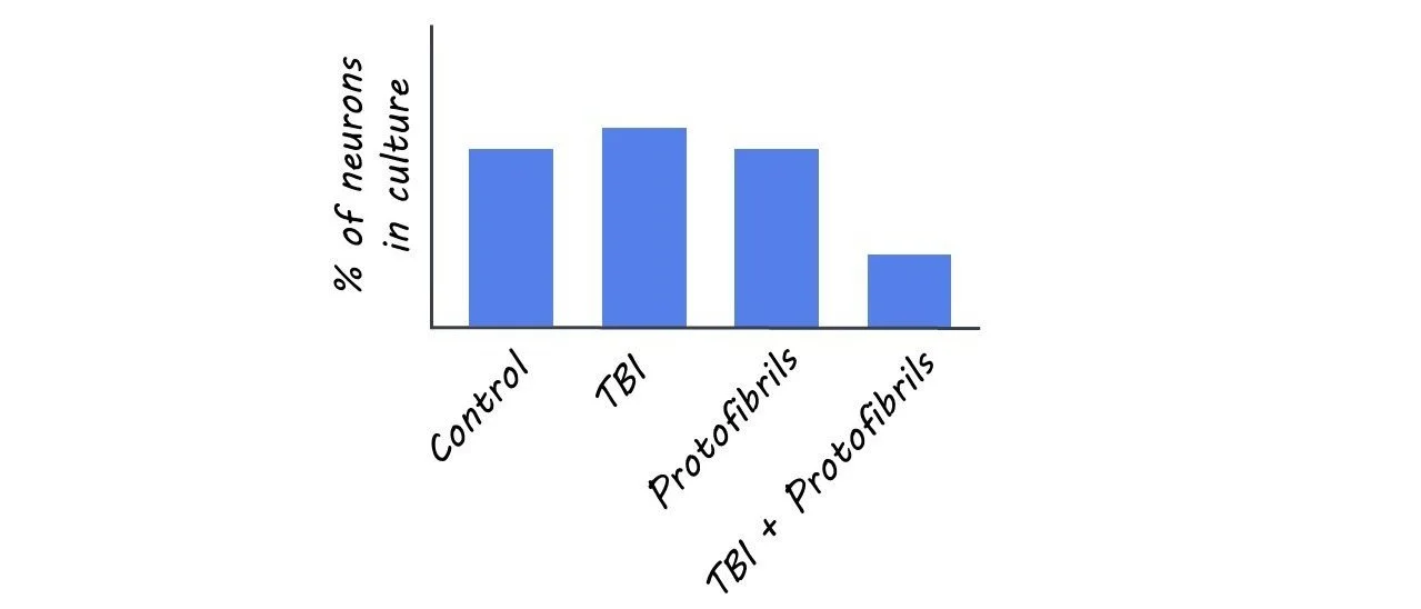
In order to investigate the effects of physical injury caused by TBI in the presence of Aß deposits, the authors exposed co-cultures of astrocytes (specialized glial cells fundamental to maintaining homeostasis and cellular function) and neurons to Aß protofibrils. After the exposure, they subjected the cultures to a “scratch injury” using a scalpel in order to mimic TBI. There were four experimental groups: untreated (control), scratch injury (TBI), Aß protofibril exposure, and the combination of both TBI and Aß.

The authors performed immunocytochemistry to examine the effect of TBI and Aß protofibril exposure on neuronal survival. They employed the same method to examine how TBI and Aß protofibril exposure affects astrocytes. To do this, they used two well characterized astrocytic markers, Glial fibrillary acidic protein (GFAP) and calcium-binding protein (S100ß). They used transmission electron microscopy to further assess changes to cellular homeostasis caused by TBI and Aß protofibril exposure by examining the function of the mitochondrial network of astrocytes. Finally, using both immunocytochemistry and immunoblotting, they also examined whether autophagic activity was impaired in response to TBI and Aß protofibril exposure.

What did they find?

The number of neurons in the experimental TBI group, Aß protofibril-exposure group, and control group remained stable. However, in the experimental group with the combination of Aß and TBI, the percentage of neurons decreased significantly. One explanation for this finding may be that the double burden of Aß deposits and TBI hindered astrocytic functions, which threw their ability to maintain homeostasis off balance, making it more difficult to clear debris and protect neurons.

Aß protofibril-exposed astrocytes displayed higher GFAP levels. In contrast, astrocytes subjected to both Aß and TBI displayed lower GFAP levels. This was a surprising finding as GFAP usually upregulates in response to TBI. The other astrocytic protein, S100ß, showed increased levels in astrocytes in all three experimental groups compared to the control group. This is in line with previous work that has shown that S100ß upregulates in response to trauma and has increased levels in the tissue cells and astrocytes of dementia patients. However, it remains to be determined whether these increased levels have positive or negative effects in pathological conditions.

Aß pathology and TBI caused mitochondrial damage in astrocytes (but no clear additive effect of the two), which may have negative consequences for vital astrocytic functions thereby disturbing homeostasis. There was an increase in autophagic activity in the Aß protofibrils exposure group and TBI group. Interestingly, when cells were exposed to a combination of Aß protofibrils and TBI, autophagy failed to upregulate, which may be indicative of an inefficient compensatory mechanism when faced with this double burden.

What's the impact?

This is the first study to show that the double burden of Aß deposits and TBI result in neuronal loss, altered astrocytic responses as indicated by changes in the expression of two key astrocytic proteins, mitochondrial disturbances, and abnormal autophagic activity. The findings of this study demonstrate the underlying cellular mechanisms involved in the relationship between TBI and the development of dementia. Overall, this study advances our understanding of geriatric TBI on the cellular level.

Deep Brain Stimulation as an Individualized Treatment for Depression

Post by Leanna Kalinowski

The takeaway

Depression is a psychiatric disorder that affects individuals differently, leading to difficulty in developing universal treatments. Researchers have successfully tested a new therapeutic strategy that uses individualized brain activity to guide treatment for depression with deep brain stimulation.

What's the science?

Deep brain stimulation (DBS) is a surgical procedure in which electrodes are implanted into the brain to deliver therapeutic electrical stimulation. It has shown success in treating movement disorders (e.g., Parkinson’s disease), which has led to a great interest in applying DBS to psychiatric disorders like treatment-resistant depression (i.e. depression that does not respond to medication). While preliminary studies on the effectiveness of DBS for treating depression showed promise, clinical trials were not as successful. This is likely due to depression being a highly heterogeneous disorder, highlighting a need for an individualized understanding of the specific brain regions contributing to a patient’s depressive symptoms. This week in Biological Psychiatry, Sheth and colleagues used intracranial recordings coupled with DBS to develop an individualized treatment strategy for depression.

How did they do it?

The researchers first surgically implanted two sets of electrodes into the brain: (1) DBS electrodes, to therapeutically stimulate the brain, and (2) stereo electroencephalography (sEEG) electrodes, to measure brain activity. The brain surgery was guided by an innovative holographic augmented reality platform to visualize the brain targets and their connections in 3D.

Following the first surgery, the researchers used the sEEG electrodes to take continuous recordings of the patient’s brain activity in an inpatient setting. During these recordings, the patient engaged in a variety of behavioral tasks designed to measure their mood state. The brain recordings, coupled with information about the patient’s mood state, then allowed for the researchers to calculate the personalized brain stimulation parameters most likely to produce an effective treatment outcome. Following these calculations, the patient underwent a second surgery to remove the sEEG electrodes and internalize the DBS electrodes. The patient then entered the outpatient phase of the trial, where they underwent eight months of DBS, after which they underwent a weekly gradual decrease in stimulation levels.

What did they find?

During the ten-day inpatient phase, the patient’s depression symptom scores rapidly improved. They reported a desired mood state on the ninth day of DBS which, in combination with computational methods, informed the stimulation parameters to be used for the remainder of the study. During the eight-month outpatient phase, the patient reported improvements in mood, increased interest in pleasurable activities, and closer emotional connections to loved ones. These subjective reports were accompanied by an improvement on depression symptom rating scales. Taken together, these results demonstrate the promising initial effectiveness of this approach in treating depressive symptoms. During the gradual discontinuation phase, the patient reported worsening mood, which was quickly reversed when DBS was reinstated. This suggests that symptom reduction was a true DBS-induced response and not a placebo effect.

What's the impact?

This study was the first to combine sEEG recordings with DBS, leading to a clinically effective personalized therapy for depression. This approach, if consistently demonstrated to be safe and effective, may be used to develop and improve future therapeutic strategies for other neurological and psychiatric disorders.

Genetic and Environmental Psychiatric Risk Factors Alter Neuronal Projections During Development

Post by Elisa Guma

The takeaway

Exposure to both a genetic and an environmental risk factor for psychiatric illness causes early developmental changes in hippocampal projections to the prefrontal cortex. This structural and functional alteration may underlie many of the cognitive deficits associated with major mental illness.  

What's the science?

Projections from the hippocampus to the prefrontal cortex form early in development and support cognitive function. Selective disruption of the connections from the hippocampus to the prefrontal cortex has been associated with poor cognitive function in numerous mental illnesses. The developmental timing of these disruptions has yet to be elucidated. This week in the Journal of Neuroscience, Song and colleagues characterize the structural and functional properties of this pathway during development in the presence of genetic and environmental risk factors in mouse models.

How did they do it?

The mouse model of gene-environment (dual-hit) risk factor was generated by exposing mice carrying a DISC1 allele (heterozygous; genetic risk) to maternal immune activation (environmental risk) early in gestation (gestational day 9, equivalent to the end of the human first trimester).  Importantly, DISC 1 mutations have been associated with increased risk for developing schizophrenia, while exposure to maternal immune activation in the womb is a well-recognized risk factor for several neurodevelopmental disorders, including schizophrenia and autism spectrum disorder. Experimental mice of both sexes were tested during neonatal development and pre-juvenile development. 

First, the authors aimed to characterize the effect of the dual-hit on the structure of this circuitry. To do so, they performed tracing experiments by injecting a retrograde tracer in the prefrontal cortex and an anterograde tracer in the hippocampus of the mice. Additionally, in a separate group of mice, they exposed embryos to a fluorescent dye that would stain the same circuits. These complementary experiments allowed them to visualize the fibers projection from the hippocampus to the prefrontal cortex at different stages of development.

In order to test the function of these circuits, the authors performed in vivo electrophysiological recording of prefrontal cortex neurons while stimulating the hippocampus optogenetically, in mice. Additionally, in vitro recordings were performed using the patch-clamp technique to record whole-cells in prelimbic and hippocampal neurons to better characterize single-unit activity, firing rate, membrane properties, and synaptic activity of these neurons.

What did they find?

The authors found that neonatal mice exposed to the dual-hit had the same overall pattern of connectivity, but with sparser projections from the hippocampus (specifically from the CA1 region) to the prelimbic area of the prefrontal cortex. Although connectivity increased overall in the pre-juvenile period relative to the neonatal one, these connectivity deficits were found to persist in the dual-hit mice who displayed less arborized (i.e. branched) connections relative to controls.

Given the structural deficits observed due to the dual genetic and environmental risk factor exposure, the authors wanted to determine whether early neuronal function was also disrupted. Overall, they found that stimulation of the hippocampus activated a smaller number of prelimbic neurons in dual-hit mice relative to controls. Further, these mice had decreased occurrence and more variability in their synaptic activity as measured by spontaneous excitatory postsynaptic currents. These results indicate that the sparser hippocampal to prefrontal cortex projections in the dual-hit mice also leads to an attenuated firing rate. As with the structural deficits, the functional deficits observed in the neonates persisted, but to a lesser extent, in pre-juveniles.

What's the impact?

This study suggests that exposure to known genetic and environmental risk factors for psychiatric illness alters connectivity from the hippocampus to the prefrontal cortex, a pathway critical for supporting cognitive function. These changes are detectable at the earliest stages of life, the neonatal period, and persist, albeit to a lesser extent, in the pre-juvenile period. Future work should investigate the direct link between this early disconnection and cognitive impairments, as well as attempt to explore the clinical validity of these developmental mechanisms.   

Access the original scientific publication here.