Overlapping Neural Circuitry of Traumatic Brain Injury and Trauma-Related Psychiatric Disorders

Post by Amanda McFarlan

What's the science?

Traumatic brain injury (TBI) occurs after sudden trauma to the brain and can result in physical outcomes including white matter degradation, neuronal loss and neuroinflammation as well as emotional and cognitive responses. TBI-related outcomes including emotion dysregulation have been shown to overlap with symptoms of posttraumatic stress disorder (PTSD), a psychiatric disorder that can occur after witnessing or experiencing a traumatic event. Indeed, neuroimaging studies have shown that TBI and PTSD are both associated with changes in connectivity and activation of prefrontal and subcortical brain areas that are involved in emotion regulation. This week in Biological Psychiatry, Weis and colleagues discussed the role of emotion dysregulation in TBI and PTSD outcomes.

What do we already know?

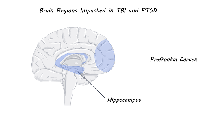
It has been shown that post-injury outcomes that follow TBI are associated with an increased risk for the development of persistent postconcussion syndrome (PCS) and psychiatric conditions including PTSD, major depressive disorder, general anxiety disorder, and substance use disorder. Researchers have proposed that this increased risk may be due to the presence of acute or chronic emotion dysregulation that occurs as a result of the physical and emotional trauma of TBI. Indeed, individuals with TBI or PTSD show significant overlap in symptoms resulting from emotional dysregulation including changes in mood and cognition, enhanced fear learning, and avoidance behaviours. These symptoms have been shown to be significantly worse in individuals with a comorbid diagnosis for TBI and PTSD compared to a diagnosis of TBI or PTSD alone, although the underlying mechanisms for this comorbidity are not well understood. The overlap in symptoms for TBI and PTSD can be explained by molecular changes that occur as a result of physical and/or emotional trauma in areas of the brain involved in emotion regulation. For example, dysregulation of the hypothalamic-pituitary-adrenal axis (involved in regulating the body’s stress response) in response to trauma can lead to maladaptive stress response and excess secretion of glucocorticoids. Heightened levels of glucocorticoids can subsequently result in increased release of glutamate in the prefrontal cortex and hippocampus (both areas involved in emotion regulation) which can be damaging to neurons and even cause cell death.

TBI_PTSD.png

What’s new?

In one study, preclinical models have shown that TBI-induced trauma results in molecular changes in subcortical structures involved in emotion regulation behaviour such as the amygdala and hippocampus. Further mild TBI has been associated with decreased resting-state functional connectivity between the prefrontal cortex and the insula. Studies using diffusion tensor imaging have shown that TBI and PTSD are both associated with white matter pathology which increases with the severity of TBI or PTSD. When looking at individuals with TBI, there is evidence of abnormalities in fronto-limbic white matter tracts (which connect the cingulate cortex to the hippocampus), while individuals with PTSD show decreased fractional anisotropy of the cingulum bundles. It is important to note, however, that TBI and PTSD were examined separately in the majority of this research. Even though worse outcomes are associated with comorbid diagnoses, structural and functional MRI studies that evaluate TBI and PTSD simultaneously are lacking. Nevertheless, the findings from these imaging studies show that white matter tracts that connect areas of the brain involved in emotion regulation are altered by both TBI and PTSD. This suggests that interventions that focus on emotion regulation may be particularly helpful for treating both conditions. In support of this, cognitive behavioural therapy, which focuses on improving emotion regulation, has been shown to be very effective in treating long-term comorbid PTSD and TBI.

What’s the bottom line?

Emotion dysregulation has an important role in the shared outcomes of TBI and PTSD. Early intervention with treatments like cognitive behavioural therapy that focus on improving emotion regulation may be helpful to minimize stress-induced molecular changes that occur following a traumatic event. In all, investigating the underlying changes in brain circuitry associated with TBI and PTSD through the lens of emotion regulation may be helpful for determining best practices for prevention and intervention when treating TBI and PTSD.


Weis et al. Emotion Dysregulation Following Trauma: Shared Neurocircuitry of Traumatic Brain Injury and Trauma-Related Psychiatric Disorders. Biological Psychiatry (2021). Access the original scientific publication here.

Misophonia: A Hatred of Specific Sounds

Post by Anastasia Sares

Characterizing a “new” disorder

Have you ever known someone who couldn’t stand the sound of people chewing? Are you that person? This isn’t just a fringe behavior or a quirk, it has a name: misophonia, literally, “hatred of sound”. The term was newly coined in 2001, describing a condition that may affect up to 20% of people despite not yet being in any official diagnostic manual. People with misophonia have aversive emotional reactions, including anger and distress, to specific sounds that do not bother other people. There are many proposed diagnostic tests for it, but no consensus on a gold standard yet.

Sensations and attention

Researchers have proposed relationships between misophonia and several other disorders: obsessive-compulsive disorder, phobias, hyperacusis (sensitivity to sound frequencies or volume), or synesthesia (when sensory stimulation produces a response in other senses). However, misophonia seems to occupy a niche all on its own. It is unlike a phobia because the primary emotion is anger rather than fear. It is different from synesthesia because sounds are associated with emotions, rather than with other sensory characteristics. People with misophonia sometimes avoid social situations, but not out of fear of judgment (like those with social anxiety)—they simply want to avoid situations where trigger sounds are likely to occur.

The brains of people with misophonia show differences in structure and function, in areas such as the anterior insula (which processes emotions such as anger and disgust), and the amygdala (involved in the fight-or-flight response). There are also differences in connectivity of the brain’s attention and salience networks. The interplay between these networks and the amygdala may be a key feature of misophonia.

What’s new?

Misophonia is rapidly garnering increased interest from researchers. There are already 25 new publications with the keyword “misophonia” this year alone (according to listings on PubMed). This year, people worked to characterize the prevalence of misophonia in different populations and refine tests for it. Though common triggers of misophonia include human oral/nasal sounds like chewing and breathing, Hansen and colleagues highlighted that not all misophonic triggers are human-produced (for example, a crow cawing or a bonfire), and called for a revision of the proposed “diagnostic” criteria. Ferrer-Torres and colleagues focused on the COVID-19 pandemic and the isolation that came with it, showing that people with misophonia experienced worse quality of life and even increased heart-rate variability, perhaps due to an inability to escape trigger sounds during confinement (trigger sounds are often associated with close family members or friends).

anastasia.jpg

What’s the bottom line?

The recognition of misophonia and its prevalence is important to better understand the condition. Further, its recognition validates the experiences of people with these symptoms. There is still a lot of scientific work to be done in characterizing misophonia before we reach a good understanding, but fortunately, research on this condition is growing more and more each year.

References

Jastreboff, M.M., and Jastreboff, P.J. (2001). Components of decreased sound tolerance: hyperacusis, misophonia, phonophobia. ITHS News Lett. 2, 5–7.

Kılıç, C., Öz, G., Avanoğlu, K. B., & Aksoy, S. (2021). The prevalence and characteristics of misophonia in Ankara, Turkey: population-based study. BJPsych Open, 7(5), e144. https://doi.org/10.1192/bjo.2021.978

Ferrer-Torres, A., & Giménez-Llort, L. (2021). Sounds of Silence in Times of COVID-19: Distress and Loss of Cardiac Coherence in People With Misophonia Caused by Real, Imagined or Evoked Triggering Sounds. Frontiers in Psychiatry, 12(June), 1–12. https://doi.org/10.3389/fpsyt.2021.638949

Ferrer-Torres, A., & Giménez-Llort, L. (2021). Confinement and the Hatred of Sound in Times of COVID-19: A Molotov Cocktail for People With Misophonia. Frontiers in Psychiatry, 12(May), 1–12. https://doi.org/10.3389/fpsyt.2021.627044

Kumar, S., Tansley-Hancock, O., Sedley, W., Winston, J. S., Callaghan, M. F., Allen, M., Griffiths, T. D. (2017). The Brain Basis for Misophonia. Current Biology, 27(4), 527–533. https://doi.org/10.1016/j.cub.2016.12.048

Eijsker, N., Schroder, A., Smit, D. J. A., van Wingen, G., & Denys, D. (2020). Structural and Functional Brain Abnormalities in Misophonia. Biological Psychiatry, 87(9), S225–S226. https://doi.org/10.1016/j.biopsych.2020.02.585

Siepsiak, M., & Dragan, W. (2019). Misophonia - A review of research results and theoretical concepts. Psychiatria Polska, 53(2), 447–458. https://doi.org/10.12740/PP/92023

Hansen, H. A., Leber, A. B., & Saygin, Z. M. (2021). What sound sources trigger misophonia? Not just chewing and breathing. Journal of Clinical Psychology, (February), 1–17. https://doi.org/10.1002/jclp.23196

Wu, M. S., Lewin, A. B., Murphy, T. K., & Storch, E. A. (2014). Misophonia: Incidence, phenomenology, and clinical correlates in an undergraduate student sample. Journal of Clinical Psychology, 70(10), 994–1007. https://doi.org/10.1002/jclp.22098

Can Gene Therapy Help Treat Brain Diseases?

Post by D. Chloe Chung

What is gene therapy?

The goal of gene therapy is to alleviate disease symptoms and ultimately cure diseases by correcting abnormal gene expression. This can be done by introducing genetic materials that express exogenous genes or suppress the expression level of endogenous genes in an effort to modify gene expression levels. Gene therapy can be also designed to directly edit gene mutations present in patients. Over the past years, development of novel gene-editing tools has resulted in improved efficacy of gene delivery to the brain. With this exciting technical advancement, gene therapies have recently gained more attention as a potential therapeutic strategy for neurodevelopmental and neurodegenerative diseases, especially for those caused by genetic mutations that disrupt the body’s usual patterns of gene expression.

Different strategies behind gene therapy

There are several ways to design gene therapy to correct aberrant gene expression in neurological diseases. One of them is to express exogenous proteins that can restore the function of faulty endogenous proteins. For this purpose, the adeno-associated virus (AAV) is a preferred viral vector because of its relative safety as well as its long-term gene expression which reduces the need for repeated administration. After being introduced into cells, AAVs can express genes that they carry by using the transcriptional and translational machinery in the host cells. In this way, AAVs produce proteins that can make up for the loss of gene function. The virus engineering field continues to refine structures of AAVs to increase their safety as well as effectiveness in delivery to the central nervous system.

DNA-editing is also an appealing approach for gene therapy as it can directly fix disease-causing mutations and modify gene expression level. The newest advancement in DNA-editing tools involves the synthetic CRISPR system (an abbreviation for ‘clustered regularly interspaced short palindromic repeats’) in which a customized guide RNA can bring a DNA-cutting enzyme (e.g., Cas9) to the specific site within the gene of interest. Once the enzyme cuts out a few base pairs from the gene, the gene sequence will eventually shift and create a premature stop codon). The cellular mechanism takes this stop codon as a sign to degrade messenger RNAs transcribed by this gene, ultimately silencing the expression of the target gene. This genetic tool has great potential to treat neurological diseases as it can be used to inactivate or activate genes of interest, or to edit precise bases within the gene and correct pathogenic gene mutations.

In addition to DNA-editing tools, the RNA-based therapy utilizing antisense oligonucleotides (ASOs) has gained much attention for its potential efficacy. ASOs are short DNA or RNA fragments that can bind to messenger RNAs based on the complementary sequence, subsequently changing the RNA expression level. As such, ASOs can be synthesized to target messenger RNAs transcribed from the disease gene of interest in hopes to regulate the protein level that could play crucial roles in neurological diseases.

How is gene therapy treatment for neurological diseases going?

In 2017, a groundbreaking study published in The New England Journal of Medicine reported the successful usage of gene therapy in young children with spinal muscular atrophy (SMA) type 1, a devastating neuromuscular disease characterized by motor neuron degeneration and progressive muscle loss. In SMA, a defective gene SMN1 reduces the amount of functional SMN1 protein, so researchers treated SMA patients with a one-time blood infusion of AAV that can express the SMN1 gene and restore protein expression level. Excitingly, most of the patients who received this gene therapy showed drastic improvement in their survival and motor functions that lasted through the 2-year follow-up assessment. Some of the patients were even able to walk with no assistance, which is a striking development considering that many SMA patients need wheelchair assistance and die at a very young age.

As of August 2021, a medication designed to increase the level of SMN protein is the only disease-modifying gene therapy approved by the US Food and Drug Administration (FDA) to treat neurological symptoms. Yet, in addition to this medication, numerous preclinical and clinical studies are actively investigating the safety and efficacy of gene therapy for a wider range of neurological diseases. For example, ASOs targeting the gene that makes tau, a protein that becomes abnormally aggregated in Alzheimer’s disease, have been tested in mouse models. After demonstrating their ability to reduce tau pathology and subsequently rescue behavioral deficits in a mouse model, the ASO-mediated tau-targeting gene therapy is being tested in a clinical trial for Alzheimer’s disease patients.

Similarly, ASO-based gene therapy has been investigated for its potential benefits in patients of Huntington’s disease, which is caused by an abnormal trinucleotide expansion in the huntingtin gene. The idea behind this therapy is to use ASOs either to globally target the total huntingtin gene level or to specifically reduce the function of the mutant allele. Based on promising results from preclinical studies, several ASOs entered clinical trials to evaluate their safety and efficacy for Huntington’s disease patients. Unfortunately, however, a couple of these clinical trials have been recently discontinued due to lack of evidence of anticipated benefits for patients.

What are the challenges and what is the hope for the future?

Perhaps the biggest challenge in utilizing gene therapy to treat neurological diseases is safety. For example, when genetic materials are being delivered at a high dosage, they can cause toxicity in patients. Also, undesirable immune responses can occur upon introduction of genetic materials. As these scenarios can lead to fatal consequences, safety is always the first aspect to consider and monitor when designing gene therapy and testing it in clinical trials. Moreover, off-target effects – unwanted changes in genes that were not the target of gene therapy – are also a potential concern. This can happen when ASOs or gene-editing tools target alternative genes based on partially complementary sequences. How to avoid off-target effects can be a major challenge when designing the reagents.

Despite these many challenges, biomedical techniques that improve our ability to utilize gene therapy to effectively treat neurological diseases continue to advance. For instance, delivery of AAVs into the central nervous system used to be highly challenging in the past, since it is not feasible to directly inject AAVs into the brain. At the same time, the blood-brain barrier firmly isolates the brain from the periphery, making it difficult for locally delivered reagents (e.g., injection into blood vessels) to bypass this physical barrier and reach the brain. Yet, characterization of various AAVs and persistent efforts in virus engineering allowed for development of a specific AAV that can be successfully and non-invasively (e.g., without surgeries) delivered to the central nervous system. This type of AAV was also used in designing the effective gene therapy for SMA patients as described above, demonstrating that technological advancement can lead to breakthrough therapeutic strategies. With rapidly evolving gene-editing tools, it's an exciting time for the development of gene therapy for neurological diseases.

References

Sun & Roy. Gene-based therapies for neurodegenerative diseases. Nature Neuroscience (2020). Access the original scientific publication here.

Martier & Konstantinova. Gene therapy for neurodegenerative diseases: slowing down the ticking clock. Frontiers in Neuroscience (2020). Access the original scientific publication here.

Bennett et al. Antisense oligonucleotide therapies for neurodegenerative diseases. Annual Review of Neuroscience (2020). Access the original scientific publication here.

Mendell et al. Single-dose gene-replacement therapy for spinal muscular atrophy. The New England Journal of Medicine (2017). Access the original scientific publication here.

DeVos et al. Tau reduction prevents neuronal loss and reverses pathological tau deposition and seeding in mice with tauopathy. Science Translational Medicine (2017). Access the original scientific publication here.

Kordasiewicz et al. Sustained therapeutic reversal of Huntington’s disease by transient repression of huntingtin synthesis. Neuron (2013). Access the original scientific publication here.

Southwell et al. In vivo evaluation of candidate allele-specific mutant huntingtin gene silencing antisense oligonucleotides. Molecular Therapy (2014). Access the original scientific publication here.

Kwon. Failure of genetic therapies for Huntington’s devastates community. Nature News (2021). Access the news article here.

Hudry & Vandenberghe. Therapeutic AAV Gene Transfer to the Nervous System: A Clinical Reality. Neuron (2019). Access the news article here.

Doudna. The promise and challenge of therapeutic genome editing. Nature (2020).Access the news article here.