Expectations of Reward and Efficacy Guide Cognitive Control Allocation

Post by Andrew Vo

What's the science?

Each day, we are faced with tasks and challenges for which we must decide whether the mental effort (in the form of cognitive control) to invest is worthwhile. The Expected Value of Control theory posits that people adjust the amount of mental effort they invest in a task by integrating information about the expected reward (the expected outcome) and the efficacy of task performance (the likelihood that investing effort will yield the desired outcome) to determine the expected value of control. The role of reward expectancy in shaping cognitive control is well-established, however, the effects and neural mechanisms of efficacy are far less studied. This week in Nature Communications, Frömer and colleagues investigated the contributions of reward, efficacy, and their interaction on behavioral and neural measures of control allocation.

How did they do it?

Across three studies, the authors tested their hypothesis that the allocation of cognitive control is based on the expected value of control. The worth of executing different types and amounts of control is determined by weighing its costs (i.e., mental effort) against its benefits. These benefits are shaped by a combination of the two incentive components: reward and efficacy.

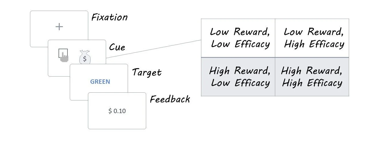
Participants completed a modified version of the color-word Stroop task that dissociated reward and efficacy effects on control allocation. On each trial, participants were initially presented with an incentive cue that disclosed whether they would receive a high or low monetary reward if successful ($0.10 vs. $1.00) and whether their efforts would be high or low in efficacy (success determined by their performance versus by the flip of a coin). They were then shown a Stroop stimulus — a color-word printed in either the same (congruent) or different (incongruent) color — to which they needed to respond with the color the word was written in rather than the word itself. Feedback was then given to indicate the value of the reward received. Correct reaction times (i.e., how fast the participant made a correct response) were used as a measure of task performance.

In Study 2, participants performed the task as their brain activity was recorded using EEG. The authors were interested in how reward and efficacy modulated the magnitude of two specific event-related potentials (ERP): the P3b (250-550 ms after cue onset that reflects incentive evaluation) and the CNV (500 ms before Stroop target onset that reflects control allocation).

What did they find?

The authors found that both reward and efficacy, as well as their interaction, modulated task performance. Participants were faster to make a correct response for higher levels of reward and efficacy. This pattern of findings was consistent whether each incentive component was varied in a binary manner (Study 1 and 2) or parametrically across four levels (Study 3). Control analyses ruled out that these effects were not simply due to speed-accuracy trade-offs, task difficulty, or practice effects.

EEG recordings (Study 2) revealed that reward and efficacy manipulations modulated the amplitude of both ERPs of interest. The P3b and CNV amplitudes were significantly larger in response to cues signaling larger rewards and efficacy. Notably, only the CNV was sensitive to the interaction of both incentive components. Examining trial-by-trial variability, the authors found that larger amplitudes in both ERPs were associated with an increased likelihood of correct responses and faster correct reaction times, with a more pronounced effect in CNV than P3b.

andrew.jpg

What's the impact?

This study provides behavioral and neural evidence supporting the critical roles of both reward expectancy and efficacy in determining the value of exerting cognitive control. The results are in line with predictions made by the EVC theory, which suggests that the integration of both incentive components (reward and efficacy) shapes how mental effort is allocated. As the authors succinctly state, “Cognitive control is critical but also costly”. This study illustrates how we may go about computing the worth of investing mental effort (in the form of cognitive control) towards achieving goals throughout our daily lives.

Hause_March2.jpg

Frömer et al. Expectations of reward and efficacy guide cognitive control allocation. Nature Communications (2021). Access the original scientific publication here.

Effects of Ketamine on Brain Function During Metacognition

Post by Lani Cupo

What's the science?

Metacognition, or thinking about the process of thought itself, is considered to be essential to the experience of consciousness.  While some studies have used neuroimaging techniques in an attempt to localize brain regions integral to metacognition, little is known about the role of specific neurotransmitter receptors underlying consciousness. Previous metacognition research has indicated the importance of the N-methyl-D-aspartate (NMDA) glutamate receptor, bound by antagonists—such as ketamine—which, at sub-anesthetic doses, can induce a psychotropic state accompanied by euphoria and out-of-body experiences. This week in Neuroscience of Consciousness, Lehmann and colleagues conducted a double-blind, placebo-controlled pharmacological functional magnetic resonance imaging (fMRI) study, in order to investigate the role of the glutamate system in metacognition and its underlying neural activity following exposure to ketamine.

How did they do it?

In the laboratory, metacognition can be measured by asking participants to reflect “trial-by-trial” on their performance on behavioral tasks, such as recall in a memory task. These tests allow researchers to assess, not only performance on the task (referred to as type 1 responses) but also participants’ insight into that performance (referred to as type 2 responses). Fifty-three adult participants (29 females) were included in the study, and 24 of them received sub-anesthetic doses (2 mg/ml) of ketamine (29 received saline) during a task-based fMRI scan. After being placed in the scanner and initiating infusion with ketamine or saline control, two phases of the study were completed. 

In phase 1, participants took part in a memory task, encoding 120 words, 60 of which they also rated on “pleasantness” leading to a deeper encoding. In the retrieval phase of the task, for type 1 responses, participants were presented with 180 words, 60 of which they had never seen before, and were asked to press a button when they recognized the word from before. For type 2 responses, they were asked to rate how confident they were in their type 1 response, either by reporting on a 6-point scale (report condition) or merely placing the cursor on a color-coded digit on the scale (follow condition). This distinction allowed the authors to study the difference between true metacognition and the “follow” control condition. 

In phase 2, participants saw 100 words, half of which they rated on pleasantness for deep encoding and half of which they merely reported the number of syllables. An hour after the scan and ketamine infusion were finished, participants performed the same retrieval task from phase 1, without the influence of current ketamine exposure. Finally, participants filled out a questionnaire designed to assess altered states of consciousness.

What did they find?

Results from the questionnaire suggested ketamine significantly altered participants’ states of consciousness, but, as expected, did not affect performance on the memory task. In phase 1, participants displayed more metacognitive sensitivity on deeply-encoded words, implying they were better at assessing their performance on the task for these words. However, ketamine reduced participants’ metacognitive sensitivity. This suggests that ketamine significantly alters metacognition, even though it did not impair memory encoding itself. When the authors analyzed the fMRI data, they found that ketamine infusion was associated with a higher blood-oxygen level dependent signal (an indirect measure of brain activity) in five clusters in the parietal and occipital lobes during type 2 ratings. The authors note that these changes were present regardless of how ratings were reported (with both the report and follow conditions) suggesting they are not specific to true metacognitive processes. Behavioral results from phase 2 memory trials were in accordance with phase 1, however, metacognitive sensitivity was not impacted by ketamine in this phase. This finding confirms that ketamine did not impair memory-encoding, and implies that an hour after termination of ketamine exposure, metacognitive self-assessment of performance on the task was no longer impaired.

Meta_March2.png

What's the impact?

This study is the first to investigate the effects of disrupting the glutamatergic system with ketamine on metacognition, showing that the pharmacological intervention may impair introspection. The authors acknowledge these results are too preliminary to attribute solely to the role of NMDA receptors, however, the evidence does suggest ketamine impacts metacognition. The present study provides a foundation for future investigations into the role of the glutamatergic system underlying metacognition and, ultimately, conscious experience.

Lehmann et al. Effects of ketamine on brain function during metacognition of episodic memory. Neuroscience of Consciousness (2021). Access the original scientific publication here.

Early Life Stress Exposure and Amygdala Reactivity Predict Symptom Improvement on Antidepressants

Post by Elisa Guma

What's the science?

Amygdala reactivity and exposure to early life stress have been implicated in the neurobiology of depression. However, not all individuals who experience early life stress develop depression, suggesting that there may be an interaction between the stressor and its effect on emotional brain circuitry (which includes the amygdala). Antidepressant treatment, shown to alter amygdala structure and function, is the standard for depression treatment, however, it only works for a subset of individuals; research suggests that those with high levels of early life stress have weaker outcomes. This week in PNAS, Goldstein-Piekarski and colleagues investigated whether a history of early life stress and amygdala reactivity to emotional faces may be predictive of antidepressant response in humans with depression

How did they do it?

Participants were enrolled in the International Study to Predict Optimized Treatment in Depression, with a confirmed diagnosis of nonpsychotic major depressive disorder. Participants were divided into three groups based on their exposure to early life stress, low (≤1 event), mid- (2–5 events), and high- (≥6 events), and evaluated using a 19-item Early Life Stress Questionnaire. Functional remission was defined as a return of symptoms to a healthy range and calculated based on a combined measure of clinician-rated depression symptom severity, self-reported symptom severity, and observer-rated functional capacity. Amygdala reactivity was measured using functional magnetic resonance imaging, while participants were shown images of happy and fearful faces as well as neutral comparison faces (drawn from a standardized series of facial expressions).

The authors used hierarchical logistic regression models to predict functional remission and used receiver operating characteristic curves to plot the performance of their regression models (this plots true and false positives). Leave-one-out cross validation was used to derive an unbiased threshold to classify remitters and non-remitters, improving the generalizability of their model. A series of successive regression models were used, starting with a simple covariate model as a baseline that included clinical and demographic variables (age, educational level, duration of MDD episode, social/occupational function, depression symptoms). Next, the authors tested the addition of early life stress level (i.e., low, mid, high) and amygdala reactivity to happy faces, followed by early life stress level and amygdala reactivity to fearful faces, and finally, an additive model with both interactions.

What did they find?

First, the authors confirmed that patient groups that achieved functional remission did not differ from those that did not achieve remission in terms of the demographic variables. Next, they found that their baseline model that included only demographic and clinical information showed a trend towards significance in its ability to classify remission. Including the interaction between early life stress and amygdala reactivity to happy faces or fearful faces as predictors in the model significantly improved the accuracy of prediction, however, both of these models had a higher probability of false positives or negatives. Finally, the additive model which included both the interaction term between early life stress and amygdala reactivity to happy faces as well as early life stress and amygdala reactivity to fearful faces further increased the ability to predict functional remission, suggesting good generalizability of this model.

elisa+%282%29.jpg

What's the impact?

The findings presented here suggest that functional remission due to antidepressant treatment may depend on an individual’s history of early life stress and the responsiveness of their amygdala to facial emotion. Further, the authors present a model which predicts, with a high degree of accuracy, those individuals who respond well to antidepressant treatment. This may be of clinical utility as a tool for screening individuals prior to initiating treatments. Finally, the results advance our understanding of how early life stress and amygdala reactivity function synergistically to predict subsequent remission from depression.  


Goldstein-Piekarski, et al. Human amygdala engagement moderated by early life stress exposure is a biobehavioral target for predicting recovery on antidepressants. PNAS (2016). The original scientific publication here.