A Coronavirus Protein Crosses the Blood-Brain-Barrier in Mice

Post by Leigh Christopher

What's the science?

COVID-19 has been associated with a number of central nervous system (CNS) symptoms including the loss of taste and smell, headaches, impaired consciousness, and even stroke. One reason for these symptoms could be that the SARS-CoV-2 virus (i.e., the virus responsible for COVID-19) enters the brain and acts directly on the CNS. Another possibility is those immune molecules known as cytokines (inflammatory molecules) associated with the virus cross the blood-brain-barrier, resulting in CNS symptoms. Yet a third possibility is that the virus sheds various proteins, which then cross the blood-brain-barrier and enter the brain. This week in Nature Neuroscience, Rhea and colleagues test whether the SARS-CoV-2 spike 1 protein (S1) can cross the blood-brain-barrier in mice.

How did they do it?

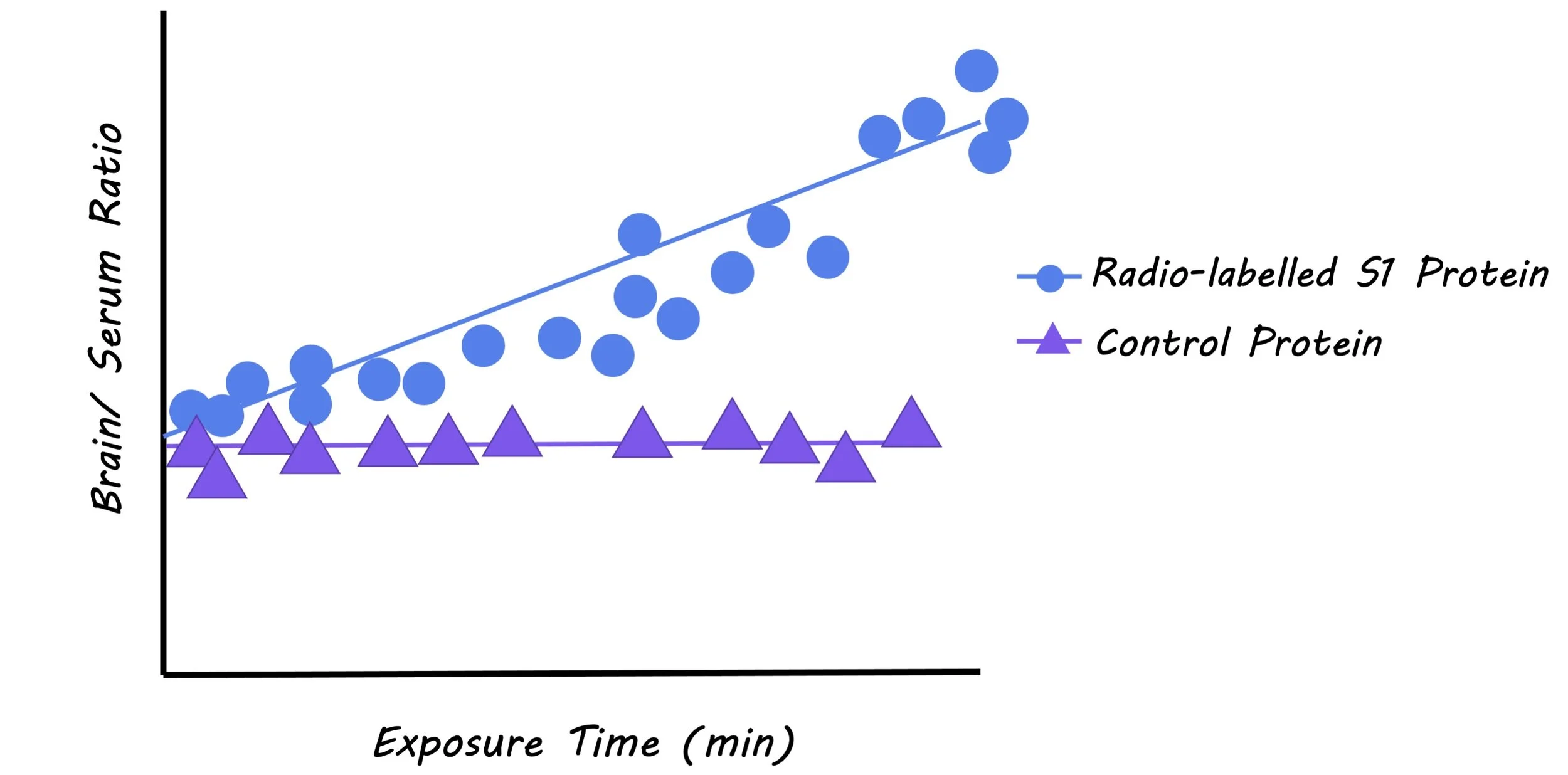
The authors radio-labelled S1 proteins from the SARS-CoV-2 virus, injected them intravenously into mice, and measured the blood-to-brain influx constant (a measure of the influx of S1 protein across the blood-brain-barriers). They did this using a method called multiple time regression analysis which allows for the measurement of protein influx to the brain while correcting for clearance of the protein out of the brain over time. They co-injected another radio-labelled protein along with the S1 protein that is known to have poor brain uptake, as a reference for how much S1 was crossing the blood-brain-barrier into the brain. The authors tested whether the S1 protein cleared the brain and entered other peripheral tissues and organs. A series of experiments were also performed to assess the mechanism of transport of the S1 protein across the blood-brain-barrier and into other tissues.

What did they find?

Radio-labelled S1 protein influx levels were significantly higher than the control protein, demonstrating that S1 does pass through the blood-brain-barrier and into the brain. They found that the virus entered a number of brain regions, and was also cleared from the brain through the blood, entering peripheral tissues, including the liver, kidney, and spleen. The authors attempted to understand the mechanism through which the S1 protein was transported across the blood-brain-barrier and into other tissues. When investigating the mechanism of transport of S1, they found evidence of blood-brain-barrier passage through adsorptive transcytosis, a mechanism where molecules bind to glycoproteins on endothelial cells and enter the brain through vesicle transport.

S1_image_dec22.jpg

SARS-CoV-2 is thought to enter cells by binding to a protein called ACE2. The authors found that co-injection of ACE2 and radio-labelled S1 protein increased the influx of S1 protein into the brain and lungs, suggesting that S1 uptake into these tissues was mediated by ACE2. They also found strong evidence of the involvement of other receptors. Next, the authors wanted to assess whether S1 uptake is increased during an inflammatory state, which is typically induced by the virus. Upon injection of lipopolysaccharide (a substance inducing an inflammatory state), the influx of S1 protein to the lungs (via adsorptive transcytosis) was higher, and the influx of S1 protein to the brain was also higher (via blood-brain-barrier disruption). These findings suggest that inflammation further increases S1 protein entry into the brain and lungs.

What's the impact?

This study is the first to show that the S1 protein of SARS-CoV-2 crosses the blood-brain-barrier and enters the brain in mice. This research sheds light on the potential mechanisms by which the S1 protein enters the brain and other tissues. Further understanding the mechanism of uptake, and whether the virus itself can also pass into the brain will be an important question for future research. As this study was performed in mice, it will also be crucial to investigate whether S1 passes through the blood-brain-barrier in humans.

Banks_Dec22.jpg

Rhea et al. The S1 protein of SARS-CoV-2 crosses the blood–brain barrier in mice. Nature Neuroscience (2020). Access the original scientific publication here.

The Default Network in the Human Brain Is Associated with Loneliness

Post by Amanda McFarlan

What's the science?

Humans have evolved to rely on socialization and, therefore, so has the human brain. What happens to the brain when an individual’s social expectations are not met? Since socialization is such a quintessential part of being human, it is thought that loneliness may result in distinct changes in the brain. Research has shown that loneliness is associated with reduced activity in reward areas of the brain as well as a greater vigilance and detection of negative social information. Still, the study of loneliness in higher-order brain regions that make up the “social brain” is lacking. This week in Nature Communications, Spreng and colleagues used a multimodal approach to investigate whether loneliness is associated with structural and functional changes in the brain.

How did they do it?

The authors used data collected for the UK Biobank initiative to investigate whether differences in brain structure and connectivity between brain regions exist between individuals who self-reported as being lonely and those that did not. In the first set of analyses, the authors used the Bayesian hierarchical approach to examine whether loneliness is associated with variations in gray matter volume, for both regional (localized) brain structure and large-scale brain networks. Next, they investigated how resting-state functional connectivity (which identifies brain regions with correlated activity, termed functional networks) relates to loneliness. Finally, the authors explored whether loneliness was associated with the integrity of white matter tracts in the brain. The authors also examined whether biological sex was associated with loneliness for all three neuroimaging approaches.

What did they find?

The authors found that gray matter volume for brain structures including the posterior superior temporal sulcus, the left temporoparietal junction, the left fusiform gyrus, the right inferior temporal gyrus, right posterior parietal lobe, and the right dorsal anterior cingulate cortex were positively associated with loneliness (high volume associated with high loneliness). Conversely, volume in several regions such as the left dorsal anterior cingulate cortex, the dorsolateral prefrontal cortex, the right central operculum, the right inferior parietal lobule, the left retrosplenial cortex, and the inferior visual cortex was negatively associated with loneliness. At the network level, the authors showed that loneliness was most strongly related to variations in gray matter volume in the default network (a network of brain regions that are typically more active when a person is not paying attention to the external world, i.e. daydreaming).

amanda.jpg

Next, the authors determined that individuals who self-reported as being lonely had greater resting-state functional connectivity between brain regions in the same network. This link between connectivity pattern and loneliness was expressed more strongly in men compared to women. Finally, the authors found that the structural integrity of white matter tracts originating from the hippocampus was greater in lonely individuals. This anatomical relationship between white matter tracts and loneliness was more prominent in men compared to women.

What’s the impact?

This study shows that loneliness is associated with distinct changes in gray matter volume, white matter tract integrity, and functional connectivity within brain regions. Notably, the authors identified the default network as the network most strongly associated with the neural expression of loneliness. Altogether, the link between loneliness and the default network may reflect increased mental simulation of inner social events in the absence of real-world social interaction.

Spreng et al. The default network of the brain is associated with perceived social isolation. Nature Communications (2020). Access the original scientific publication here.

The “Learning Layer” of the Cortex

Post by Anastasia Sares

What's the science?

The hippocampus is known to play a central role in learning and memory. The brain areas immediately surrounding the hippocampus, like the perirhinal cortex, are also involved. However, mature memories don't rely on the hippocampus —they are stored in a distributed way throughout the cortex (outer layer of the brain). The exact locations and mechanisms for memory storage in the cortex have been a long-standing mystery since the early 20th century. Nevertheless, it is generally assumed that auditory memories will be stored predominantly in the auditory cortex, or a movement memory will be stored in the motor cortex, and so on. The exact location of memories in the cortex is difficult to pin down.

This week in Science, Doron and colleagues demonstrate a new way of locating memory in the cortex. Instead of treating the cortex like a 2D map, they looked at the brain in 3D space, like a forest of trees, and showed that memories are actually stored in a specific sublayer of the cortex, layer 1, atop the forest. They were able to demonstrate that messages crucial for long-term memory formation are sent from the hippocampus to layer 1.

How did they do it?

The authors first traced the paths of neurons from the perirhinal cortex, showing that neurons starting here signal to other parts of the cortex, in layer 1. Next, they taught mice and rats to respond to a small electrical stimulation applied directly to their brain, specifically, the somatosensory cortex (the part of the cortex that processes touch information from the body). When they licked a small apparatus right after receiving stimulation, the animals were rewarded. With this design, the authors could pinpoint exactly where in the cortex the long-term memory would be made: it would be in the same place as the stimulation. In some animals, the authors disrupted the connection between perirhinal cells and layer 1 of the somatosensory cortex, either during or after training on the task. Other animals' connections were left intact as a control.

What did they find?

Compared to the controls, animals with disrupted connections between the perirhinal cortex and layer 1 of somatosensory cortex showed impaired learning, but only if the disruption happened during the learning phase. Expert animals were not affected by this disruption, since their memory for the task had already been formed. For all of the animals, the neurons in the perirhinal cortex fired in response to the stimulus when the animal responded correctly. However, activity in the somatosensory cortex was less robust in the animals whose connections were disrupted. The authors reasoned that the real targets for these signals from perirhinal cells were the large pyramidal neurons in layer 5 of the cortex because their dendrites stretch all the way up to layer 1, where they should capture the signal from the perirhinal cells. In the mice and rats that learned normally, these layer 5 cells changed their firing pattern after a correct response, similar to the perirhinal cells. In the animals with a disrupted layer 1 connection, layer 5 cells were much less responsive.

anastasia+%281%29.jpg

What's the impact?

This study highlights the central role of layer 1 of the cortex in storing memory. Human memory is extremely complex, and we are growing in our understanding of it every year. Unlocking the mechanisms behind memory formation will aid in the fight against human diseases like Alzheimer's, and may also inform the way we make artificial neural networks, helping machines to learn in a more biological way.

Larkum_dec22.jpg

Doron et al.Perirhinal input to neocortical layer 1 controls learning. Science (2020). Access the original scientific publication here.