
Blood-Brain Barrier Gene Expression and Function Changes with Neuronal Activity

Post by D. Chloe Chung

What's the science?

The blood-brain barrier (BBB) is a semi-permeable structure that tightly controls the movement of molecules between the brain and neighboring blood vessels. Several studies have described the dynamic communication between the brain vasculature and neuronal activity. For example, blood volume and flow increase following neuronal activity (likely for oxygen and nutrient provision). However, it is not yet well understood whether changes in neuronal activity can directly influence the properties of BBB. This week in Neuron, Pulido and colleagues report that neuronal activity in the mouse brain, either chemically or behaviorally stimulated, can regulate efflux via BBB transporters (i.e. for waste removal) by altering its gene expression profile.

How did they do it?

The authors used transgenic mouse models in which glutamatergic neurons can be either activated or silenced using specific drugs. Three hours after drug injection, the authors collected endothelial cells, which form blood vessel walls, from the mice and performed RNA sequencing to examine gene expression changes in the BBB. These mice were also injected with fluorescent-labeled substrates of one of the BBB transporters to test whether functional properties of BBB, such as molecule transport, changed with neuronal activity. As an additional approach to stimulate neuronal activity, the authors capitalized on the knowledge that whisker movement typically leads to neuronal firing in a specific brain area. They designated two groups: mice with intact whiskers were made to explore their cage with many things to interact with using their whiskers, while mice with their whiskers trimmed off were left in an empty cage. Similar to the transgenic mouse models, these mice were analyzed for the gene expression changes in their endothelial cells. To evaluate the relationship between BBB efflux and neuronal activity-regulated circadian rhythm, the authors made an additional mouse model in which circadian genes can be deleted specifically in endothelial cells.

What did they find?

The authors found that changes in glutamatergic neuronal activity can dramatically regulate the expression level of hundreds of genes in endothelial cells, including multiple transporters and efflux pumps that can control the transfer of molecules across the BBB. In particular, several BBB efflux transporters were upregulated when neuronal activity was silenced, and downregulated when neuronal activity was higher. These gene expression changes led to transporter substrates (but not the non-substrates) entering the mouse brain more readily when neurons were activated. 

In addition to transporters, several genes involved in circadian rhythm were also differentially regulated upon neuronal activity. They showed a similar pattern to transporters: gene expression was upregulated with neuronal silencing and downregulated with neuronal activation. In the context of circadian rhythm, the authors observed that the gene expression level of a BBB efflux transporter and its permeability rhythmically fluctuated throughout the light-dark cycle and generally correlated with net neuronal activity throughout the day. Interestingly, this rhythmic fluctuation and acute neuronal regulation were completely abolished when one of the circadian genes was deleted in the endothelial cells, indicating that circadian genes, regulated by neuronal activity, can impact the expression and function of the BBB efflux transporter. Of note, the same transporters and circadian genes were also downregulated in mice in which neurons were activated via whisker stimulation, further demonstrating that neuronal activity can modulate these genes in the BBB.
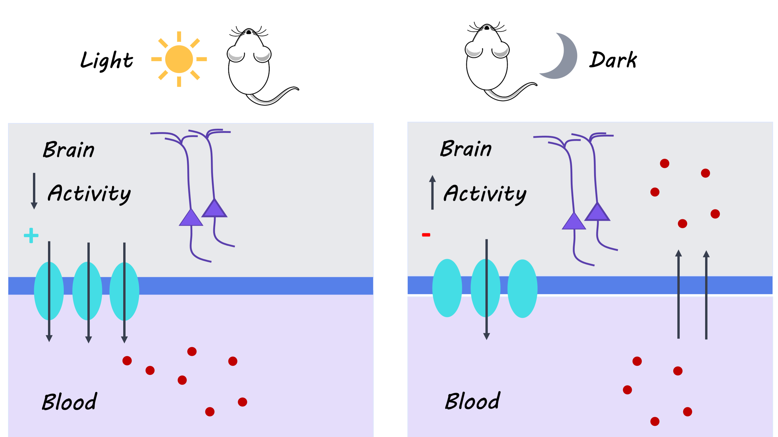
BrainPost_images_Oct12.png

What’s the impact?

This work revealed that the gene expression profile and functional properties of the BBB can be regulated by neuronal activity, in contrast to the previous belief that the BBB remains largely static. As the authors suggest, future studies examining activity-dependent gene expression changes in the endothelial cells across different time points will expand our understanding of the dynamic nature of the BBB. From a clinical standpoint, this study can be useful in optimizing drugs to cross the BBB with higher efficiency, as these BBB efflux transporters pose a major obstacle for drug delivery into the brain. Finally, findings from this work may be applied to investigate the relationship between dysregulated BBB efflux and neurological disorders.
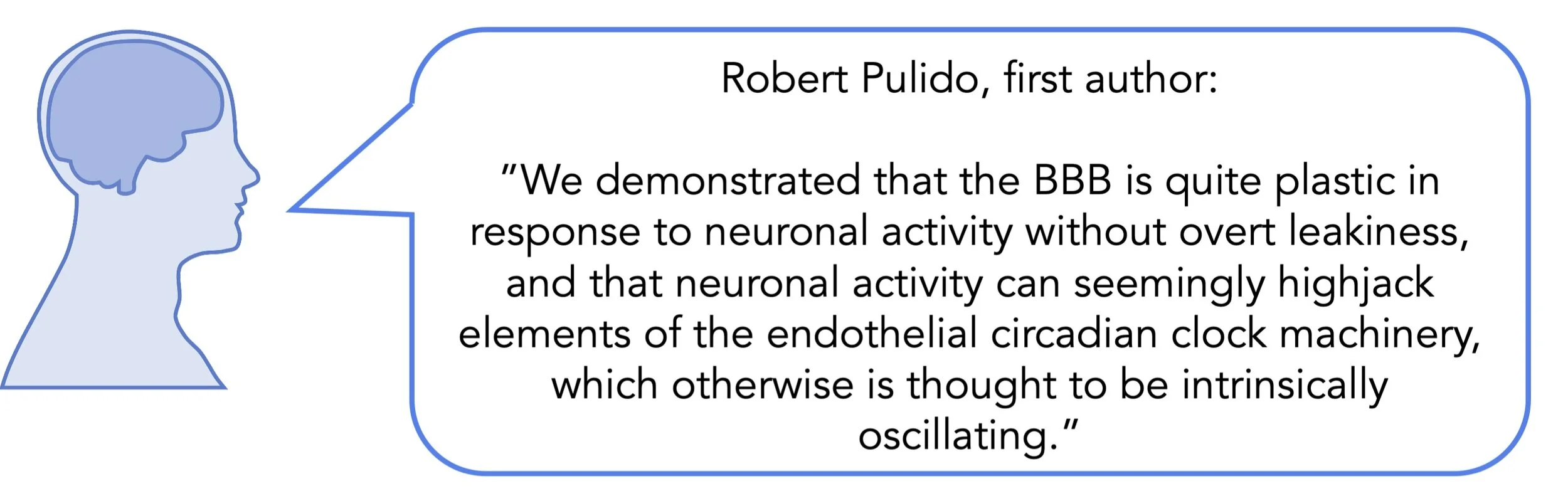
BBB_quote_oct12.jpg

Pulido et al. Neuronal Activity Regulates Blood-Brain Barrier Efflux Transport through Endothelial Circadian Genes. Neuron (2020). Access the original scientific publication here.
Multidimensional Encoding of the Prefrontal Cortex During Decision Making

Post by Elisa Guma

What's the science?

The prefrontal cortex plays a critical role in context-dependent decision making. It receives sensory, cognitive, and motor signals, which must all be integrated to make decisions. Understanding the prefrontal cortex’s role in this process is confounded by the fact that neurons within this structure respond to multiple types of input signals in a heterogeneous manner, making it difficult to understand the neural representation of different sensory and cognitive variables. This week in Nature Neuroscience, Aoi and colleagues applied novel modeling techniques to dissect the specific context and temporal evolution of neural activity in the prefrontal cortex during decision making.

How did they do it?

The authors acquired electrophysiological recordings of neural activity from the frontal eye-fields (a region of the prefrontal cortex) of two adult male monkeys while they performed a context-dependent, forced-choice decision making task. In each trial of the task, monkeys were cued to either make a choice based on the colour or the motion of moving dots by focusing their gaze on one of the two targets; thus, if the colour was cued, they were required to ignore motion, and vice versa.

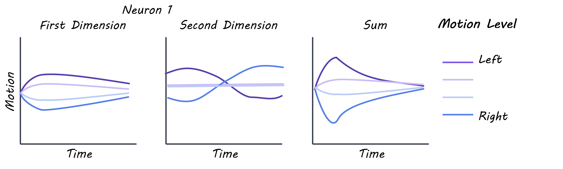
The authors analyzed the neural activity recorded from the prefrontal cortex during stimulus presentation using a model-based targeted dimensionality reduction (mTDR) technique. This allowed them to identify the dimensions of neural activity that encode information about different task variables over time (i.e. response to sensory or motion cues). They used colour strength, motion strength, context and choice, and absolute values of colour and motion strength as their dimensions. They then applied a new data dimensionality reduction technique to the outputs of the mTDR — sequential principal component analysis (seqPCA) — to decompose the data into a set of axes ordered by the time at which each variable becomes available. This allowed them to determine if and when a neuronal population became active during the task period. Finally, given that neurons exhibit heterogeneous activity, and response to both motion and color, the authors then tested the “rotational dynamics” of the neural activity, i.e. does the neural response shift from one stimulus to another over time.

What did they find?

The authors found a heterogeneous tuning to the different variables (colour, motion) when examining the pre-stimulus time histograms, suggesting that the neural population recorded encodes several task variables, not just a single one. The authors found that the mTDR technique was able to accurately model the heterogeneous neural response, as most neurons had their variance differently distributed across components (e.g. color strength, motion strength, context, etc).

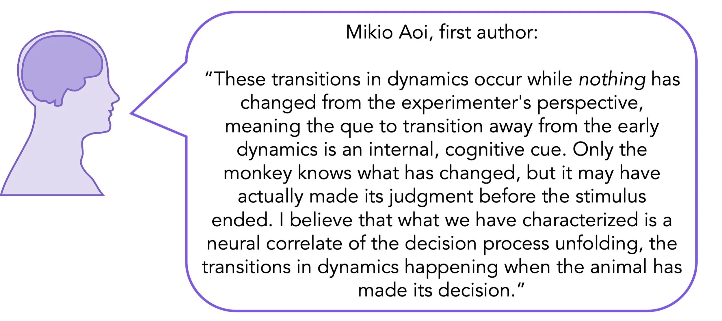
Using seqPCA to decompose neural activity into different temporal axes, the authors found that most of the variance in neural trajectories occurred in the time immediately after stimulus onset (early axis). They also identified a middle and late axis within the stimulus viewing window. Interestingly, neurons did “rotate” through this multidimensional space, (from early to middle or late axes) all during the stimulus viewing session; this process may reflect an internal, cognitive cue and may also reflect the moment in which the monkey has to make a judgment (before the end of the stimulus). Further, they found that the differences in neural activity did not arise from a distinct population of neurons, but from a single population with broad tuning characteristics. A rotation was indicative of a commitment to a decision, and not an accumulation of further sensory information, suggesting that the PFC maintains information about a choice as well as relevant and irrelevant sensory information over the course of a single trial.  

Elisa.png

What's the impact?

This study applied novel modeling techniques to more accurately decode prefrontal cortex neural activity in response to a decision making task. This work highlights that the prefrontal cortex has a multidimensional code for types of stimuli involved in decision making, that these different codes may change as a result of the decision-making process, and that these codes are not neuron-specific.

Aoi_quote_oct12.jpg

Aoi et al. Prefrontal cortex exhibits multidimensional dynamic encoding during decision-making. Nature Neuroscience (2020). Access to the original scientific publication here.
Modeling Human Movement Perception with a Computer Algorithm

Post by Anastasia Sares

What's the science?

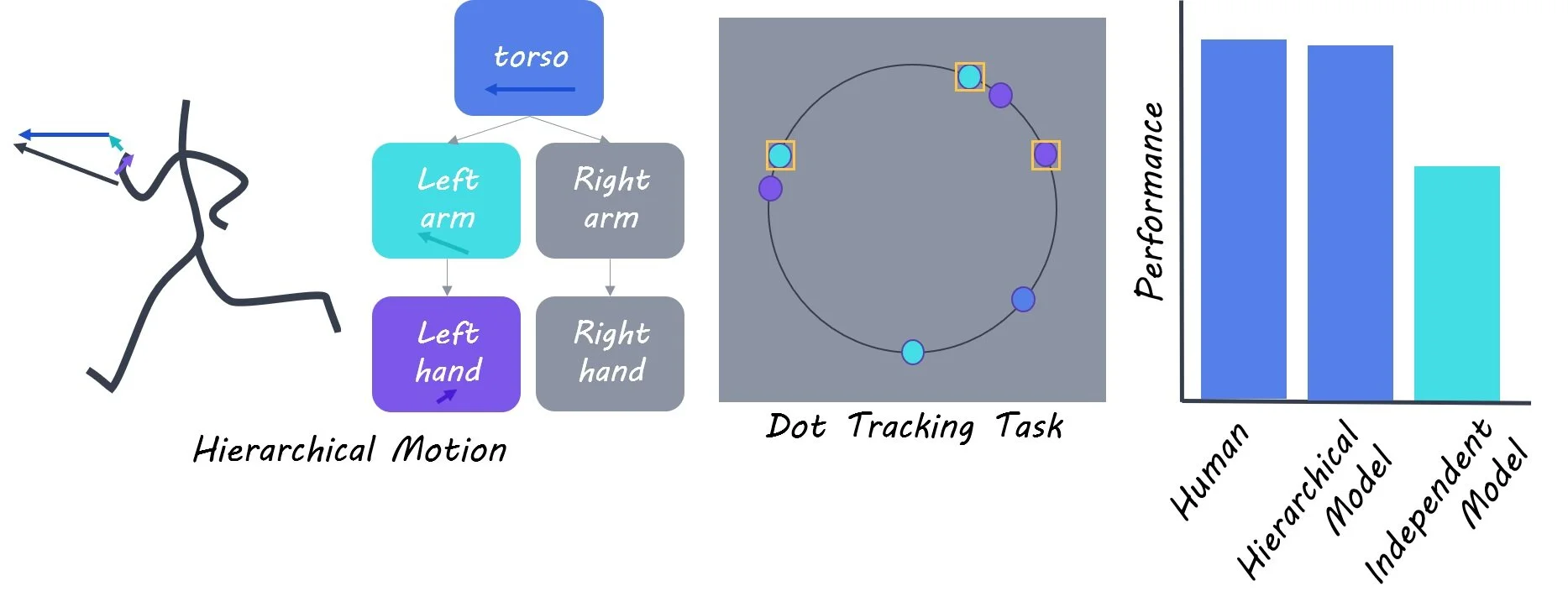
Picture a person dancing. How can you recognize all of those body parts as belonging to one individual, even when they aren’t all moving together? Our brains have an ability to track and predict complex movement, and this could be because we use a hierarchical structure to organize everything. In a hierarchical structure, the hand has its own movements, but it also inherits the motion of the arm, which in turn inherits the motion of the torso, and all of these movements are added up to produce the motion we see. If we are aware of these relationships, we can track and predict the movement more accurately. This week in PNAS, Bill and colleagues showed that giving hierarchical information to a movement-tracking algorithm allowed it to mimic human performance levels.

How did they do it?

Modeling is an increasingly popular approach that differs from classic experimental science. The logic goes like this: if we can construct a computer algorithm that successfully mimics something in the real world (human movement perception, for example), then the characteristics of that computer algorithm might help us understand how the real process works.

The authors first brought human participants into the lab to test their motion perception. The participants had to track a few target dots moving around a circle while ignoring other distracter dots. In some conditions, the moving dots moved independently, and in other conditions they had a hidden hierarchical structure (You can find video examples of the task here). The humans performed significantly better when there was a hierarchical structure.

After the data from the human participants were collected, the authors created a computer algorithm (a Bayesian observer model)  that had human-like performance only on the non-hierarchical stimuli. They then tested what would happen before and after they improved the model by giving it access to hierarchical structures.

What did they find?

At first, when the algorithm was exposed to the hierarchical condition, it did not improve as the humans had, indicating that it did not have enough information. However, when it had access to hierarchical information, the algorithm’s performance resembled human data much more closely. This suggests that the addition of hierarchical information was the missing element necessary for better motion tracking.

Anastasia.jpg

What's the impact?

Getting a computer algorithm to reproduce human behavior doesn’t prove that the human and the algorithm work the same way, but it does give us an idea of the minimum amount of information necessary, and a plausible mechanism for how it might work. The next step would be to actually find this mechanism at work in the human brain.

bill-quote-oct6.jpg

Bill et al. Hierarchical structure is employed by humans during visual motion perception. Proceedings of the National Academy of Sciences (2020). Access the original scientific publication here.