Polygenic Risk and Orbitofrontal Cortex Volume Predict Smoking Behavior in Adolescents

Post by Elisa Guma

What's the science?

Tobacco smoking is the leading cause of preventable illness and death in the world. Smoking behavior is thought to be heritable, with contribution from a large number of genetic variants. These genetic variants can be used to calculate a polygenic risk score, which has also been shown to predict brain structure, particularly cortical volume, another heritable feature that is altered in chronic smokers. This week in Psychological Medicine, Li and colleagues investigated the association between the polygenic risk score for smoking, and brain structure in a large population-based European dataset of 14-year old adolescents (IMAGEN cohort).

How did they do it?

The authors analyzed data from the IMAGEN project, a longitudinal imaging genetics study conducted across eight European cities. Blood samples from 1774 secondary school-aged adolescents (50.68% female, mean age 14.43±0.4 years) were collected for DNA extraction from which 477245 single nucleotide polymorphisms (markers of genetic variation) were extracted following quality control. To calculate the polygenic risk score for smoking, the authors used publicly available data from a genome-wide meta-analysis of smoking initiation from the Tobacco and Genetics Consortium (n=74053). Smoking behavior was captured using a self-report questionnaire from which the authors classified participants into 3 categories based on the frequency of smoking: ‘never users’, ‘users on a few occasions’ (1-9 occasions), and ‘users on more occasions’ (10 or more).

Cortical volume was calculated using FreeSurfer from T1-weighted structural magnetic resonance imaging scans acquired across the 8 different sites (on 3T MRI scanners from various manufacturers, but the same scanning protocol. Linear regression models were used to investigate the association between smoking polygenic risk score and smoking behavior (controlling for gender, age, site). Next, a generalized linear model was used to analyze the association between vertex-wise cortical volume and smoking polygenic risk score (controlling for gender, site, and total brain volume). Finally, a mediation model was used to test the effect of brain anatomy on the relationship between smoking polygenic risk score and smoking behavior.

What did they find?

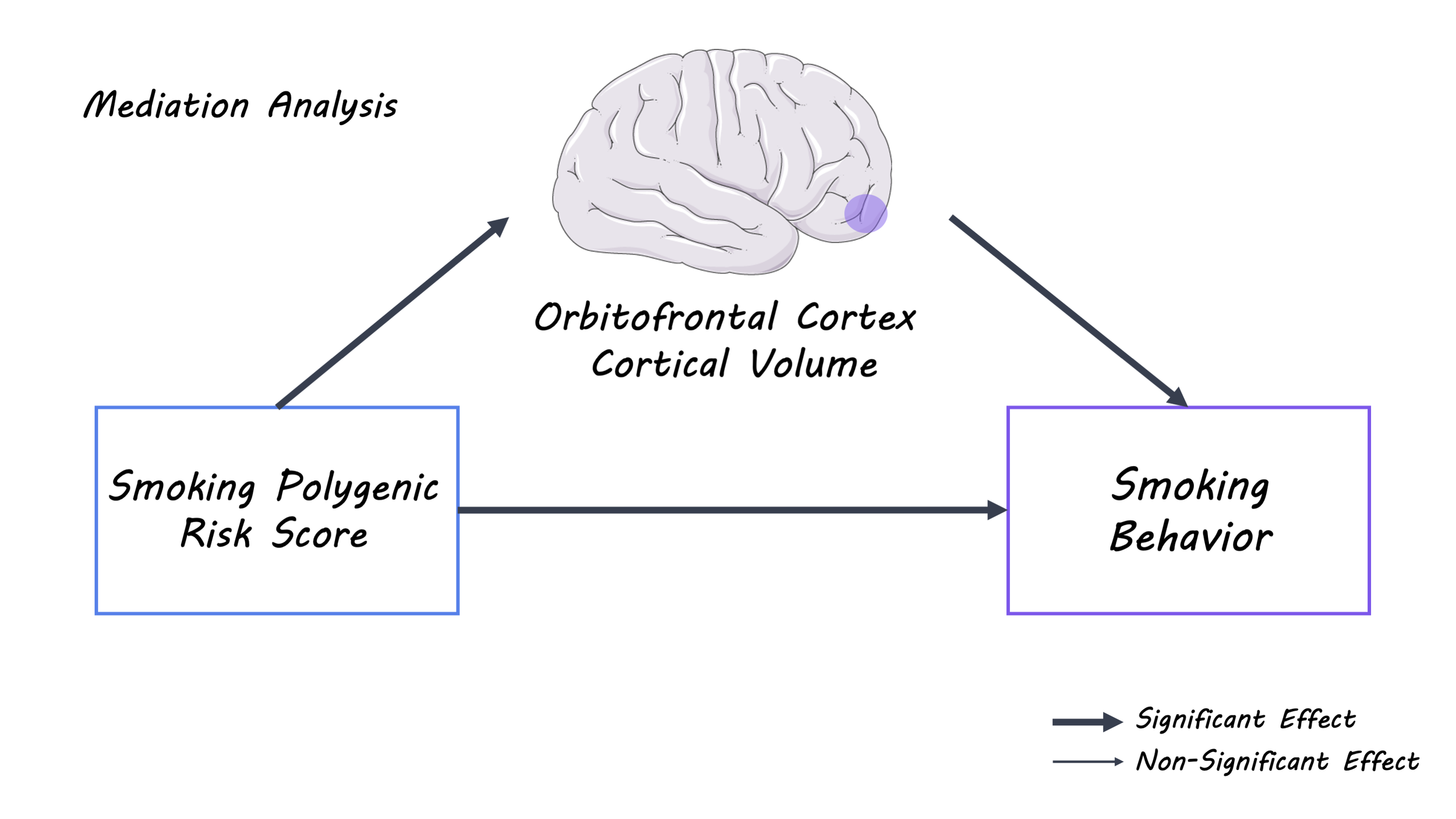
The authors identified a nominally significant association between smoking polygenic risk scores and smoking behaviors. Next, they observed an inverse correlation between smoking polygenic risk score and cortical volume of the right orbitofrontal cortex. The volume of this region was significantly correlated with the number of occasions of tobacco use. This indicated that adolescents with a higher smoking polygenic risk score had a smaller volume of the right orbitofrontal cortex and that adolescents with smaller volume in the right orbitofrontal cortex were more likely to consume cigarettes. Finally, the mediation analysis suggested that variations in the volume of the right orbitofrontal cortex partially accounted for the relationship between the smoking polygenic risk score on smoking behavior in this adolescent sample.
Smoking_img_sep7.png

What's the impact?

This study found that higher smoking polygenic risk scores were associated with an increase in the number of cigarette smokes, and smaller cortical volume of the right orbitofrontal cortex in a large adolescent population-based sample. Interestingly, this brain structure has previously been associated with risk-taking behavior and impulsivity. This strategy of combining polygenic risk scores with brain imaging to investigate the etiology of tobacco smoking can be extended to other disorders or behaviors. Finally, further investigating the role of the orbitofrontal cortex in smoking behavior may be an interesting avenue of future research.

Li et al. Orbitofrontal cortex volume links polygenic risk for smoking with tobacco use in healthy adolescents. Psychological Medicine (2020). Access the original scientific publication here.
Neuropeptides and Astrocytes Regulate Adult Neural Stem Cell Activity

Post by Lincoln Tracy 

What's the science?

The dentate gyrus is part of the hippocampus, an area of the brain that plays a critical role in memory formation. The dentate gyrus contains neural stem cells (brain cells that develop into other cell types) as well as a diverse range of other cell types, referred to as niche cells, such as granule cells, mossy cells, and astrocytes. These niche cells can release signals that act on neural stem cells to regulate neurogenesis, the process of generating new neurons. While previous research in the Song lab has identified that certain local niche cells can directly act on neural stem cells and regulate their behavior, it remains unknown if and how these niche cells interact within the dentate gyrus to regulate neural stem cells. This week in Neuron, Asrican and colleagues sought to address a range of challenges associated with determining the role of cholecystokinin signaling in regulating neural stem cells and neurogenesis within the hippocampus. 

How did they do it?

First, the authors selectively activated cholecystokinin neurons to examine the effect of endogenous cholecystokinin release on neural stem cells. To do this, they injected transgenic mice with adeno-associated viruses to selectively target cholecystokinin neurons within the dentate gyrus. Once the neurons had successfully been labeled, these neurons were activated via a chemogenetic approach. Next, they sought to address whether the cholesystokinin induced any direct or indirect effects on the neural stem cells, by recording their membrane potentials after GABAergic and glutamatergic signaling had been blocked. In addition, calcium imaging was performed to identify the effects of cholesystokinin on other cell types in the dentate gyrus, such as granule cells, local interneurons, and mossy cells. After these effects were identified, in vivo analysis of neural stem cell behavior was performed using immunohistology and stereological counting of neural stem cells and their progeny. Finally, adeno-associated viruses were used to reduce cholecystokinin levels in the dentate gyrus to assess the effect on neural stem cell behavior. RNA sequencing of the dentate gyrus was undertaken to explore the mechanisms by which reduced cholecystokinin levels impairs neural stem cell behavior. 

What did they find?

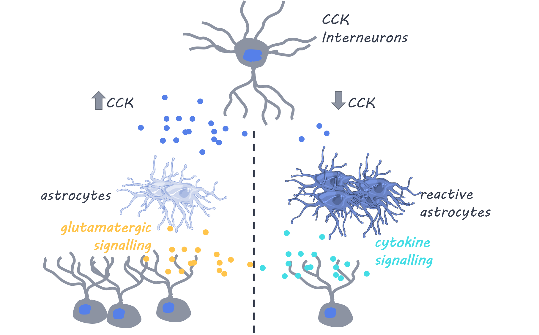
The authors showed that stimulating cholecystokinin neurons in the dentate gyrus induced neural stem cell depolarization. Interestingly, they found that it was the cholecystokinin that induced the depolarization, not GABA or glutamate transmission from these neurons. Moreover, cholecystokinin-induced neural stem cell depolarization requires the activation of glutamate receptors on the neural stem cells. The cholecystokinin induces astrocytes to release glutamate, which acts on receptors on the stem cells. In addition, the authors found that the cholecystokinin also excites parvalbumin neurons, which provide inhibitory inputs onto granule cells and mossy cells. Finally, the authors found that decreasing cholecystokinin release induces astrocyte reactivity and increases innate immune system activity. The resulting neuroinflammation leads to decreased proliferation of neural stem cells, most likely through proinflammatory cytokine signaling.  

lincoln (1).png

What's the impact?

This study shows that promoting cholecystokinin release in the dentate gyrus supports neurogenesis through an astrocyte-mediated glutamatergic signaling pathway. In contrast, reducing cholecystokinin release in the dentate gyrus induces neuroinflammation which impairs the neurogenic potential of neural stem cells. The generation of new neurons in adult life also plays a critical role in cognitive functioning. Therefore, increasing cholecystokinin release to stimulate neurogenesis while also reducing inflammation may be utilized as a novel treatment strategy to treat conditions where abnormal neurogenesis and neuroinflammation occur.

Neurogenesis_quote_sept7.jpg

Asrican et al. Neuropeptides modulate local astrocytes to regulate adult hippocampal neural stem cells. Neuron (2020). Access the original scientific publication here.

Decoding the Neural Representation of Pain Using a Brain-Machine Interface

Post by Amanda McFarlan

What's the science?

The lack of adequate treatments to safely manage acute and chronic pain is a serious public health issue. Recent advances in machine-learning technology have shown that it may be possible to decode a neural representation of pain by analyzing brain responses. Using this technology, researchers may be able to identify potential biomarkers for pain that could be used to design new therapeutic treatments. This week in Current Biology, Zhang and colleagues built a machine-learning based decoder to investigate whether brain activity measured with functional magnetic resonance imaging (fMRI) and electroencephalography (EEG) could be used to predict pain in real-time.  

How did they do it?

The authors recruited healthy participants for their study. On the first experiment day, the participants’ brain activity was measured using fMRI while they received a painful electrical stimulus of high or low intensity to their left hand. The blood oxygen level-dependent (BOLD) responses from the insula (known to play a role in pain encoding) were used to build a multivoxel-pattern analysis decoder that would later be used to predict the intensity of a painful stimulus. On the second day of the experiment, participants’ brain activity was measured using fMRI while receiving a painful stimulus of either high or low intensity (a ‘high intensity stimulator’ and a ‘low intensity stimulator’). Based on real-time fMRI responses, the control computer used the decoder (built from the participants' fMRI data on the first day) to predict whether the participant perceived the stimulus to be of high or low intensity, with the goal of identifying the stimulator that was associated with the low intensity stimulus so that it would be chosen for the next trial. The participants were encouraged to attend to the painful stimuli and use their brain responses to help the computer learn so that it could administer lower pain stimuli.

Next, the authors had participants complete a similar adaptive control task in which they applied a painful electrical stimulus to participants’ lower backs while their brain activity was measured using EEG (experimental group) or while listening to a podcast (control group). Both the experimental and control groups also completed a temporal contrast enhancement task before and after the main experimental task, in which they applied and modulated the intensity of a tonic heat pain stimulus before and after the main experimental task in both the control and experimental groups. Contrast enhancement is a known phenomenon in which small changes in tonic pain result in unexpectedly large effects on pain ratings.

What did they find?

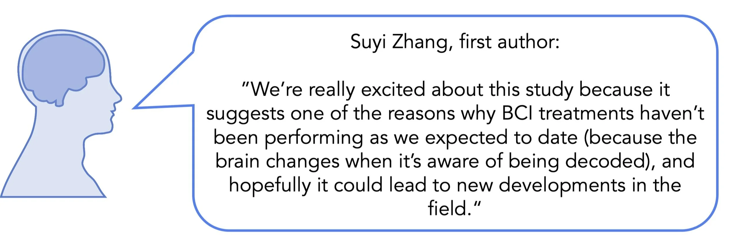
The authors showed that the decoder’s accuracy for predicting the participants’ perception of pain intensity was above chance. Because of this, the control computer was able to deliver significantly more pain stimuli of low intensity than of high intensity, suggesting that it is possible to use brain activity to decode and predict pain. The authors found that the decoder’s accuracy for predicting pain level was higher in the pregenual anterior cingulate cortex and lower in the left anterior insula. They also determined that the level of uncertainty (which quantifies the amount of new acquired information that will be used to improve learning) was positively correlated with BOLD responses from the pregenual anterior cingulate cortex and participants’ pain ratings. Finally, the authors found enhanced responses of the periaqueductal gray (which is known to mediate pain control) to high pain intensity on the second day of the experiment. This suggests that participants adaptively controlled their brain response to pain. 

Anastasia (2).png

In the EEG experiment, uncertainty was also positively correlated with participants’ pain ratings. In the temporal contrast enhancement task, pain ratings were lower in the experimental group after they performed the adaptive control task — evidence of endogenous pain control system engagement. Together, these findings suggest that the participants’ attention to pain stimuli may change the neural representation and encoding of pain in the brain by engaging endogenous pain modulation mechanisms.

What’s the impact?

This study shows that a multivoxel-pattern analysis decoder can use brain activity to identify an individual’s perceived pain intensity and use that information to reduce pain in real time. The authors found that the neural encoding of pain is subject to change when the brain’s activity is being decoded. Notably, pain encoding in the insula was disrupted, which suggests this region would not likely make a good biomarker for pain. Together, these findings provide insight into how brain-machine interfaces may be developed to help alleviate pain.   

pain_quote_sep1.jpg

Zhang et al. Pain Control by Co-adaptive Learning in a Brain-Machine Interface. Current Biology (2020). Access the original scientific publication here.