Uncovering Proteins That Interact with a Disease Form of Tau

Post by D. Chloe Chung

What's the science?

Tau is a microtubule-binding protein that normally contributes to the stability of the central nervous system. In multiple neurodegenerative diseases including Alzheimer’s disease, tau becomes abnormally phosphorylated and detaches from microtubules, leading to synaptic dysfunctions and neuronal death. Many studies have investigated the exact mechanisms of how tau phosphorylation contributes to the disease course, but there is a gap in knowledge of what types of proteins interact with phosphorylated tau (pTau). This week in Brain, Drummond, Pires, and colleagues use complementary proteomics approaches to discover previously unknown protein interactors of pTau.

How did they do it?

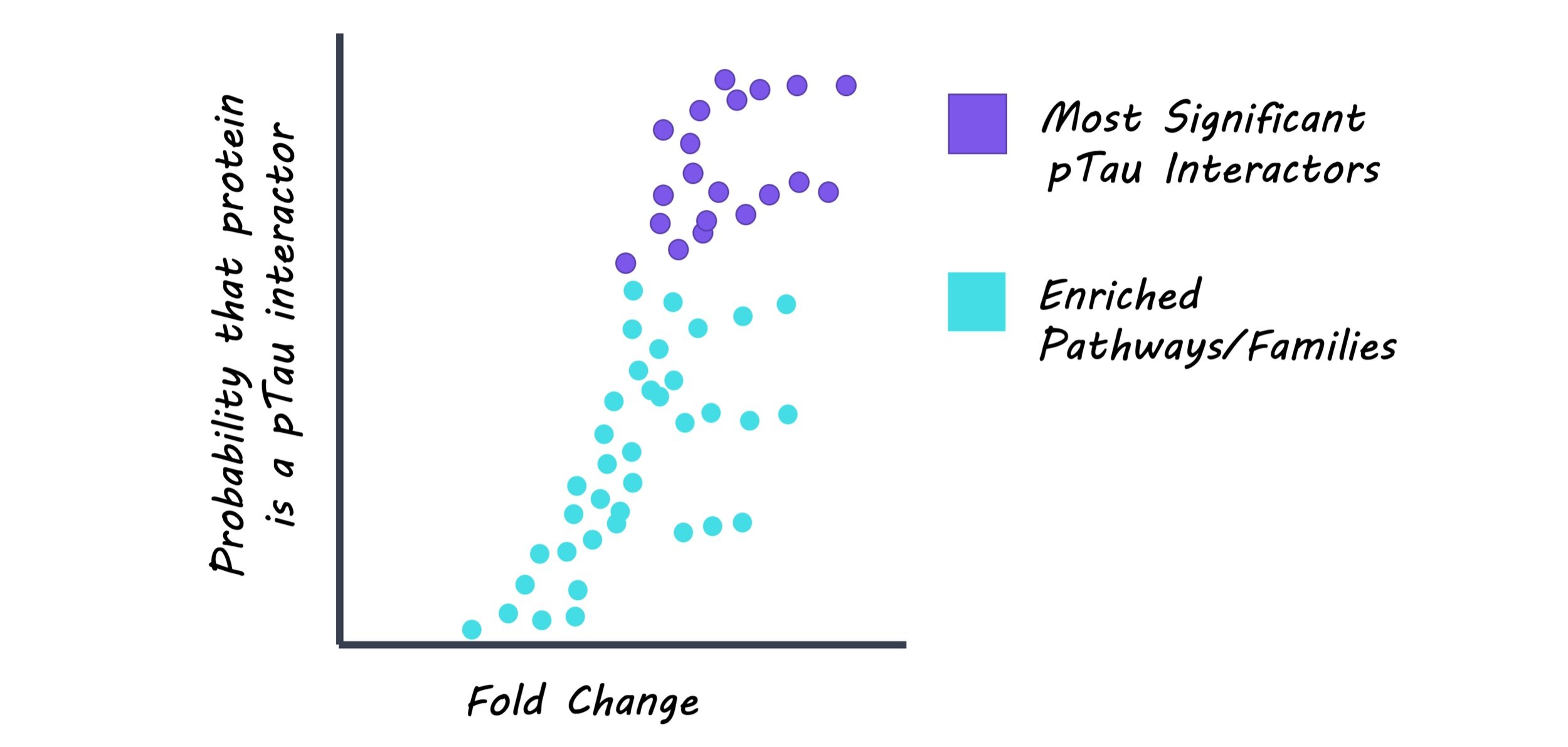
Neurofibrillary tangles (NFTs), composed of pTau, were dissected and isolated from brain samples of Alzheimer’s disease patients using a laser-capture microdissection technique. These samples were analyzed to identify proteins other than pTau that are present in NFTs. pTau was extracted along with its protein interactors from the Alzheimer’s disease brain tissue. Here, an antibody that can detect a specific phosphorylation site on tau was used to isolate the pTau-interactor complex. Using these samples, the profile of pTau and its interacting proteins was compared to the list of NFT-associated proteins. Also, pTau-interacting proteins were categorized based on related biological pathways or protein families to better understand which biological aspects are closely related to pTau.

What did they find?

From the first proteomics analysis, the authors found 542 proteins to be present in NFTs, including tau and other proteins that have been well-documented for their association with NFTs. From the second proteomics analysis, the authors found 125 proteins that interact with pTau. Interestingly, many of pTau interactors were associated with two major protein degradation pathways: one involved ubiquitin that tags proteins to be degraded by proteasomes, and the other involved lysosomes that break down proteins with digestive enzymes. These findings suggest an important pathological link between pTau and impaired protein degradation, which has been largely hypothesized in the field. Importantly, the authors compared two proteomics datasets and found that 75 out of 125 pTau-interacting proteins were also present in NFTs. Among them, 12 proteins have never been reported to interact with pTau before, meaning that the authors found new pTau-interacting proteins from their analysis. The type of these new pTau interactors varied, ranging from proteins that bind to DNA or RNA to subunits of enzymes or ribosomes.

tau_image_aug25.jpg

What’s the impact?

While there have been several studies that have reported protein interactors of tau, this study is the first to find proteins that interact specifically with phosphorylated Tau in the human brain. In particular, this work identified novel proteins that have never been reported to be associated with tau before, opening new areas to investigate tau biology. Findings from this study will elevate our understanding of how tau exerts its toxic effects in the brain upon phosphorylation. Also, this study will be useful in identifying potential drug targets to treat Alzheimer’s disease and other neurodegenerative diseases in which tau is abnormally phosphorylated.

Brain_quote_Aug24.jpg

Drummond, Pires et al. Phosphorylated tau interactome in the human Alzheimer’s disease brain. Brain (2020). Access the original scientific publication here.

Impact of COVID-19 on the Nervous System

Post by Amanda McFarlan

What's the science?

The spread of the SARS-CoV-2 virus, better known as COVID-19, has become a global public health concern. Infection with COVID-19 is known to affect the respiratory system, causing fever, dry coughs, and in more serious cases, shortness of breath and chest pain. Although it is less common, some individuals infected with COVID-19 also display neurological symptoms such as headache, dizziness, and a loss of taste and smell. However, it remains unknown exactly what the COVID-19 virus is doing in the body to cause these neurological symptoms. This week in Cell, Iadecola and colleagues summarized the recent data describing the effects of COVID-19 on the nervous system.

What do we already know?

Neurological abnormalities, ranging from a headache to total delirium, have been observed in ~30% of hospitalized individuals infected with COVID-19. In some cases, COVID-19 has also been shown to have delayed neurological symptoms post-infection affecting both the central and peripheral nervous systems. Studies have shown that infection with COVID-19 increases an individual’s risk of having a stroke. Indeed, the incidence of stroke in hospitalized patients with COVID-19 is ~7 times higher compared to patients with influenza, which suggests that the COVID-19 virus is likely to be involved in causing vascular brain injuries. Additionally, the majority of studies testing the cerebrospinal fluid (CSF) and brain tissue samples of COVID-19 patients with neurological abnormalities did not show evidence of viral invasion, however, small quantities of the COVID-19 virus were detected in the CSF of at least two cases of hospitalized individuals with severe infectious encephalopathy (altered mental state due to damage to the brain). The peripheral effects of COVID-19 could potentially explain the occurrence of encephalopathy without evidence of brain invasion by the virus. For example, COVID-19 is known to act on the respiratory system and in severe cases, can cause lung damage which can lead to hypoxia (lack of oxygen). In line with this, post-mortem studies from COVID-19-related deaths revealed that there was brain damage in regions most susceptible to hypoxic brain injury: the neocortex, hippocampus, and cerebellum.

What’s new?

One major question to ask when investigating the effects of COVID-19 on the nervous system is: can the COVID-19 virus enter the brain, and if so, how? The main entry point for COVID-19 into human cells is through an enzyme that is located on the membrane of cells in the lungs, kidneys, arteries, heart, and intestines called the angiotensin converting enzyme-2 (ACE2). Recent data has shown that ACE2 was also expressed in the choroid plexus (brain cells that produce CSF) and in neocortical neurons. In support of these findings, it was shown in studies using organoids and transgenic mice expressing the human ACE2 gene that the COVID-19 virus can infect neurons and cause cell death in an ACE2-dependent manner. Since one of the common neurological symptoms associated with COVID-19 is a loss of smell and taste, the olfactory system may be a possible route for the virus to enter the brain. Indeed, studies have shown that olfactory epithelial cells had detectable levels of ACE2 protein and RNA, although it is unknown whether the virus could reach the olfactory neurons to enter the brain. Additionally, since COVID-19 has been described as being blood-borne, it could potentially enter the brain via the blood-brain-barrier or infected immune cells. Thus, there are many possible ways in which the COVID-19 virus could invade the brain, however, more conclusive data from CSF and post-mortem tissue is required to determine the exact route of entry. 

COVID_image.png

What’s the bottom line?

The evidence to date suggests that the COVID-19 virus can result in acute and prolonged neurological abnormalities that range in level of severity. Most of this data, however, is derived from case studies, which means that the acute and long-term neurological effects of COVID-19 at the population level are still unclear. The development of adequate experimental models as well as the collaboration between clinical and basic scientists around the world will be critical for answering these questions quickly and efficiently.

Cell_quote_Aug24.jpg

Iadecola et al. Effects of COVID-19 on the nervous system. Cell (2020). Access the original scientific publication here.

Coactivating Large Neuronal Populations Enhances Cortical Excitability

Post by Elisa Guma

What's the science?

Our brain’s ability to structurally and functionally remodel in response to sensory inputs is critical to our survival. Repetitive exposure to the same sensory stimulus can induce long-term changes in underlying neural circuits, which in turn can alter future responses to the same stimulus. Importantly, exposure to sensory stimuli typically coactivates large groups of cortical neurons, leading to correlated neuronal activity and increased efficacy of synaptic transmission and neuronal excitability. This week in PNAS, Zhang and colleagues used optogenetic methods to directly activate populations of cortical neurons in the mouse cortex to determine the mechanisms underlying enhanced neuronal excitability in response to repetitive stimulus exposure

How did they do it?

The authors injected the adult mouse primary visual cortex with adeno-associated virus vector that would transfect pyramidal neurons with channelrhodopsin 2 (ChR2), a blue light sensitive channel, and implanted them with multi-unit electrodes in layers 2 and 3 of the visual cortex. With this set up, the authors were able to stimulate the visual cortex (using a laser to activate ChR 2) and measure neuronal activity in discrete response areas (RAs) at each electrode in anesthetized mice.

In their first experiment, the authors examined whether repetitive synchronous coactivation (compared to random stimulation) of multiple neuronal populations in 4 identified RAs of the visual cortex could alter the spiking rate of the neurons in these areas in response to a test (light) stimulus. The authors characterized the duration of the enhancement of evoked spiking following neuronal coactivation, as well as the dependence of the enhancement of the number of coactivation stimuli, and the size of the neuronal population coactivated. The experiments were repeated in the motor cortex (to understand whether they were generalizable or specific to the visual cortex. To better understand how synaptic transmission might affect neuronal response to synchronous coactivation, the authors infused the visual cortex with an antagonist for the AMPA or NMDA subtype of glutamate receptors and measures stimulus-evoked synaptic activity using in vivo whole cell recording.

Next, the authors wanted to know if the enhancement of neuronal excitability could be achieved at a longer range. First, they examined cross-cortical enhancement between the visual cortex and somatosensory cortex, and then examined whether the cross-hemispheric spread of spiking enhancement could occur between the right and left visual cortices. The size of coactivated populations and distance between regions were considered to be modulating factors. Finally, for contralateral communication to occur between hemispheres, neuronal activity passes through subcortical regions. To understand the underlying mechanisms of this pathway, the authors also chemogenetically inhibited neuronal activity in the midline/intralaminar thalamus, as there is evidence for it being connected to the visual cortex and measured whether spiking enhancement between hemispheres could still be achieved.

What did they find?

First, the authors confirmed that repetitive activation of neuronal populations caused enhancement of neuronal spiking rates in all RAs recorded, which was not observed if the sequence was random, suggesting that the synchronous coactivation of cortical neurons is required for the observed enhancement of neuronal firing. The enhancement induced by synchronous coactivation was long-lasting (persisted up to 100 minutes following the coactivation), saturable (plateaued after 3 episodes of coactivation), and dependent on the size of the coactivation area (smaller area only show enhancement of closer RAs). Similar coactivation was observed in the motor cortex suggesting that enhancement is a general property of the cerebral cortex. AMPA receptor blockade led to a reduction or elimination of spiking of neurons, whereas NMDA receptor blockade abolished enhanced test stimulus-evoked spiking highlighting the critical role these glutamate receptor subunits play in inducing neuronal excitability. Many of the features observed thus far are reminiscent of long-term potentiation.

spiking_image_aug25.png

To achieve cross-cortical neuronal enhancement, the size of coactivated neuronal populations needed to be increased; no enhancement was observed if only one region within the visual and somatosensory cortices were stimulated, however, there was a significant elevation of test-stimulus-evoked spiking if two visual RAs and one somatosensory RA (or vice versa) were coactivated. Finally, the authors found that repeatedly coactivating four RAs in the left visual cortex led to enhanced neuronal spiking in both hemispheres. This was markedly reduced by silencing the midline/intralaminar thalamus, suggesting that it plays a critical role in mediating contralateral coactivation.

What's the impact?

This study shows that repetitive coactivation of large populations of cortical neurons results in persistent enhancement of neuronal spiking evoked by optogenetic stimulation. This effect was dependent on NMDA receptor activity and could be observed in local cortical areas, distance cortical areas, and even in different hemispheres. Finally, neuronal activity in the thalamus may be involved in the enhancement of cortical excitability. This work elucidates some of the mechanisms underlying cortical excitability and may help in understanding disorders in which cortical excitability is aberrant, such as epilepsy.

Zhang et al. Global enhancement of cortical excitability following coactivation of large neuronal populations. PNAS (2020). Access the original scientific publication here