
New Experiences Help Strengthen Old Memories

Post by D. Chloe Chung

What's the science?

Long-term memories that are already stored in our brains can be retrieved and reactivated via memory reconsolidation, which is distinct from memory consolidation: encoding recent information into new memory. Several studies have suggested that memory of a weak experience can be strengthened by an associated experience that produces proteins related to plasticity (“behavioral tagging”). Indeed, new experience and information can be simultaneously acquired as pre-existing memories get reactivated, but how new experiences mediate memory reconsolidation is not fully understood. This week in Proceedings of the National Academy of Sciences, Orlandi and colleagues use rat models and multiple behavioral tests to show how memory reconsolidation can be mediated by an accompanying new experience.

How did they do it?

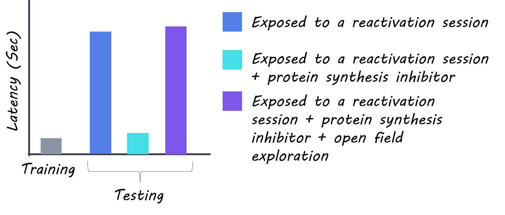
Rats were subject to the weak avoidance test in which they receive an electric foot shock when they step down on the lower platform during the training session. Twenty-four hours later, rats were returned to the test chamber without an electric foot shock as part of the “reactivation session” to retrieve their long-term memory from the earlier training session. The following day, rats were placed in the chamber again and the authors measured how long it took for rats to step down to the lower platform. If rats took a longer time to step down to the platform, they were thought to have a better memory of the foot shock. Different types of inhibitors were given to rats to explore the importance of new protein synthesis and second messenger pathways in memory reconsolidation and behavioral tagging. Also, to introduce a novel experience adjacent to this memory, rats were made to freely explore a new open field in addition to the weak avoidance test. The same experimental scheme was also applied to another assay called the object recognition task to expand understanding of how reconsolidation works for different types of memory. Here, two identical objects were placed in a chamber and one of the two was later moved to a different location. Rats were considered to remember the original locations of objects if they spent more time interacting with the object at a new location.

What did they find?

During the weak avoidance test, rats normally waited for about 2 minutes on average before descending to the lower platform, showing that they possess a long-term memory of the previous electric foot shock. However, rats failed to remember their earlier experience when the new protein synthesis was prevented right after the reactivation session, indicating that proteins related to plasticity are required for memory reconsolidation. Interestingly, forgetfulness was resolved when rats underwent the open field exploration close to the reactivation session (up to 60 minutes before or 30 minutes after the session). This effect disappeared when protein synthesis was inhibited right after the open field exploration, suggesting that a novel experience facilitates memory reconsolidation by providing proteins that could be used to re-solidify pre-existing memories. The authors also tested different protein quinase inhibitors and further dissected that different pathways are responsible for specific aspects of the behavioral tagging process during memory reconsolidation. For the second behavioral test of object recognition, rats showed similar results as they failed to remember the location of objects upon inhibition of protein synthesis, which was resolved by open field exploration (that provides plasticity proteins).
Chloe.png

What’s the impact?

This study presents evidence that behavioral tagging is critical in memory reconsolidation. Specifically, the authors showed that new experiences can contribute to updating old memories by providing newly synthesized proteins that can be used to mediate plasticity. These findings can be potentially useful in developing effective ways to alleviate traumatic memories in individuals who suffer from their negative effects. Conversely, these findings can also help us find ways to strengthen long-term memory for educational purposes.
Orlandi et al. Behavioral tagging underlies memory reconsolidation. PNAS (2020). Access the original scientific publication here.
Lipid Biomarkers of Psychosis in Clinically High-Risk Individuals

Post by Shireen Parimoo 

What's the science?

Psychotic disorders like schizophrenia can disrupt daily functioning and reduce quality of life, but early detection and intervention has the potential to mitigate some of these effects. Patients often experience rapid weight gain and develop insulin resistance due to changes in their metabolism, leading researchers to hypothesize that metabolic abnormalities might precede the development of psychosis. In particular, the concentration of lipids – a class of metabolites found in fats and oils – might be altered in individuals who are at risk of developing psychosis. This week in Biological Psychiatry, Dickens and colleagues performed lipidomic analysis and used machine learning to identify lipid biomarkers that predict clinical outcomes of psychosis in clinically high risk (CHR) individuals.

How did they do it?

The authors recruited 263 individuals identified as being at clinical high risk (CHR) for psychosis from a large-scale longitudinal study along with 51 healthy controls (HC). At the beginning of the study, they recorded clinical symptoms and obtained serum samples from both groups; they then followed up with the participants 1, 2, and up to 5 years later. At their follow-up clinical assessment, patients were categorized as (i) being in remission if their symptoms improved and they no longer met the diagnostic criteria for being at risk for psychosis (CHR-remission), (ii) having persistent symptoms, or (iii) having transitioned to psychosis.

The authors identified serum concentrations of lipids using a combination of liquid chromatography and mass spectrometry, which are techniques used in lipidomic analysis to separate and characterize different types of molecules based on their molecular properties. They performed a clustering analysis to identify different lipid clusters in each group and compared them between the groups at baseline. Lipids belonging to clusters that differed the most between the two groups were then used to predict clinical outcomes for HC and individuals in each CHR group. For a more fine-grained understanding of how lipid concentrations at baseline and demographic variables might be related to later outcomes, the authors used predictive logistic regression (a machine learning technique) to distinguish between the each CHR sub-group (e.g., CHR-remission vs. other CHR individuals) and between HC and all CHR individuals.

What did they find?

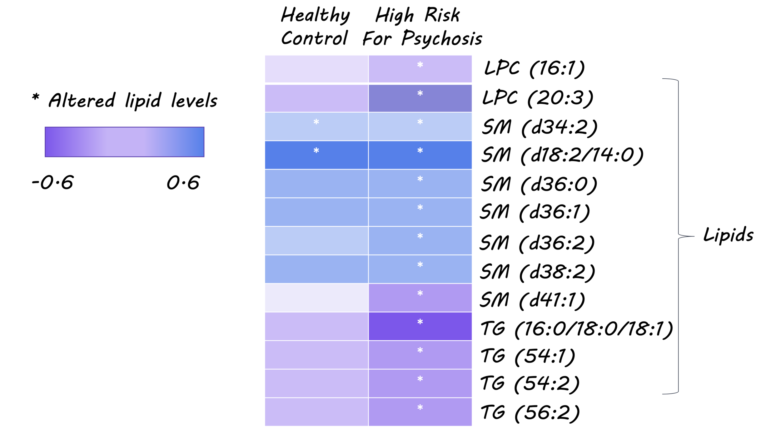
At baseline, CHR patients generally had higher serum lipid levels than HC. The authors identified 12 clusters of lipids differed between the HC and CHR individuals, which included triglycerols, ether phospholipids, and sphingomyelins, among others. Triglycerol levels most reliably distinguished between the two groups based on their structural properties: triglycerols with lower number of carbon atoms and fewer double bonds were higher in the CHR group, whereas HC had higher levels of triglycerols with longer, polyunsaturated fatty acid chains. Lipid profiles also differed between males and females. In both the HC and CHR groups, triglycerol levels were higher in males whereas sphingomyelin levels were higher in females. Serum lipid concentrations at baseline also predicted clinical outcomes at follow-up. Specifically, individuals who developed psychosis had lower ether phospholipids levels at baseline compared to other high-risk individuals. On the other hand, lipid profiles also differed between those who went into remission and those whose symptoms persisted. Importantly, there were no differences in the lipid profiles between HC and CHR-remission individuals. Overall, these results indicate that serum lipid levels in high-risk individuals are a sensitive marker of both the progression and regression of clinical symptoms of psychosis.

psychosis_img.png

What's the impact?

This study demonstrates that serum lipid levels can be used to predict clinical outcomes in individuals at high risk of developing psychosis. Although further work needs to be done to validate the predictive model, these findings have exciting implications for predicting the symptom trajectory of high-risk individuals and for providing early and personalized intervention to mitigate the progression of symptoms.

Dickens et al. Dysregulated lipid metabolism precedes onset of psychosis. Biological Psychiatry (2020). Access the original scientific publication here.

How Do Different Brain Regions Communicate?

Post by Anastasia Sares

What’s the science?

Scientists have been mapping out the brain and its connections for over a century. Still, we are very far away from a complete understanding of how the brain functions. Understanding the brain on a deeper level will require us to understand how physically distant groups of neurons can interact. This week in Trends in Neurosciences, Kohn and colleagues reviewed four possible mechanisms for communication between brain regions.

What do we already know?

Though new discoveries are still being made, we have a decent understanding of the architecture of the brain. Chemical stains and even viruses can be used to trace neural circuits forwards and backward. However, making a simple circuit diagram won’t explain what is really going on, just like having a map of a building doesn’t tell you how the people inside are interacting. We must measure the working brain.

Scientists can use electrodes inserted into the brain to measure the firing activity of small groups of neurons, giving us a zoomed-in picture of brain activity. MRI, when combined with advanced statistical techniques, can reveal large patterns of activity that tend to occur on average—a zoomed-out picture. What we don’t understand as well is the middle-range: communication between different local brain regions is complex, and very flexible, changing from one moment to the next.

What’s new?

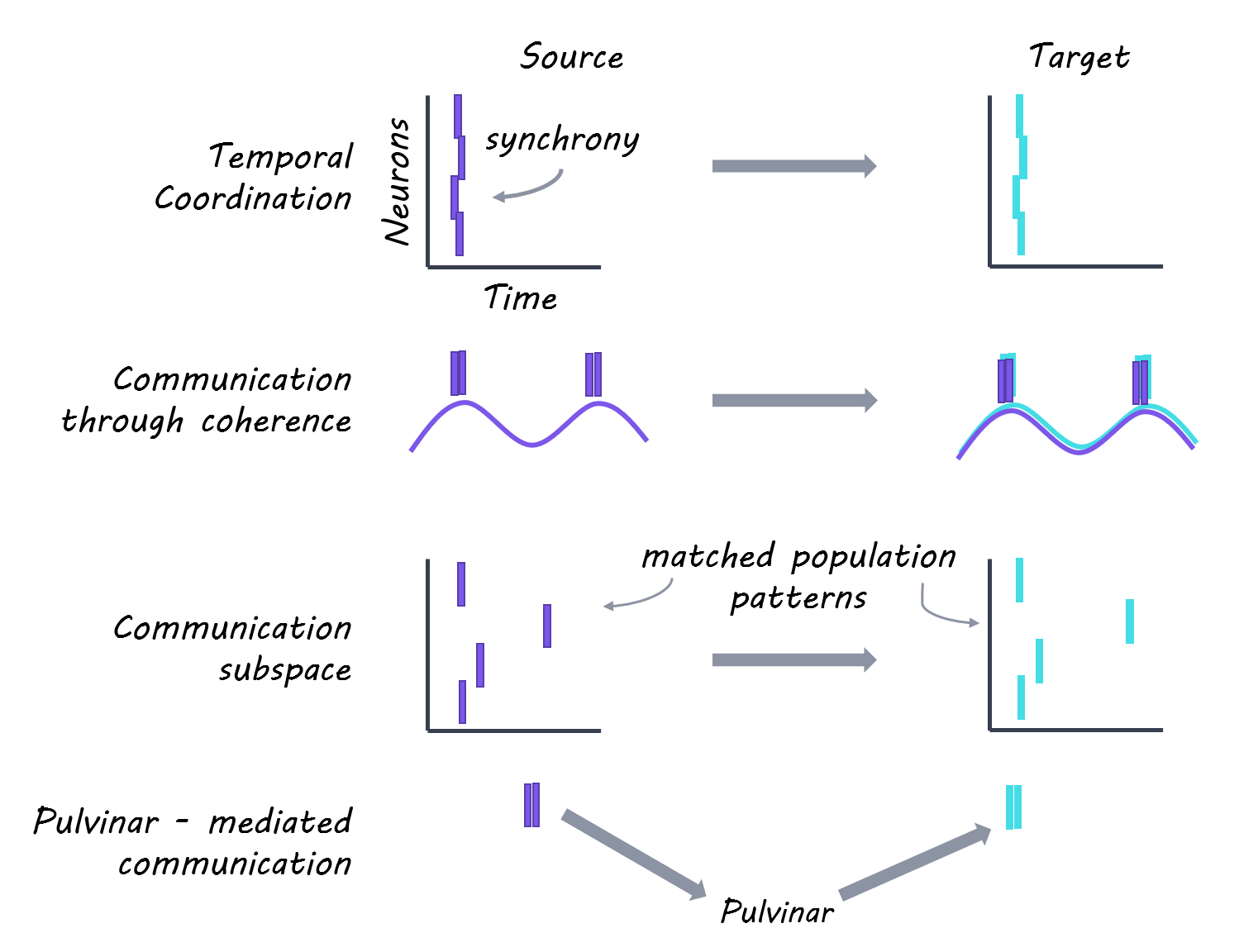
The authors highlighted four different theories that might help explain how different brain regions communicate:

1.     Synchronized neurons in the source region— an organized firing of neurons from a source region is much more likely to get picked up by downstream neurons. The same amount of firing, if disorganized, will have less of an effect.

2.     Coherence—neurons go through cycles of potential, being more or less likely to fire at different points in this cycle. If a signal arrives from a different area during a point of low firing potential, it probably won’t get picked up. But when two distant areas have coordinated cycles of low and high firing, communication between them will be strong. However, it takes some time to synchronize oscillations, which might not be fast enough to account for some of the brain’s more quick and flexible communication.

3.     Communication subspace— this one has to do with the specific pattern of neurons that fire. Like a pin lock on your phone, it doesn’t matter if any of the individual numbers is right; all of them have to be right at the same time in order to get access. This behavior has been seen in the motor cortex, which is often active without actually causing movement. However, when the pattern of neurons in the motor cortex is just right, it sends a signal to a specific muscle to move.

4.     Pulvinar-mediated communication— the pulvinar is a deep brain structure that receives input from many different regions of the cortex. It is possible that this structure is responsible for regulating communication strength between different areas of the brain. It could do this by way of one of the methods mentioned above (for example, changing synchrony or coherence between the regions), or by activating key messenger neurons in the source area. 

Anastasia (1).png

After establishing a few of the ways that neural populations might communicate, it is important to account for one more factor. Signals from “lower” regions (basic perception) send strong signals up to the next level of the brain’s hierarchy: we can think of these as feedforward signals. Information from “higher” regions (attention, context, etc.) is also sent back down the hierarchy, but these feedback connections are more diffuse. Feedforward and feedback connections may behave in very different ways.

What's the bottom line?

Brains are wonderfully complex, and though we have made great strides in understanding how they work, there are still a lot of questions about how distant neurons communicate. Moving forward, we will need to determine which of the theories of brain communication above are the most realistic and consistent with experimental data, keeping in mind that they may not be mutually exclusive.

Kohn et al. Principles of Corticocortical Communication: Proposed Schemes and Design Considerations. Trends in Neurosciences (2020). Access the original scientific publication here.