
Pupillary Responses Linked to Neuronal Activity in the Insula

Post by Lincoln Tracy

What's the science?

Much of our knowledge about activity in the brain has been obtained from studies of “task-evoked activations”—or looking at how groups of neurons respond to external events (such as lights or sounds) or experimental conditions. These types of studies have predominantly used functional neuroimaging to examine activity between different areas of the brain. However, due to the limitations surrounding the temporal resolution and signal-to-noise ratio of neuroimaging data, it is challenging to draw conclusions about the profiles and relevance of neuronal activity at the population level. This week in The Journal of Neuroscience, Kucyi and Parvizi developed a combined intracranial electroencephalography (iEEG)-pupillometry paradigm that reliably characterizes the functional significance of spontaneous neuronal activity.

How did they do it?

The authors recruited three patients with refractory focal epilepsy who had electrodes surgically implanted in their brain (specifically, the dorsal anterior insula) to monitor epileptic activity. After surgery, patients completed two tasks: a gradual-onset continuous performance task (where images of cities and mountains were displayed and transitioned from one to the other), and a resting-state task (where patients focused on a white cross displayed over a dark background for five to seven minutes). During the tasks, the authors recorded neuronal activity via the iEEG and changes in pupil diameter via an eye-tracking device.

What did they find?

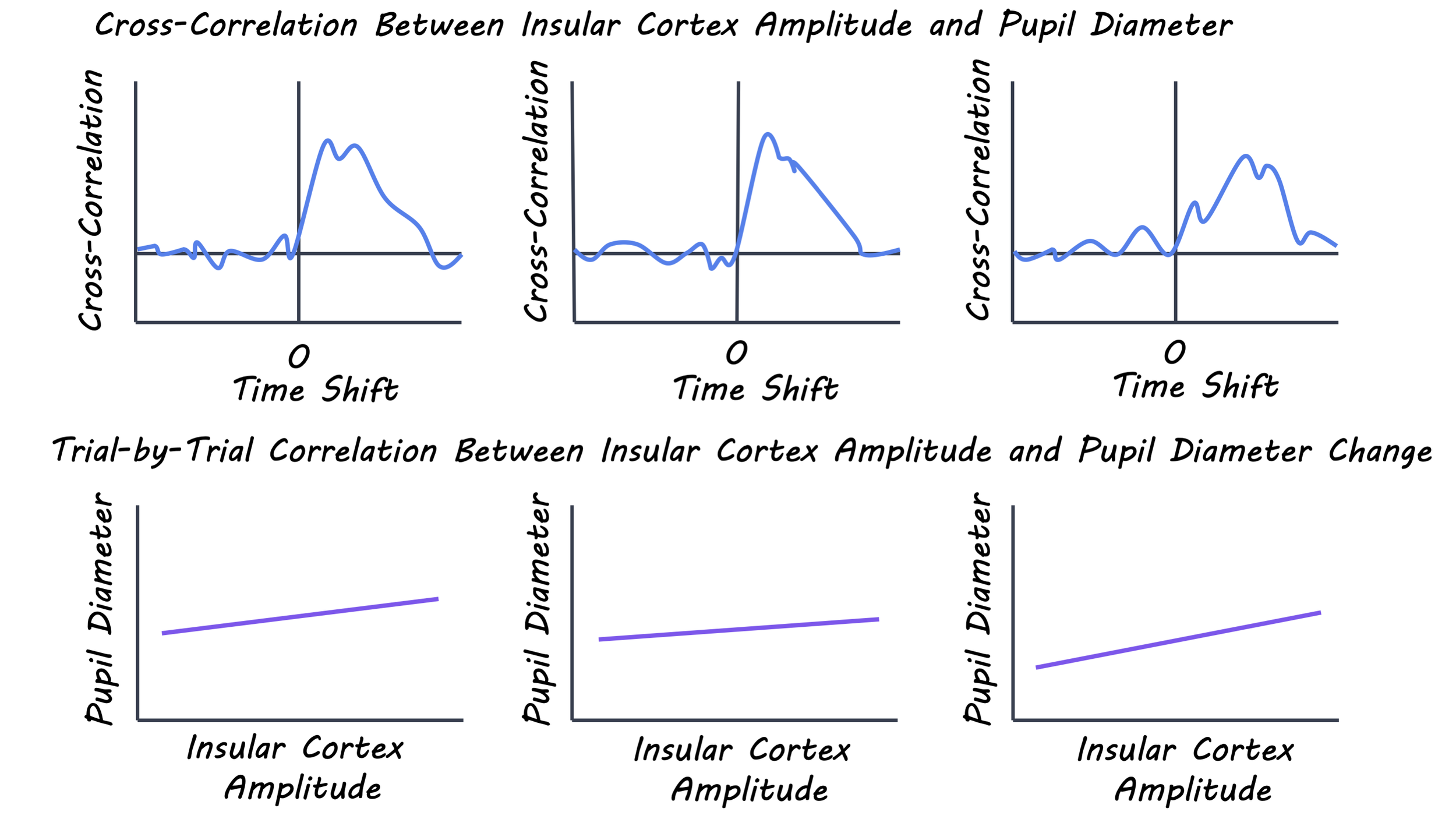
Insular activity and pupil size both increased following the presentation of the target stimulus during the continuous performance task. The authors examined the specific timing of the increases in insular activity and pupil size to determine whether the task-evoked insular activity was associated with the increase in pupil dilation. They found that the pupil dilation consistently occurred after the task-evoked insular activity. Furthermore, the magnitude of insular activity was positively correlated with how much the pupil increased in size (i.e., more insular activation was associated with a larger increase in pupil size).
Kucyi_image_Aug11.png

The authors also found that there was less frequent and more spontaneous neuronal activity in the insula during the resting-state task compared to the continuous performance task. Although there was less activity during rest, the electrophysiological properties of the activity were similar to the properties of activity during the active task. Finally, the authors further examined the relationship between neuronal activity and pupillary responses at rest. They found that the pupil response was time-locked to the insular activity, regardless of the task.

What's the impact?

This study found that spontaneous or task-induced activity in the insula was linked with brief dilations of the pupil. Such an association may arise from coordination between the insula and a network of cortical and subcortical areas that integrate with autonomic and neuromodulatory systems. While these findings shine the spotlight on a novel link between neural activation and autonomic function, additional work is required to determine whether the insula has a causal role in pupil control.
Kucyi_quote_Aug11.jpg

Kucyi & Parvizi. Pupillary dynamics link spontaneous and task-evoked activations recorded directly from human insula. Journal of Neuroscience (2020). Access the original scientific publication here.
Astrocytes Contribute to Hyperexcitable Neurons in a Mouse Model of Rett Syndrome

Post by Deborah Joye

What's the science?

Rett syndrome is a rare X-linked neurological disorder that occurs in 1 in 10,000 - 15,000 children, resulting in mild to severely disabling symptoms including seizures and breathing, speech, and movement impairments. Rett syndrome is caused by loss-of-function mutations in the Mecp2 gene, which makes a protein that interacts with DNA to change how genes are expressed. Loss of Mecp2 in mice is associated with changes in brain cells that express the important inhibitory transmitter GABA. In healthy brain networks, GABA-expressing cells inhibit the excitability of neurons by releasing GABA when activated (called phasic inhibition) but also by maintaining levels of GABA in the extracellular space, creating a constant inhibitory signal known as tonic inhibition. But exactly how mutations in Mecp2 alter GABA transmission resulting in Rett syndrome symptoms is not entirely clear. This week in Journal of Neuroscience, Dong and colleagues demonstrate that a decrease in extracellular GABA in the hippocampus of a Rett syndrome mouse model reduces tonic inhibition resulting in neuronal hyperexcitability that could be targeted to slow down Rett syndrome disease progression.

How did they do it?

To compare how neural signaling might be different in Rett syndrome, the authors used a mouse model of the disease that had the Mecp2 gene knocked out and compared them to wildtype mice. To test whether tonic inhibition was altered in the absence of Mecp2, the authors used electrophysiology to measure electrical currents caused by ambient GABA in hippocampal neurons. Since altered GABA currents could be due to changes in extracellular GABA or changes in GABA receptor expression, the authors next recorded electrical currents in the presence of exogenous GABA (i.e. not produced in the brain), or with drugs that block or activate GABA receptors.

Changes in extracellular GABA could be the result of altered clearance of GABA from the extracellular space or differential release of GABA from synapses. The authors used electrophysiology to test whether neurons from Mecp2 knockout mice exhibited altered GABA release or clearance. To directly test if GABA transporter function was altered, the authors also included a condition with a specific blocker for GAT3, a GABA transporter that is preferentially expressed by astrocytes. To test whether changes in tonic GABA currents in Mecp2 knockout mice were the result of alterations to GABA transporter expression in astrocytes, the authors used a technique that specifically knocks out Mecp2 only in astrocytes and recorded electrical currents caused by tonic GABA. Finally to test whether inhibiting the astrocyte-specific GABA transporter could alleviate Rett syndrome-like symptoms in Mecp2 knockout mice the authors recorded electrical currents and behavioral measures of wildtype mice, Mecp2 knockout mice, and Mecp2 knockout mice in the presence of a GAT3 blocker.

What did they find?

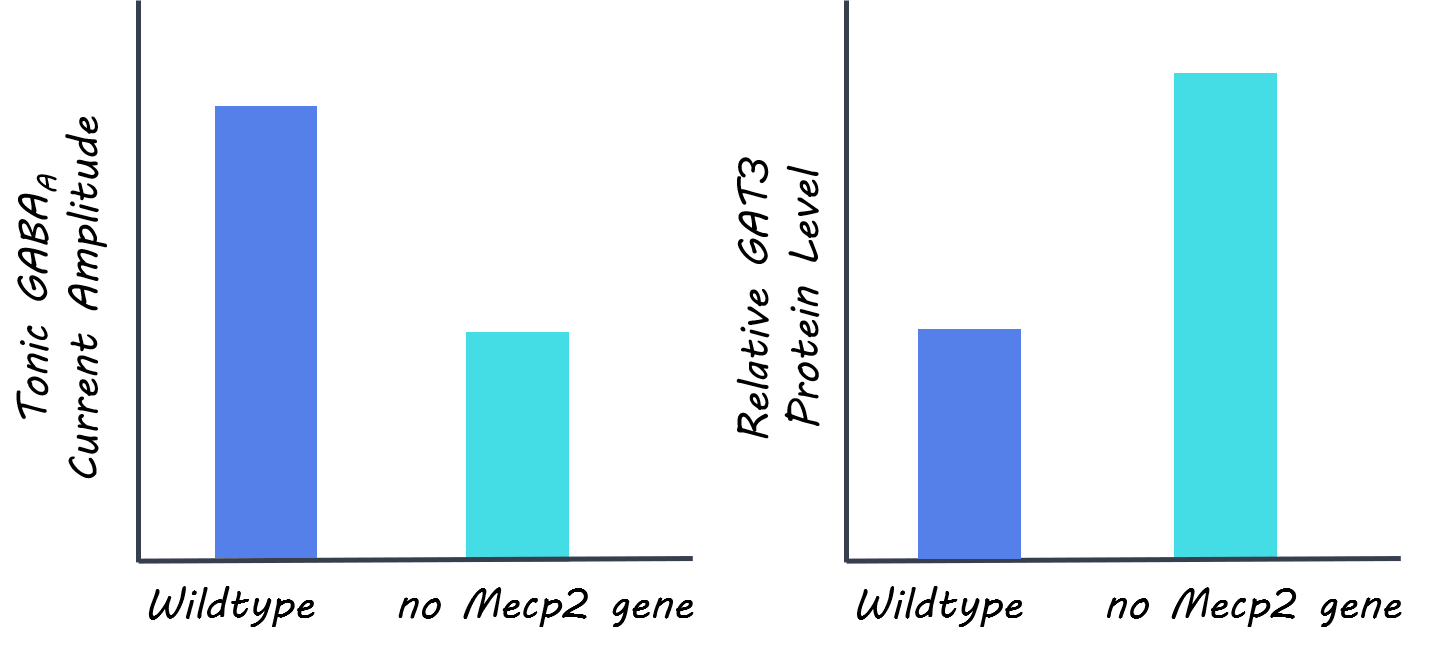
The authors found that knockout of the Mecp2 gene results in reduced tonic GABA currents in hippocampal neurons. Interestingly, they found that this difference in tonic GABA currents is not present in young mice, only manifesting around 8 to 10 weeks of age. Next, the authors found that the decrease in tonic GABA currents was due to a reduction in extracellular GABA in Mecp2 knockout mice and not a difference in GABA receptor expression. The authors next demonstrate that enhanced activity of the astrocyte-specific GABA transporter GAT3 results in the reduced tonic GABA currents seen in Mecp2 knockout mice. This suggests that since the GABA transporter is clearing GABA from the extracellular space at a faster rate, there is less ambient GABA resulting in less tonic inhibition and more hyperexcitable neurons. Notably, this upregulation of GABA transport appears specific to GAT3, since the authors show that GAT1 is not altered. The authors also reveal decreases in tonic GABA current when Mecp2 is knocked out specifically in astrocytes, further suggesting that the increase in GABA clearance is due to increased uptake by astrocytes. Finally, the authors demonstrate that blockade of GAT3 function returns neuronal excitability to wildtype levels, decreases the severity of Rett syndrome-like symptoms, and lengthens survival. This suggests that targeting of the hyperexcitable phenotype by blocking increased GABA uptake by astrocytes may have therapeutic value in the treatment of Rett Syndrome

deb.png

What's the impact?

This study provides a potential therapeutic mechanism for Rett syndrome by revealing that blockade of astrocyte-specific GAT3 can decrease the severity and slow the progression of Rett syndrome symptoms in mice. This important finding could have significant impacts on the clinical understanding of how Rett syndrome progresses and may result in better clinical outcomes for those living with this debilitating disease.

Dong et al., An Astrocytic Influence on Impaired Tonic Inhibition in Hippocampal CA1 Pyramidal Neurons in a Mouse Model of Rett Syndrome, Journal of Neuroscience (2020). Access the original scientific publication here.
Unraveling the Role of REM and Non-REM Sleep in Visual Learning and Brain Plasticity

Post by Elisa Guma

What's the science?

Sleep is important for learning and memory, amongst many other vital functions. However, there are still some open questions regarding the underlying neural mechanisms of sleep: (1) how do different phases of sleep such as non-rapid eye movement (NREM) or REM sleep, influence learning and memory? (2) is learning acquired before sleep enhanced or stabilized after sleep, or does sleep protect what was learned from being “overwritten” by learning something new? (3) is the facilitation of learning learning-specific or learning-independent? This week in Nature Neuroscience, Tamaki and colleagues addressed these questions by measuring the degree of plasticity and stability in neural signals during different phases of sleep after a visual perceptual learning task.  

How did they do it?

The authors trained participants on two different visual texture discrimination tasks (designed to interfere with each other), one before and one after sleep, and then tested performance on both after sleep. During sleep, they recorded glutamate and GABA (the main excitatory and inhibitory neurotransmitters in the brain) in visual areas using magnetic resonance spectroscopy (MRS) (a non-invasive technique for measuring brain metabolites). They simultaneously acquired polysomnography, a method used to record brain waves to identify sleep stages and cycles. Simultaneous MRS and polysomnography were also repeated in a separate session with no learning in order to investigate the E/I balance during different sleep phases without learning.

The ratio between GABA and glutamate was used to calculate an excitatory/inhibitory (E/I) balance, which is thought to be a reliable index of plasticity and stability in the brain. Thus, a high E/I balance indicates increased plasticity, and a low E/I balance indicates increased stability. This allowed authors to compare E/I balance (i.e. plasticity) in different phases of sleep (NREM and REM relative to wakefulness), and in individuals who had both NREM and REM or just NREM sleep. Further, the authors wanted to know if enhancement in plasticity for pre-sleep learning is followed by a less plastic period of stabilization and whether learning of one task interfered with the learning of another. They also examined whether these changes were specific to learning or simply innate to sleep.

What did they find?

Plasticity — based on E/I balance — of visual areas was increased during NREM sleep, and predicted improvements in performance on the learned task. Conversely, the E/I balance decreased in REM. The authors also examined whether pre-sleep learning stabilization occurred during sleep and found that individuals who had both NREM and REM sleep were able to maintain their learning from the pre-sleep training without interference from the post-sleep training on the new task relative to those who only had NREM sleep. The degree of resilience was also negatively correlated to E/I balance. 

REM_sleep_img.jpg

The authors then compared E/I balance in the different sleep phases when pre-sleep learning had occurred, to when no pre- or post-sleep training was performed. They found that E/I balance during NREM sleep was increased in both scenarios, irrespective of pre-sleep learning. Conversely, REM sleep E/I balance was only decreased from baseline after pre-sleep learning; if no learning had occurred, it was no different than baseline. This suggests that REM sleep played a stabilizing role, protecting the learning before sleep from interference by new learning after sleep, whereas NREM sleep increases plasticity in a learning-independent manner. After taking a closer look at the neurotransmitters, GABA decreases seemed to be driving changes in E/I balance during NREM sleep, whereas glutamate decreases seemed to drive E/I balance decreases during REM sleep after learning.

What's the impact?

This study found there are two distinct mechanisms for NREM and REM sleep, with complementary patterns of neurochemical and functional processing, involved in learning facilitation. During NREM sleep, plasticity increases independent of whether or not learning occurred, whereas stabilization occurs during REM sleep only if pre-sleep learning occurred. While these findings aid in our understanding of the relationship between sleep and learning, future work may be extended to understanding how sleep abnormalities may interfere with learning and memory.

REM_sleep_quote_Aug4.jpg

Tamaki et al. Complementary contributions of non-REM and REM sleep to visual learning. Nature Neuroscience (2020). Access the original scientific publication here