Non-Deceptive Placebos Alter Neural Markers of Emotion Regulation

Post by Shireen Parimoo

What's the science?

There is a lot of debate and controversy surrounding the placebo effect because placebos – an inactive form of medication or treatment – generally improve symptoms like emotion and pain (also known as the ‘placebo effect’). Yet, the effect relies on the patient’s belief that the treatment is real, as they are often unaware of receiving a placebo rather than an active medication. This has raised several ethical concerns about administering placebos without the patient’s knowledge. To address this, emerging research has investigated the efficacy of ‘non-deceptive placebos’ (NDP) on subjective outcomes such as pain, anxiety, and well-being, but it is not known whether their effects extend to biological outcomes as well. This week in Nature Communications, Guevarra and colleagues used electroencephalography to examine whether non-deceptive placebo effects extend to neural markers of emotional distress.

How did they do it?

Two experiments were conducted to investigate the impact of NDPs on (i) self-reported emotional distress (N = 62), and (ii) neural markers of emotional distress (N = 198 females). Participants were randomly assigned to a non-deceptive placebo group or a control group and were educated about the effectiveness of placebos and how they can still work even without deception (non-deceptive placebo group) or about the neural basis of pain (control group). A saline nasal spray was administered to both groups; the non-deceptive placebo group was informed that the spray was a placebo but would reduce their experience of emotional distress if they believed it would, whereas the control group was told that the spray would improve the quality of physiological recordings.

Participants then completed an image viewing task in which they saw highly negative and neutral images. In the first experiment, they rated how much distress they felt in response to each image, which provided a subjective measure of the effectiveness of NDP. In the second experiment, electroencephalography recordings were obtained while participants completed a similar image viewing task. The authors were interested in the late positive potential (LPP), which is an event-related potential associated with emotional distress. The LPP consists of an early attentional component that occurs between 400-1000ms following image presentation, and a sustained component between 1000-6000ms that is thought to reflect conscious appraisals. This sustained LPP component is reduced by cognitive reappraisal strategies.

What did they find?

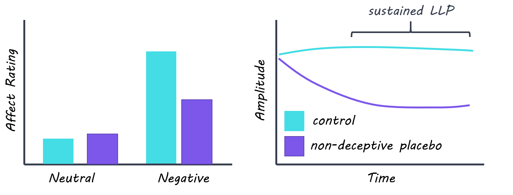
Emotional distress ratings for neutral images were similar across participants in both groups. The non-deceptive placebo group provided lower ratings for negative images compared to the control group, which is consistent with past research on the effect of NDPs on self-reported outcomes. The early component of the LPP did not reliably differentiate between the two groups, suggesting that NDPs do not affect the early attentional processing of emotional stimuli. In contrast, there was a widespread reduction in the sustained component in the non-deceptive placebo group that occurred around 2000-3000ms after the images were presented. This means that the immediate processing of the images did not differ between the two groups, whereas the NDP seemingly influenced the appraisal stages of emotional processing of the images. Interestingly, this reduction occurred for both the neutral and negative images in the placebo group, indicating that the NDP had a general effect on emotional distress rather than a specific effect toward processing negative images.
shireen (2).png

What's the impact?

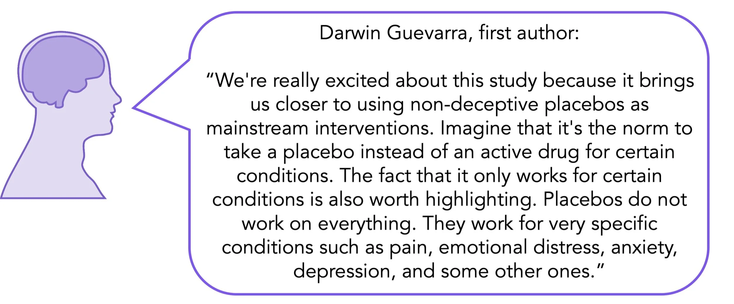
This study is the first to demonstrate the neural and subjective impact of non-deceptive placebos on emotional distress. These findings are exciting because they open up the possibility of using NDPs as a treatment approach for managing psychiatric disorders characterized by emotional distress, such as depression. Further research is needed on the longer-term efficacy of NDPs and their potential generalizability to other populations and domains like pain management.
placebo_quote_Aug4.jpg

Guevarra et al. Placebos without deception reduce self-report and neural measures of emotional distress. Nature Communications (2020). Access the original scientific publication here.
 

The Effect of Bilingualism on Brain Development

Post by Cody Walters

What’s the science?

While it is well-known that bilingual brains are structurally different from monolingual brains, it is less clear how these structural changes unfold over time. This week in Brain Structure and Function, Pliatsikas et al. explored how the brains of over 700 monolingual and bilingual individuals differentially developed through childhood and adolescence. 

How did they do it?

The authors analyzed an existing neuroimaging dataset (from the Pediatric Imaging, Neurocognition, and Genetics project) that consists of monolingual and bilingual individuals aged 3-21. The dataset includes structural MRI and DTI data which the authors used to quantify cortical thickness, surface area, and volume as well as white matter fiber tract integrity (as measured by fractional anisotropy and mean diffusivity). In order to examine the relationship between age and brain development, the authors used generalized additive models (GAMs). GAMs use a basis set of smooth functions, where each function has an associated coefficient that can be adjusted to fit the data. The coefficients were estimated using penalized regression in order to prevent overfitting.

What did they find?

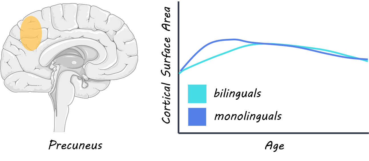
The authors found that monolinguals (as compared to bilinguals) had increased cortical thickness and volume across several parieto-frontal structures during early childhood. However, bilinguals had increased cortical thickness and volume across several regions in late childhood and early adolescence, with this increase converging back to monolingual levels in many cortical areas. Interestingly, the precuneus was the only region that showed a difference in cortical surface area between monolinguals and bilinguals, with monolinguals having greater precuneus surface area in early childhood. Altogether, bilinguals generally had lower cortical metrics (e.g., thickness and volume) than monolinguals in early childhood, but higher metrics in late childhood and early adolescence that either persisted in some regions or converged back to monolingual levels in others. No differences were observed between monolinguals and bilinguals in subcortical structures or the cerebellum. Only one white matter tract (connecting the striatum to the inferior frontal cortex) showed differences between monolinguals and bilinguals, and this difference followed the same trend as the cortical data, with the fiber tract density being lower in bilinguals during early childhood but higher in late adolescence. 

cody (2).png

What’s the impact?

This study provides new insight into how the bilingual brain develops over a nearly 20 year period. These data largely highlighted differences in striato-frontal connectivity as well as frontal and parietal structures. These findings suggest that, during development, bilingualism affects brain structures underlying the parieto-frontal dorsal language stream (which is involved in mapping speech sounds to articulatory representations); basal ganglia-based procedural memory (which underlies the implicit learning of sequences and plays important roles in syntax, morphology, phonology, and articulation); and executive function (which allows bilinguals to appropriately alternate between their two languages).

language_quote_Aug4.jpg

The effect of bilingualism on brain development from early childhood to young adulthood, (2020). Access the publication here.

How Does the Brain Map Our Increasingly Complex Social World?

Post by Flora Moujaes

What's the science?

When navigating the world around us it is imperative we keep track of our increasingly complex social network: who our family, friends, and co-workers are, and how they relate to each other. Developing a map of our social environment is useful as it allows us to make inferences in novel situations or based on sparse information. We know from previous research that when we encounter a new physical environment, such as a new city, we first sample the environment, building up small and separate representations. Then, as we get to know the physical environment better, we integrate these representations into a coherent internal map. So is the same process used to represent abstract relationships, such as social networks? This week in Neuron, Park and colleagues use fMRI to show that the brain builds maps of social networks the same way it builds maps of physical space.

How did they do it?

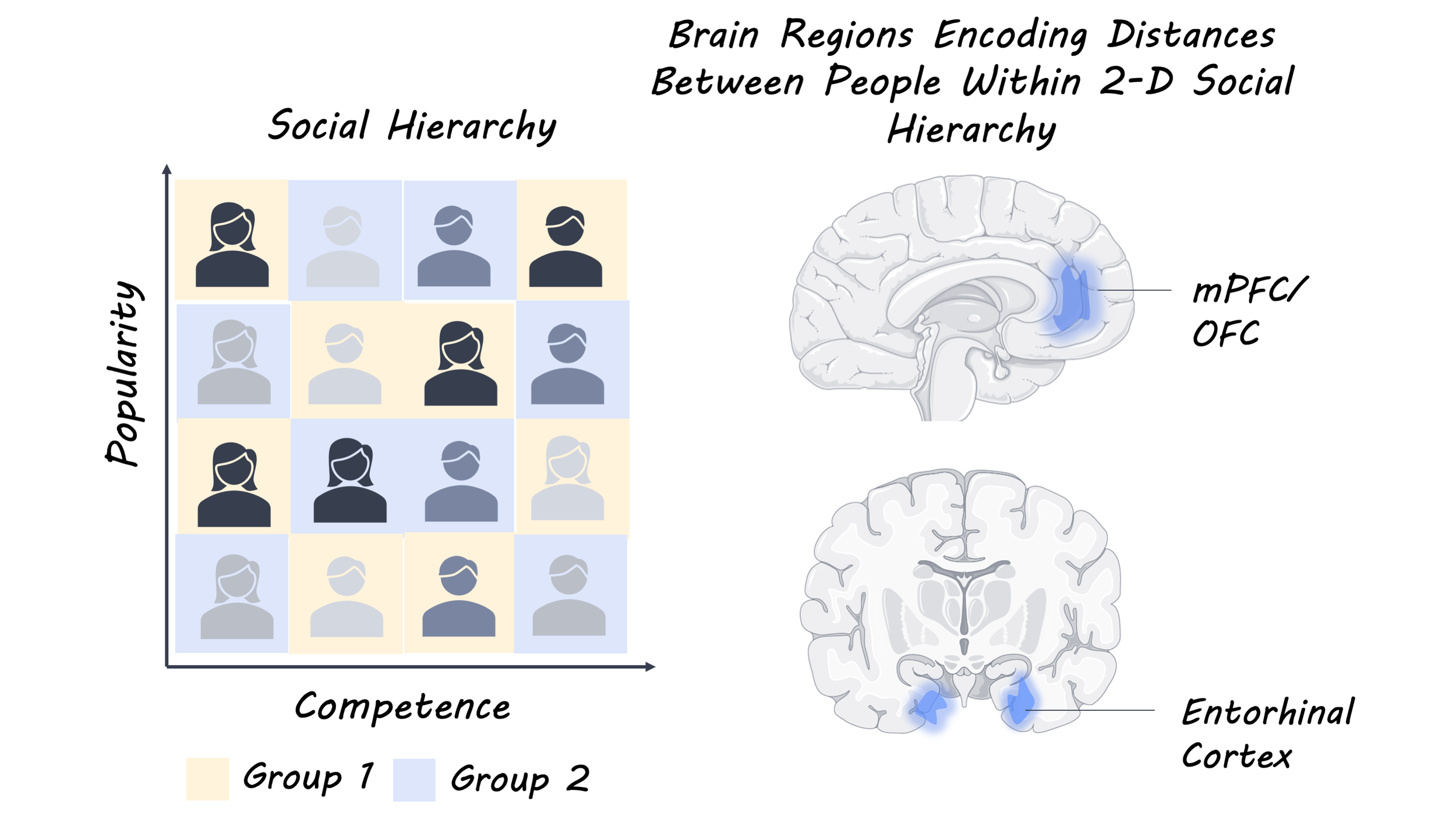
To investigate how the human brain constructs maps of social hierarchies, 27 participants were trained on a task where individuals ranked in two social hierarchies: popularity and competence. The training involved the participants being given relational information about the two dimensions, on different days. For example, the participant may be presented with two individuals, Alice and Bob, and informed that Alice is more popular than Bob. The true social hierarchy could thus be mapped as a two dimensional grid defined by the two hierarchies: popularity and competence. 

To explore whether the human brain represents social hierarchies as a one dimensional or multidimensional map, and what brain regions are involved in the representation, the training period was followed by an fMRI experiment. This experiment examined whether participants represent social hierarchies in a single dimension (e.g. they have separate maps for competency and popularity) or in two dimensions (e.g. competency and popularity are represented in the same map). To do this participants were required to make inferences about the relative competency and popularity of novel pairs of individuals. Neural activity was examined in various regions of the brain including the hippocampus and entorhinal cortex, which organize both spatial and non-spatial relational information into a reference map, and the orbitofrontal cortex, which is theorized to represent a goal or the current state in a task structure to guide goal-directed decision making. 

What did they find?

The researchers found that the brain spontaneously represents individuals’ status in social hierarchies in a map-like manner in 2-D space, as participants were able to generalize to both social hierarchies (popularity and competence) when presented with novel pairs. They also found that distances between people in the 2-D grid were related to neural activity, as the pattern similarity between faces represented in the hippocampus, entorhinal cortex, and the medial orbitofrontal cortex was related to the distance between faces in the social network grid.  This result is particularly striking, as the grid itself was never shown to participants, demonstrating that participants instinctively built up this grid-style map of social hierarchy from relational information they received about pairs of participants.

Social_image_Jul28.png

What's the impact?

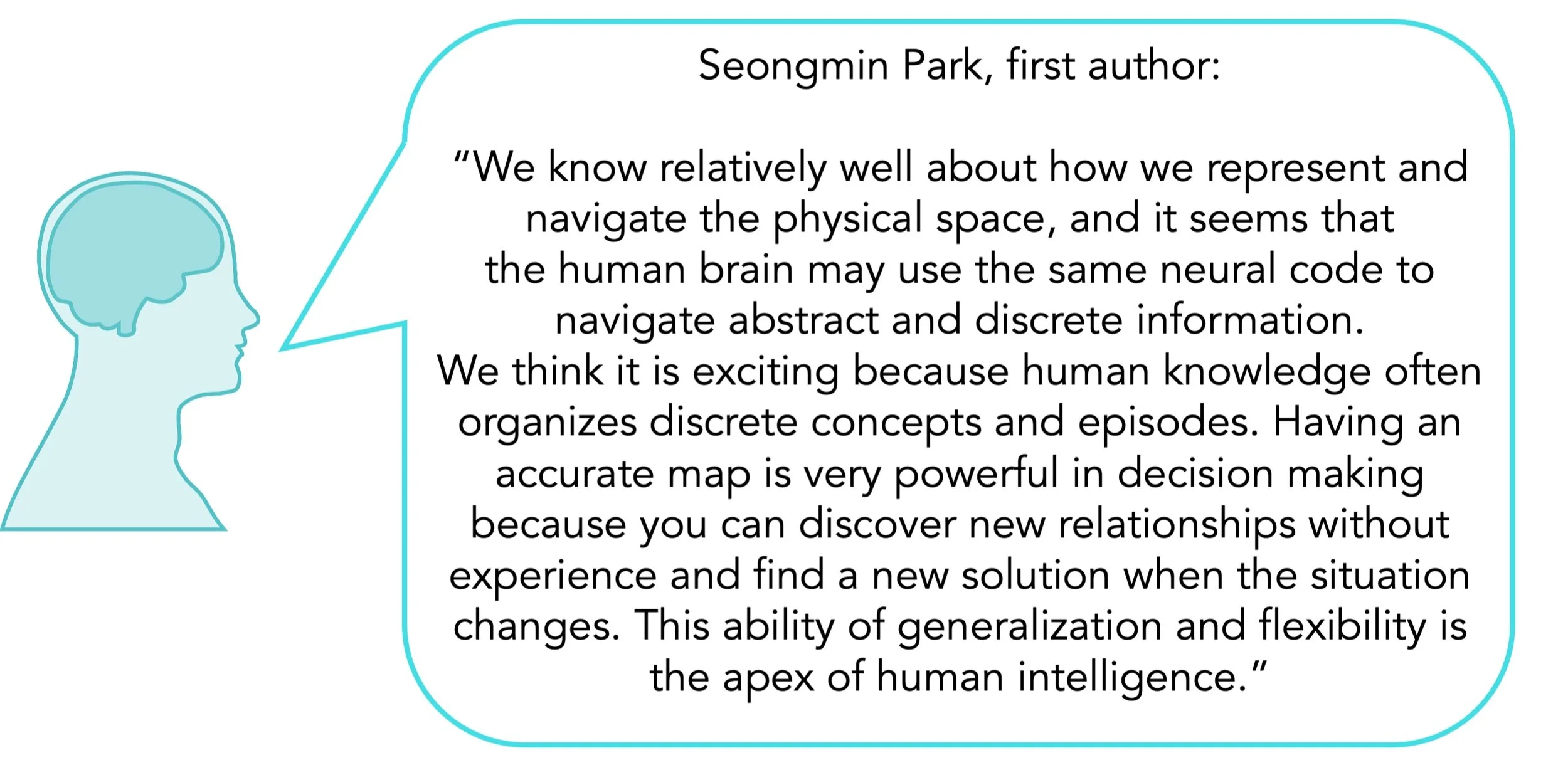
Overall this study suggests that the brain utilizes the same neural system for representing our physical space and our social network. It shows that by building a social network map, participants are able to make accurate inferences about novel situations. Further, these results support the theory that the hippocampus and entorhinal cortex play a key role in constructing a global map from local experiences, whether physical or social.

social_quote_Jul28.jpg

Park et al. Map making: Constructing, combining, and inferring on abstract cognitive maps Neuron (2020). Access the original scientific publication here.