Replay of Spatial Paths in the Medial Prefrontal Cortex Facilitates Strategy Shifting

Post by Cody Walters 

What’s the science?

Planning and decision-making often require remembering specific routes and locations. The hippocampus and prefrontal cortex have been shown to replay task-relevant spatial trajectories following learning, and this reactivation of behavioral sequences has been viewed as a possible neural mechanism for memory consolidation and retrieval. However, how hippocampal and prefrontal cortex replay relate to one another and whether they play dissociable roles in learning and memory remains unclear. This month in Neuron, Kaefer et al. identified novel properties of prefrontal replay in rats navigating a rule switching task. 

How did they do it?

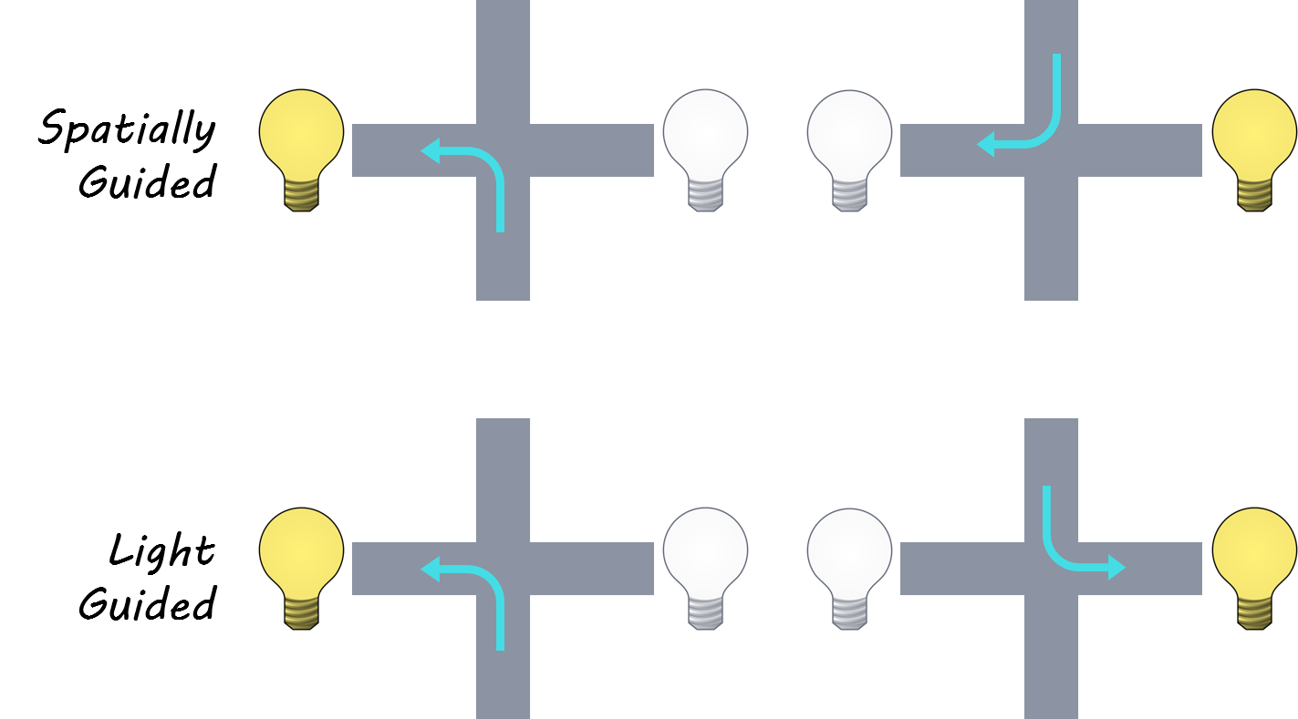
The authors trained four rats to perform a rule switching task on a plus maze (a maze with four arms, shaped like a plus sign). Rats were placed at the end of either the north or south arm and then had to navigate to either the east or west arm to receive a food reward. Importantly, there were two rules: under the spatial rule, one of the two horizontal arms was consistently rewarded, while under the visual rule rats had to go to the arm that had a light cue to receive food. Each session started off with one of the two rules in play until the animal reached a set performance criterion, at which point there would be an unannounced rule switch. They recorded neural activity from the medial prefrontal cortex (mPFC) and the dorsal hippocampus (HPC) of the rats as they performed the task. This allowed the authors to explore the differences and similarities between how the HPC and mPFC encode information about space, replay spatial paths, and respond to rule changes.

March2_Cody.png

What did they find?

They found that, unlike hippocampal place cells, most mPFC neurons had symmetrical spatial representations of the environment (e.g., an mPFC neuron that responded to the middle location on the north arm of the maze also responded to the middle location on the south arm of the maze). They also observed both forward (start arm to goal arm) and backward (goal arm to start arm) spatial trajectory replay in the mPFC. Importantly, these mPFC replay events did not significantly co-occur with HPC replay. At the goal location (where the rats received food), the rate of mPFC replay was positively correlated with rule switching performance (i.e., the number of laps it takes before shifting over to the correct strategy following a rule switch). On the other hand, the rate of HPC replay at the goal location was negatively correlated with rule switching performance. Interestingly, when rats were at the center of the plus maze (just prior to making a choice to either go left or right) there were more mPFC forward replays on error laps and more backward replays on correct laps (whereas the HPC exhibited an increase in both forward and backward replays on correct laps relative to error laps at the center of the maze).  

What’s the impact?

Previous work has shown that mPFC task-relevant replay occurs during sleep, but this study suggests that mPFC replay 1) occurs in awake states, 2) facilitates behavioral flexibility in a dynamic environment, and 3) might be largely independent of HPC replay. These findings advance our understanding of how different networks respond to the challenge of shifting environmental contingencies and highlight replay as potentially being a more general neural computation with structure-specific function.

cody_quote_Mar3.jpg

Kaefer et al. Replay of Behavioral Sequences in the Medial Prefrontal Cortex during Rule Switching. Neuron, (2020). Access the publication here.

Natural Rhythms of Periodic Temporal Attention

Post by Stephanie Williams 

What's the science?

Temporal attention allows us to flexibly allocate attentional resources to specific points in time. We don’t fully understand the limits of our temporal attention resources, or whether different sensory modalities may be constrained in different ways. This week in Nature Communications, Zalta, Petkoski, and Morillon show that temporal attention is constrained within specific frequency ranges specific to each sensory modality and modulated by overt motor activity. 

How did they do it?                             

The authors performed a set of six behavioral experiments across two sensory modalities; auditory and visual. They designed the experiments to characterize the optimal frequencies of, and the limits of, periodic temporal attention during visual and auditory perception.  The authors played pure tones or displayed a visual grating to participants at different isochronous tempi, and then asked participants to report whether the last stimulus of the sequence was on or offbeat. They used the accuracy of participants in this task to estimate their optimal beat frequency. The authors added distractors to both the auditory and visual tasks to adjust for difficulty at the individual level and achieve similar performance across modalities. They found that they had to add fewer distractors to the visual task to achieve a similar level of difficulty as in the auditory task. To test how overt motor activity could contribute to temporal attention, the authors asked participants to either sit completely still while performing the task (the “passive” session) or to tap their index finger on beat with the target stimuli (the “tracking” session). They compared the behavioral results across the two sessions to understand the motor contribution to temporal attention. Finally, they asked participants to tap their finger (“free tapping”) at a comfortable rate for 60 seconds to estimate the natural rate of rhythmic movements of each participant.   

The authors then built a model for sensory-specific periodic temporal attention. The model was made up of three time-delayed coupled oscillators comprising the periodic stimulus, a sensory-specific temporal attention oscillator, and a motor oscillator. In their model, periodic temporal attention was modeled as an ongoing intrinsic oscillatory process with an optimal sampling rate. 

What did they find?

The authors found that there was an optimal beat frequency for temporal attention in both the auditory and visual perception tasks. In experiment one, the authors found that performance across the eight different auditory tempi investigated had an inverse U-shape profile. The peak of the inverse U sat at the tempo for which participants showed their peak performance, which was different for the auditory and visual tasks. The authors found that the optimal tempo of auditory temporal attention was obtained for a rhythmic sampling frequency of 1.4 Hz. In contrast, the authors found that visual temporal attention had an optimal sampling frequency of approximately 0.7 Hz (an optimal beat frequency approximately two times lower than the auditory rhythm).

March2_Stephanie.png

When the authors compared the passive and motor tracking sessions from the experiments investigating motor contributions, they found different effects in the auditory and visual conditions. In the auditory task, having participants tap their index finger could improve performance of up to 10%, but only within a specific frequency range, between 1.3 to 2.2 Hz. The authors calculated the optimal beat frequency to be around 1.5 Hz in both the passive and tracking sessions. During the fastest tempi, participants tended to tap too slowly. Similarly, during the slowest tempi, participants tended to tap too fast. In line with previous findings, the authors, therefore, suggest that overt motor activity optimizes auditory temporal attention. Specifically, the benefit of the motor activity is rate-restricted and has a maximum effect at 1.5 Hz. In the visual perception task, motor tapping significantly decreased performance at specific frequency ranges (between 1.7 and 2.2 Hz). The authors observed that in this visual task participants tapped faster and later than the actual beat. Trials that participants scored correctly on were associated with lower sensorimotor simultaneity than trials scored incorrectly. When participants closely tracked the beat with the motor task, they performed worse on the visual trials, especially at 1, 1.7 and 2.2 Hz. In short, motor tracking improved performance accuracy in the auditory perception task, but negatively impacted accuracy in visual perception task, due to the constraints in the temporal alignment between motor and attention fluctuations. When the authors asked subjects to free tap, they found that the subject tapped at a rate of 1.7 Hz, on average.

The authors’ model reproduced the results they found in both the visual and auditory sessions of the “passive” and “tracking” sessions (the model’s fit quality to the behavioral data was .92 for auditory and 0.95 for visual tasks). The model showed that the time delay between stimuli and the motor oscillator was critical for estimating the impact of motor tracking on performance across different sensory modalities. The authors found that a slight time delay (100 ms) had a positive impact on the quality of periodic temporal attention, while a longer delay had a disruptive effect, respectively compatible with auditory and visual periodic temporal attention. 

What's the impact?

The authors quantify the optimal sampling frequencies of auditory and visual temporal attention and show that periodic temporal attention may be optimal at these frequencies. They build a model that captures their behavioral results and further shows that the impact of motor activity on temporal attention depends upon the temporal alignment between motor acts and attention fluctuations.

Steph_quote_mar3.jpg

Zalta et al. Natural Rhythms of Periodic Temporal Attention. Nature Communications. (2020). Access the original scientific publication here.

Nucleus Accumbens Inhibition is Required for Appetitive or Fearful Behaviors

Post by Lincoln Tracy 

What's the science?

The neurotransmitter glutamate plays a key role in causing neurons to fire action potentials. These impulses are required for neurons in different areas of the brain to communicate. DNQX is a peptide that inhibits neurons from firing. Injecting DNQX into the nucleus accumbens, the part of our brain that mediates reward, can produce either motivating (such as increased eating and food intake) or fearful/avoidance behaviors (such as digging to escape predators) in rats. The type of behavior caused by the injection of DNQX depends on which part of the nucleus accumbens it is injected into. It is hypothesized that inhibiting the medium spiny neurons of the nucleus accumbens leads to motivating behaviors. However, there is no evidence that preventing neurons of the nucleus accumbens from firing is required for DNQX to produce either type of behavior. This week in the Journal of Neuroscience, Baumgartner and colleagues sought to directly test whether local neuronal inhibition is required for DNQX injected into the nucleus accumbens to elicit motivating behaviors. 

How did they do it?

First, a group of rats underwent brain surgery. The surgery involved implanting a cannula (a thin tube) and optic fibers (which could be stimulated with lasers) into the nucleus accumbens. The authors also injected one of two viruses into the nucleus accumbens. One virus contained channelrhodopsin, a light-sensitive protein the authors could optogenetically stimulate. The other virus was an inactive control virus. Rats were placed in a chamber for an hour while they were observed for appetitive (e.g. eating) and defensive (e.g. digging) behaviors. Each rat completed the behavioral testing four times: the four conditions were (a) a baseline condition, where saline was injected into the nucleus accumbens and optogenetic laser stimulation was not applied, (b) a laser alone condition, where saline was injected but the nucleus accumbens was stimulated with the laser, (c) a DNQX alone condition, where the rats received DNQX but not laser stimulation, and (d) a combined condition where rats were injected with DNQX and had laser stimulation applied. Additionally, half the rats completed the behavioral testing in a ‘high stress’ environment – with Iggy Pop’s 1977 Hippodrome Paris gig playing in the background. The remaining rats were tested without the music being played. The authors then compared the appetitive and defensive behaviors between the different testing conditions.

What did they find?

The authors first found that injecting DNQX into the nucleus accumbens in the normal laboratory environment made the rats eat more food and eat for longer compared to when they were injected with saline. However, stimulating the channelrhodopsin in the nucleus accumbens with light reversed the DNQX-induced increase in food intake in rats where the tips of the cannula and the optic fibers were close together. The lasers had no effect on eating behaviors if the two tips were separated by more than 1mm. Few defensive behaviors were observed under any of the testing conditions in the normal laboratory environment. When rats were subjected to the stress of listening to Iggy Pop, injecting DNQX resulted in an increase in defensive behaviors. The increased defensive behavior in the ‘high stress’ environment was counteracted by combining the DNQX injection with the laser stimulation. The effects of DNQX and optogenetic laser stimulation on feeding behaviors in the normal lab environment were replicated in the ‘high stress’ environment of listening to Iggy Pop.

Lincoln_img_Feb25.jpg

What's the impact?

This study demonstrates that neuronal inhibition in the nucleus accumbens is required to generate either motivating or avoiding behaviors. Combining optogenetic stimulation and DNQX administration at the same site within the nucleus accumbens simultaneously prevents the activation of additional brain regions and the resulting appetitive or fearful behaviors. These findings shed light on the role of the nucleus accumbens in psychiatric conditions of pathological motivation, such as addiction or paranoia.  

Baumgartner et al. Desire or dread from nucleus accumbens inhibitions: Reversed by same-site optogenetic excitations. Journal of Neuroscience (2020). Access the original scientific publication here.