Surprise and Decision Making in the Anterior Cingulate Cortex

Post by Anastasia Sares

What's the science?

The anterior cingulate cortex, situated in the frontal lobe at the midline where the two hemispheres meet, is an important region of the human brain. Previous studies have connected it to error detection, cognitive control, and decision making. However, there are a few different hypotheses about what drives activity in this area. The “choice difficulty” hypothesis (CD) says that the anterior cingulate tracks difficulty—tougher decisions generate more activity. The “expected value of control” hypothesis (EVC) tracks net gain—how much reward will I get for the effort I put in? EVC predicts more activity for high reward, especially if the brain has to gear up and make an effort in order to get the reward. Finally, the “predicted response outcome” hypothesis (PRO) tracks surprise—events that violate expectations generate more activity, regardless of whether the results are perceived as “good” or “bad.” This week in Nature Human Behaviour, Vassena and colleagues pitted these three hypotheses of anterior cingulate function against each other in a speeded decision-making task.

How did they do it?

Before they started the experiment, participants learned to associate random fractal images with a certain number of points (picture 1 = 30 points, picture 2 = 80 points, etc.). They were told that the points they accumulated in the task would translate into extra money at the end of the experiment. In each trial of the task, four of the fractal images were shown on the screen. The participants had three-quarters of a second to choose either the set of images on the left or the set of images on the right. Of the images they chose, one would be randomly selected, and they would receive the equivalent amount of points. To complete this task, therefore, the participants had to be good at quickly estimating the value of the options on the right versus the options on the left. The task was completed in MRI so the response of the anterior cingulate could be measured.

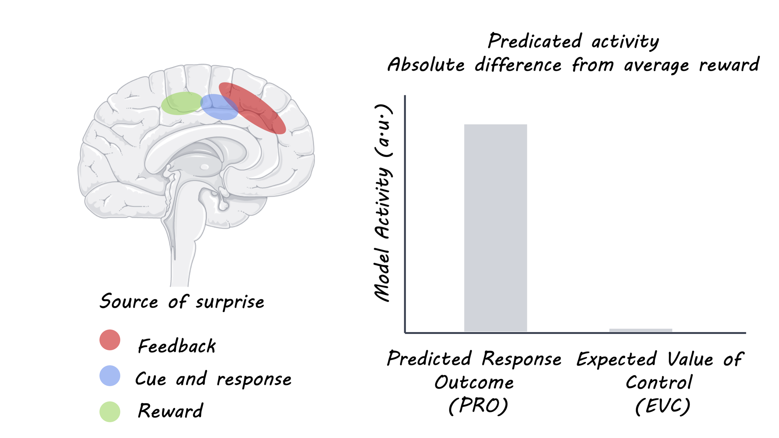
Each of the hypotheses presented earlier (choice difficulty, expected value of control, and predicted response outcome) should show a different pattern of activity in the anterior cingulate during this task. CD predicts the most activity when the choice is difficult; that is when the amount of points on the left is similar to the amount on the right. EVC predicts the opposite: the anterior cingulate should be least active when the options are similar because the difference in potential reward is small compared to the effort required to distinguish between them. PRO predicts two peaks of activity, because with a large difference in value, one option is surprisingly bad, and with a small difference in value it is uncertain which option you will choose (making the choice itself “surprising”).

 What did they find?

The authors compared the activity in the anterior cingulate cortex to the three different predictions and found that the PRO model (predicted response outcome) matched most closely. This is in line with the idea that the anterior cingulate reacts to unlikely or unpredicted events (surprise). During a task like this, people are quickly able to figure out how difficult the average trial is, so when a surprisingly bad option or two very similar options appear, the anterior cingulate’s activity increases. What’s more, an extremely low or high reward at the time of feedback also activated the same area.

acc_img_Jan21.png

What's the impact?

This study tested predictions from three different hypotheses and found one to be the clear winner: the main function of the anterior cingulate is processing surprise. This is a crucial step in the scientific enterprise; after some hypotheses are generated about the natural world, they must be tested against one another in carefully designed experiments that allow us to determine which hypothesis is stronger.

ACC_quote_Jan21.jpg

Vassena et al. Surprise, value and control in anterior cingulate cortex during speeded decision-making. Nature Human Behavior (2020). Access the original scientific publication here.

The Time Of Day We Eat Is Associated with Diet-Induced Obesity

Post by Flora Moujaes 

What's the science? 

Worldwide obesity has nearly tripled since 1975: currently over 13% of the world’s adult population is obese. This increase in obesity is correlated with the more widespread availability of highly processed energy-dense rewarding foods that encourage snacking outside of regular meal times. However, it is not just the number of calories consumed that are important for understanding weight gain, but also when they are consumed. Proper maintenance of energy throughout the day requires that meals are synchronized with daily metabolic rhythms. For example, even if two mice consume the same number of calories, eating food at different times (e.g. snacking) could lead to obesity in one mouse but not the other. This issue is particularly prevalent in modern society as the central pacemaker is under constant dysregulation by artificial light. This week in Current Biology, Grippo et al. investigate the mechanisms through which the increased availability of energy-dense food and feed times lead to diet-induced obesity. 

How did they do it? 

To explore the mechanisms underlying diet-induced obesity, mice were either fed a diet comparable to that eaten in the wild, or had unlimited access to a high fat, high sugar diet. To examine the involvement of dopamine in diet-induced obesity, the cre-lox recombinase enzyme (an enzyme that allows you to knock out genes solely in subsets of cells e.g. the brain) was used to knock out the Drd1 gene in the brain. This gene encodes the D1 subtype of the dopamine receptor, which is the most abundant dopamine receptor in the central nervous system. These mice are referred to as the ‘knockout’ mice. Finally, to explore exactly where in the brain dopamine is involved in diet-induced obesity, the researchers selectively restored Drd1 expression in (1) the nucleus accumbens or (2) the suprachiasmatic nucleus (SCN). The nucleus accumbens was chosen as it is the reward processing center of the brain. The SCN was chosen as it is the main biological clock: the SCN receives light cues from the eyes and interprets them as the time of day, as well as cues when the body consumes and metabolizes food.

What did they find?

Researchers first showed that unlimited access to energy-dense food led to obesity. While mice fed a diet akin to that eaten in the wild maintained normal eating and exercise schedules and proper weight, mice with unlimited access to energy-dense food rapidly developed obesity, diabetes, and metabolic diseases. However, knockout mice with impaired dopamine D1 receptor functioning were resistant to weight gain following exposure to unlimited energy-dense food. Researchers also found that unlimited access to energy-dense food led to eating at irregular times. As nocturnal animals, mice usually eat 80% of their food during the night when exposed to a healthy diet, however mice with unlimited access to energy-dense food only ate 60% of their food during the night. In contrast, knockout mice with impaired dopamine D1 receptor functioning did not change their feeding times following exposure to unlimited energy-dense food. Taken together, these data suggest that D1 is important for the overconsumption of energy-dense food, predominantly during rest, leading to obesity.

Obesity.jpg

Mice with restored D1 dopamine receptor functioning in the nucleus accumbens did not gain weight when exposed to unlimited energy-dense food - while they did increase their consumption of food during rest, they did not increase their overall calorie intake and therefore, did not become obese. In contrast, mice with restored D1 dopamine receptor functioning in the central circadian clock (SCN) did gain a substantial amount of weight when exposed to unlimited energy-dense food. Both their consumption of food at rest and overall calorie intake was significantly increased. Overall, this indicates that dopamine D1 receptor functioning in the central circadian clock (SCN) is crucial for diet-induced obesity. 

What's the impact? 

This study uncovered a novel mechanism for understanding how energy-dense diets lead to obesity, defining a connection between the reward and circadian pathways in the regulation of pathological calorie consumption. The authors demonstrate that dopaminergic signalling within the central circadian clock (SCN) disrupts the timing of feeding, resulting in an overconsumption of food, which leads to obesity, diabetes, and metabolic disease. This research not only has significant clinical implications by furthering our understanding of the mechanisms that underlie obesity but also helps to explain the growing popularity and effectiveness of diets that involve time-restricted feeding (e.g. intermittent fasting).

eating_quote_Jan14.jpg

Grippo et al. Dopamine Signaling in the Suprachiasmatic Nucleus Enables Weight Gain Associated with Hedonic Feeding. Current Biology (2020). Access the original scientific publication here.

Astrocytes Detect Changes in Intracranial Pressure and Maintain the Homeostatic Control of Brain Blood Flow

Post by Amanda McFarlan 

What's the science?

Cerebral perfusion pressure, determined by the difference between mean arterial blood pressure and intracranial pressure (pressure inside the skull), is responsible for driving blood flow and delivering oxygen to brain tissue. Cerebral blood vessels within the brain are wrapped with the endfeet of astrocytes, a type of glial cell important for the metabolic and structural support of neurons. Blood vessels respond to changes in intracranial pressure by dilating or constricting, which likely causes structural changes to the astrocyte endfeet, and thus makes astrocytes ideally located to act as pressure sensors in the brain. This week in Nature Communications, Marina and colleagues tested the hypothesis that astrocytes in the brain act as physiological sensors that detect changes in blood flow. 

How did they do it?

The authors investigated how decreasing cerebral perfusion pressure affects cerebral blood flow. To induce acute decreases in cerebral perfusion pressure, the authors increased intracranial pressure in adult rats by infusing saline into the lateral cerebral ventricle. Then, they recorded changes in calcium levels from cortical astrocytes in response to increased intracranial pressure using in vivo 2-photon imaging. Next, the authors studied how astrocytes in the brainstem (close to the sympathetic nervous system control circuits) respond to changes in cerebral perfusion pressure. To do this, they used confocal microprobe imaging to record the frequency and duration of calcium signals from astrocytes of the ventrolateral medulla oblongata (part of the brainstem) in response to changes in cerebral perfusion pressure. Finally, the authors investigated the role of brainstem astrocytes in mediating homeostatic mechanisms that are initiated with increased intracranial pressure. To accomplish this, they interrupted the signalling between astrocytes and sympathetic nervous system neurons by virally expressing either the light chain of tetanus toxin (TeLC), the dominant negative SNARE (dnSNARE), or an ATP-degrading enzyme transmembrane prostatic acid phosphatase (TMPAP) in astrocytes of the ventrolateral medulla oblongata. They then measured arterial blood pressure, heart rate and sympathetic nerve activity in response to increased intracranial pressure.

What did they find?

The authors found that increasing the intracranial pressure by 10-15 mmHg (i.e. what would be expected to occur in response to an acute change in posture) caused a reduction in cerebral blood flow in the brain by 40% as well as increased systemic arterial blood pressure and heart rate, which facilitated oxygen delivery to the brain. Then, using 2-photon imaging, they revealed that cortical astrocytes had robust calcium signals in response to increased intracranial pressure, suggesting that the astrocyte activity was elevated. They also showed that the dilation of cortical blood vessels (that are wrapped by astrocyte endfeet) in response to increased intracranial pressure preceded the increased calcium signals in astrocytes. This suggests that astrocytes may play a role in mediating blood vessel dilation. Next, the authors found that, similar to astrocytes in the cortex, the frequency and duration of astrocyte calcium signals in the ventrolateral medulla oblongata (a region of the brainstem) was increased. Finally, they determined that control rats with intact astrocyte signalling showed increased levels of arterial blood pressure, heart rate and sympathetic nerve activity in response to increased intracranial pressure, while rats expressing either TeLC, dnSNARE or TMPAP in brainstem astrocytes did not show any change. Together, these findings suggest that brainstem astrocytes may use calcium-dependent signalling to activate sympathetic control circuits in response to changes in intracranial pressure.

Astrocytes_img_Jan14.png

What’s the impact?

This is the first study to show that both cortical and brainstem astrocytes sense changes in cerebral perfusion pressure in the brain. Moreover, brainstem astrocytes use calcium-dependent signalling to activate compensatory mechanisms that maintain blood flow and oxygen delivery to the brain. Together, these findings suggest that astrocytes may be important physiological sensors in the brain, responding to changes in pressure and activating sympathetic control circuits that help to maintain homeostatic control of cerebral blood flow. 

blood_flow_quote_Jan14.jpg

Marina et al. Astrocytes monitor cerebral perfusion and control systemic circulation to maintain brain blood flow. Nature Communications (2020). Access the original scientific publication here.