How Do Pathogens Invade the Brain?

Post by Anastasia Sares

What's the science?

Infections of the central nervous system can be life-threatening. Their dynamics are changing, influenced by globalization, climate change, and other factors. Meningitis alone affects 1.2 million people, and certain subtypes can be extremely deadly. This week in Neuron, Cain and colleagues reviewed the multiple ways that pathogens (bacteria, fungi, viruses, toxins, and parasites) can make their way into the brain.

What have we learned?

Our body has many lines of defense against would-be invaders, and the brain is one of the most protected parts of the body. First, there are the skin and the skull, which provide formidable physical barriers to entry. Below the skull is a leathery layer called the dura mater. Below this is the spongy arachnoid mater, housing a number of blood vessels. Finally, there is the pia mater, a thin and delicate layer that is almost like shrink-wrap around the brain. Unlike in the rest of the body, blood nutrients do not migrate freely into brain tissue. Blood vessels are tightly sealed, forming what we call the “blood-brain-barrier.” The cells surrounding the blood vessels get to pick and choose what nutrients they want and transport them selectively across their membranes. Even some of our own immune cells are not allowed past this barrier. Instead, the brain and spinal cord have a separate, dedicated system for pumping nutrients around, the cerebrospinal fluid (CSF), and in-house immune cells. At this point, you might think the brain is extremely well protected, however, there are surprisingly still ways for invaders to penetrate these defenses.

So, how do some pathogens still manage to get inside the brain? Some only need to get through one or two layers of the brain’s protection to be dangerous. Meningitis is a swelling of the membranes that protect the brain: it typically happens when a pathogen gets in between those layers and multiplies there. Even bacteria that commonly exist in some people’s noses and throats can be life-threatening if they make their way into these spaces. Another point of entry is through the peripheral nerves. If a pathogen manages to get inside neurons somewhere else in the body, it can make its way back along the nerves up to the spinal cord and brain. This is called retrograde transport and allows the virus to bypass the blood-brain barrier completely (used by rabies, poliovirus, and herpes viruses including chickenpox).

pathogens_image.png

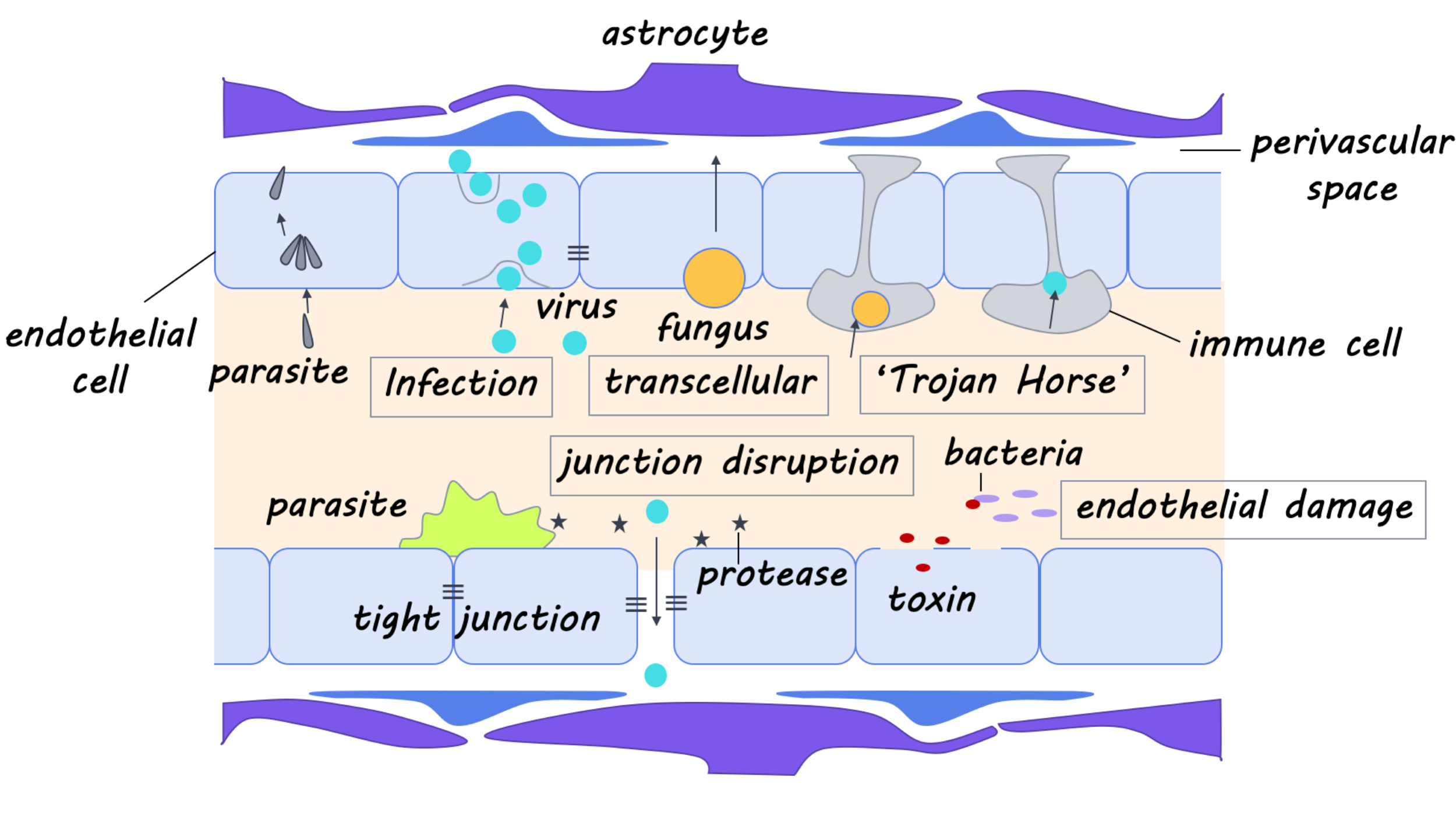
Other pathogens confront the blood-brain barrier head-on. This means dealing with the endothelial cells surrounding blood vessels, which can be thought of as “gatekeeper cells,” tightly restricting access to brain tissue. One way to overcome this is to hijack the mechanisms usually involved in nutrient transport (one of the many strategies of West Nile Virus). Then, there is the “Trojan Horse” method: hitch a ride inside the few immune cells that are allowed to squeeze through the blood-brain barrier (another strategy for West Nile and many other viruses). Still, other pathogens specifically infect the gatekeeper cells themselves (Toxoplasma gondii in mice). If an infected cell ruptures, the pathogens can then cross into the brain itself. Finally, invaders can also disrupt the function of gatekeepers or other cells that help maintain the blood-brain barrier so that it gradually degrades. Or, they can cause an inflammatory response in the brain, which has a similar effect (used by S. pneuomniae).

What else is new?

New technologies have allowed us to better understand the different ways that pathogens enter the central nervous system. For example, we are able to grow and compartmentalize individual neurons in different chambers and see their networks more clearly; we can also image live, fluorescent viruses as they move. The ability to sequence and modify genomes has allowed us to determine which genes and proteins are used by these pathogens and what happens when we turn them on or off. All of these advances have allowed us to understand things like how viruses can move up nerves, co-opting cellular transport machinery along the way.

What’s the bottom line?

The more we know about how pathogens overcome our body’s natural defenses, the better we can combat them with medicine. With some work, we might also be able to repurpose the mechanisms used by neuro-invasive pathogens to deliver life-saving treatments past the blood-brain barrier. 

Cain et al. Mechanisms of Pathogen Invasion into the Central Nervous System. Neuron (2019).Access the original scientific publication here.

How Amyloid, Vascular and Resilience Factors Interact to Associate with Cognitive Aging

Post by Deborah Joye

What's the science?

In general, our cognitive abilities decline as we get older. Sometimes cognitive decline is related to a disease state, such as Alzheimer’s Disease or other dementias, but many non-disease factors likely contribute as well. To better prevent or treat age-related cognitive decline, we must first understand the many intersecting processes that contribute to it and how they interact. For example, systemic vascular issues such as rapid increases in blood pressure in mid-life can negatively impact brain health later in life. Declining vascular health also contributes to small vessel disease and white matter hyperintensities which can decrease the structural integrity of brain fibres. However, some loss of brain tissue integrity is also a normal part of aging, so consideration of normal age-related brain changes is also warranted. There may also be factors that increase the brain’s resilience to aging, such as intellectual enrichment. This week in Annals of Neurology, Vemuri and colleagues model the complex process of cognitive aging and examine several possible causal mechanisms including systemic vascular health, cognitive abilities, brain health indicators such as amyloid build-up (amyloidosis), cortical thickness, integrity of fibers in the corpus callosum, and resilience effects of education and occupation. 

How did they do it?

The authors selected 1230 participants enrolled in the Mayo Clinic Study of Aging, aged 50 years or older who had undergone brain scans and had at least two clinical follow-ups. The authors also calculated a composite cardiovascular health score for each participant that considered cardiovascular and metabolic conditions such as hypertension, diabetes, and stroke prior to the brain scans. The authors analyzed brain scans of participants to quantify average cortical thickness in brain regions known to be affected by Alzheimer’s Disease pathology and created a single measure as a proxy for aging- and Alzheimer’s-related neurodegeneration. They also computed a measure of amyloid levels in each participant’s brain (high levels can indicate pre-clinical stages of Alzheimer’s disease). To quantify brain tissue integrity, the authors measured the fractional anisotropy of a region of the corpus callosum. Fractional anisotropy quantifies the directionality of a diffusion process on a scale from 0 to 1, where a value of 0 means particles can diffuse in equally in all directions such as in a liquid (or cerebrospinal fluid), whereas a value of 1 means that particles can diffuse in only one direction, usually indicating the presence of dense fibre bundles. Finally, the authors estimated a cognitive score for each participant based on their performance on tests of executive function, language, memory, and visuospatial performance.

What did they find?

The authors found that cognition generally worsened with age, and that reduced brain tissue integrity predicted later decreases in cortical thickness. The authors also found that older age was associated with lower education (younger generations tend to be more educated), worse cardiovascular and metabolic condition, and reduced brain tissue integrity. Older women had the most risk of amyloid deposition, though older men had the most risk for cortical thinning. The authors also found that the strongest predictors of lower cognitive performance were old age, male sex, high amyloid, and decreased brain tissue integrity and cortical thickness. Factors that increased resilience of the brain included higher levels of education and occupation. Interestingly, higher education was associated with better systemic vascular health, which contributed to better brain tissue integrity, increased cortical thickness, and ultimately better cognition and reduced risk of dementia, suggesting that early-life exposures had an impact on life-long health. Finally, the authors found that increased amyloid levels predicted accelerated cognitive decline and increased risk of dementia. Notably, the authors describe that cerebrovascular, amyloid/Alzheimer’s, and resilience factors have converging effects on cortical thickness and cognitive decline. Amyloid/Alzheimer’s factors generally result in neuronal loss and a decrease in grey matter integrity, but cerebrovascular and resilience factors affect white matter integrity which can subsequently impact grey matter changes.

deb (5).png

What's the impact?

This is the first study to demonstrate that resilience and vascular factors contribute to the degradation of white matter, which then contributes to shrinkage of grey matter regions as seen in Alzheimer’s and aging. This suggests an important effect of vascular health on cognitive decline in the aging brain. This study expands our knowledge of the complex and dynamic processes of age-related cognitive decline and has broad implications for improving therapies and preventative treatments for cognitive impairment.

vemuri_quote.jpg

Vemuri et al., Amyloid, vascular, and resilience pathways associated with cognitive aging, Annals of Neurology (2019). Access the original scientific publication here.

How Does the Brain Understand Language From Letters?

Post by Flora Moujaes

What's the science?

For skilled readers, the process of reading comes so naturally that its complexities are often overlooked. In order to read at a reasonable level, the brain is required to recognise arbitrary visual symbols irrespective of their case, font, size, position in a word, or other irregularities caused by variations in handwriting. The brain must then instantaneously link the visual form of the letters with the stored meaning they represent. Neuroimaging research has suggested that the brain’s ability to abstract from letters to meaning is achieved by the ventral occipito-temporal (vOT) cortex. When a visual stimulus is encountered by the brain, the first signals reach the primary visual cortex — part of the occipital cortex. The information is then relayed through the occipital lobe towards the temporal lobe to recognize objects or symbols. However, the literature is still unclear about exactly how the vOT supports the ability to abstract from letters to meaning. This week in PNAS, Taylor and colleagues combined artificial language learning and neuroimaging to reveal how the brain represents written words.

How did they do it?

Researchers first trained twenty-four adults to read two sets of 24 novel words, written using two different alphabets of specially created symbols. They used pseudo words as this allowed the researchers to manipulate word form, sound, and meaning in a manner that would be hard to achieve in natural languages. Each pseudo word had a distinct meaning and was comprised of four symbols, three that contributed to the sound of the word and a final silent symbol. The words were similar to each other in one of three ways: (1) they contained some of the same symbols (they were from the same alphabet), (2) they sounded similar, or (3) they had a similar meaning but were written in different alphabets. This enabled researchers to examine how the brain encodes the visual stimuli itself as well as its associated sound and meaning. After two weeks of training, participants read the trained words while neural activity was measured using functional MRI.  The researchers then used representational similarity analysis to analyze the response similarity between evoked fMRI responses to similar words in selected regions-of-interest.

Flora (1).png

What did they find?

Representational similarity analysis of words from the same alphabet revealed that: right vOT and posterior left vOT represented written words in terms of their low-level visual form, and are thus sensitive to basic visual similarity. Posterior to mid -left vOT represented written words in terms of their letters. In mid-vOT these letters had similar representations even when they occurred in different positions within a word. Representational similarity analysis on words from different alphabets revealed that: The anterior left vOT had similar neural patterns for words with similar sounds or meanings, even though they were written differently with no letters in common.

Overall these results show that as you move from the posterior to the anterior vOT, representations of letters become transformed from visual inputs to meaningful linguistic information. There is thus a hierarchical gradient in the vOT where letters are transformed from merely containing visual information to having more abstract meanings in order to convey spoken language information.

What's the impact?

These findings advance our understanding of how the brain comprehends language from arbitrary visual symbols. By examining the relationship between how visual form, sound, and meaning are encoded in the occipito-temporal cortex, this study provides strong empirical support for a hierarchical, posterior-to-anterior gradient in vOT that represents increasingly abstract information about written words. Given that learning to read is the most important milestone in a child’s education, it will be important for future studies to specify how linguistic influences on vOT change over time; both in the short term while reading a word and during reading development.

rastle_quote.jpg

Taylor et al. Mapping visual symbols onto spoken language along the ventral visual stream. PNAS (2019). Access the original scientific publication here.