Spatial Attention Quenches Neural Variability Within the Visual System

Post by Sarah Hill

What's the science?

Uncovering the underlying neural processes related to attention and alertness is currently an active area of research in neuroscience. Recordings of neural activity during tasks in which subjects were prompted to pay attention suggest that attention is marked by an increase in neuronal firing rate, as well as a decrease in firing rate variability, both within individual neurons and between pairs of neurons. In other words, attention appears to improve the neural signal-to-noise ratio by increasing and stabilizing neural activity. While many studies have modeled attention-related neural signal enhancement, much less is known about how attention reduces or 'quenches' variability in neural activity. This week in The Journal of Neuroscience, Arazi and colleagues report that neural variability within the visual system is selectively quenched by spatial attention.

How did they do it?

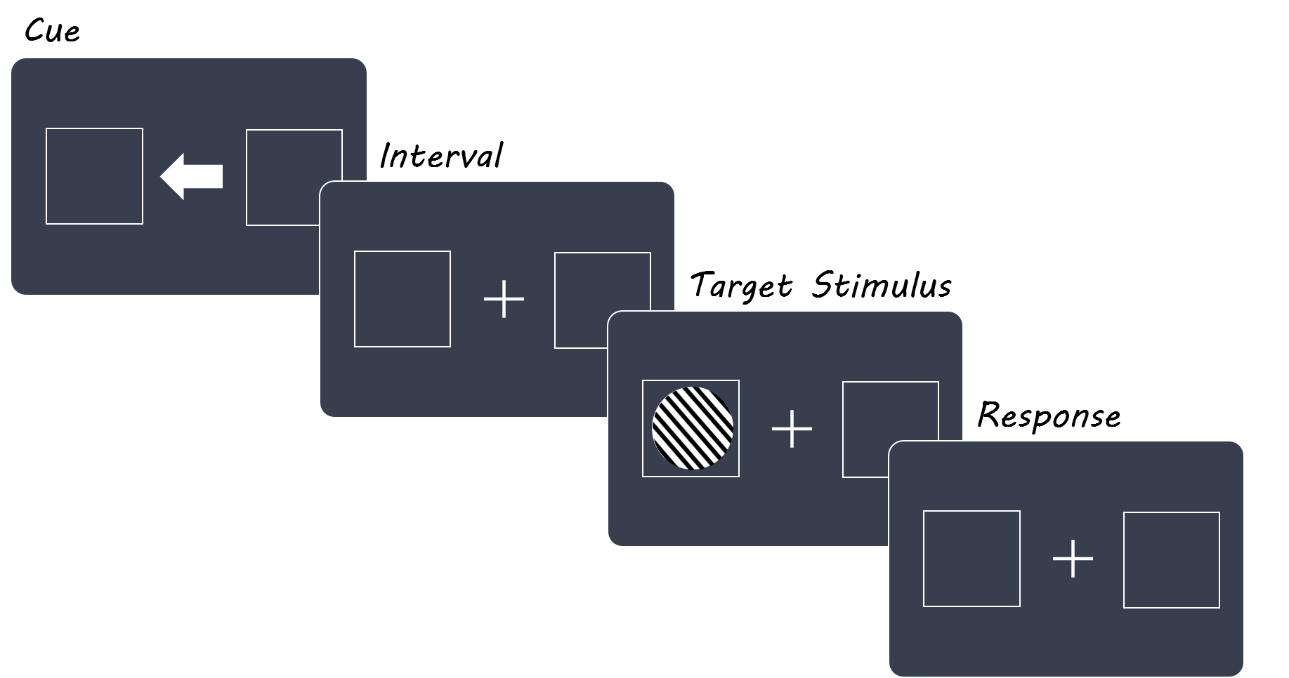
To study the effects of two different components of attention - alertness and spatial attention (or the focus of attention on a particular area of space) on neural activity, the authors used electroencephalography (EEG) to record and compare neural variability across two experiments. In the first experiment (termed the 'discrimination experiment'), human participants were briefly shown a cue (in this case, a white arrow on a computer screen) that was then followed by a target stimulus (a black and white striped circle appearing on the screen). In 60% of trials, the arrow pointed toward the location where the circle appeared shortly after, thereby directing participants' attention to a specific area of the computer screen. In the remaining 40% of trials the arrow pointed in either the opposite direction or in both directions. The subjects were then told to report which direction the circle's stripes tilted - left vs. right. In a second experiment (control), participants were presented with the arrow cue, but were not shown the circle stimulus (and were therefore not asked to report on the direction the circle's stripes). Consequently, participants in the control experiment were not prompted to pay attention to any particular location on the computer screen. In addition to having neural activity recorded through EEG, subjects' eye movements were tracked using electrooculography (EOG). The EEG and EOG recordings were then processed and used to compare neural variability between the two experiments.             

diagonallines.png

What did they find?

First, for trials of the discrimination experiment in which the arrow cue correctly indicated the location of the circle stimulus, participants had higher rates of accuracy in reporting the direction the circle's stripes tilted towards. This suggests that participants were focusing their attention on a particular spatial area of the computer screen following presentation of the arrow cue. Next, the authors observed a significant reduction in neuronal firing variability following presentation of the arrow cue in both the discrimination and control experiments, though quenching of neural variability was greater in the discrimination task than in the control task. This result demonstrates that alerting the subjects to an upcoming stimulus reduces neural variability. To confirm this, they measured neural variability across intervals directly before and after cue presentation, finding that the magnitude of variability was significantly smaller in the discrimination experiment compared to the control experiment following cue presentation, but not before. Intriguingly, quenching of neural variability in the discrimination experiment appeared restricted to electrodes placed near the visual cortex, suggesting that spatial attention specifically modulates activity within the visual system. To further examine the effects of spatial attention, the authors noted the degree of variability quenching exhibited by each electrode when the cue pointed left vs. right in the discrimination task, observing that quenching was greater in electrodes placed contralaterally to the field of view being concentrated on. Finally, subjects with the largest reductions in neural variability following cue presentation also benefited the most from directed spatial attention, as indicated by comparing each subject's accuracy rates when the cue pointed in the correct direction versus when it pointed in the opposite direction.              

What's the impact?

This is the first study to examine the effects of spatial attention on neural variability in humans. The authors showed that in addition to increasing neural activity, spatial attention and alertness minimize neural noise, thereby optimizing the neural signal-to-noise ratio specifically within the visual system. This study also provides compelling evidence that the benefits of attention may not be the same for all individuals, which is particularly relevant in terms of developing interventions for disorders such as ADD and ADHD.  

Attention_June11_Quote.jpg

Arazi et al. Neural variability is quenched by attention. The Journal of Neuroscience (2019). Access the original scientific publication here.

Thalamic Activity During Embryonic Development Helps Guide the Formation of Somatotopic Maps

Post by Shireen Parimoo

What's the science?

In sensory regions of the brain, like the primary somatosensory cortex (S1), sensory inputs from adjacent regions on the body are represented in adjacent populations of neurons (barrels). In mice, for example, whiskers are somatotopically represented in the cortex in barrels, which means that stimulating neighboring whiskers activates neighboring barrels in the mouse S1. Sensory input from the body travels through the thalamus (part of the forebrain) before reaching the cortex, and these connections are formed early in development. Interestingly, there is evidence of co-occurring spontaneous activity in the thalamus and S1 before the cortical barrels have fully developed. It is currently unclear whether this spontaneous co-activation of the thalamus and S1 facilitates the development of somatotopically organized cortical barrels or whether cortical barrel organization is dependent on external sensory input after birth. This week in Science, Antón-Bolaños and colleagues investigated the functional characteristics of thalamocortical connections in developing and newborn mice.

How did they do it?

First, the authors electrically stimulated the ventral posteromedial nucleus (VPM) of the thalamus in mouse embryos and used calcium imaging to measure the change in the response of S1 neurons. To determine how specific the projections were from the thalamus to S1, they also stimulated three other regions adjacent to the VPM and measured the cortical response. They then used a transgenic mouse line “Kir” to examine the effect of altered spontaneous thalamic activity on barrel development. Control mice have high-amplitude and synchronized neural activity in the VPM, whereas Kir mice have asynchronous and low-amplitude activity in 10-day old embryos (E10). They stimulated the VPM and adjacent thalamic regions and compared the cortical response in the Kir and control mice at 17.5 and 18.5 days.

To determine the longer term effect of altered thalamic activity on barrel development, the authors stimulated the VPM in Kir and control mice four days after birth (P4) and recorded cortical activity. They also stimulated the whiskers to determine if providing external sensory input could facilitate the development of cortical barrels. They used multichannel electrodes to measure extracellular activity in S1 of Kir and control mice at postnatal days 2 amd 3. To further examine the mechanism underlying cortical activity in Kir mice, a glutamate receptor antagonist was applied to S1 and change in cortical response was measured. Finally, axon tracing and immunostaining were used to detect cortical barrels and axons projecting between the thalamus and S1.

What did they find?

Stimulating the VPM resulted in a large cortical calcium response at day 17.5, but a smaller and more localized response at 18.5 days. Stimulation of VPM-adjacent regions at 18.5 days activated a population of neurons altogether. In transgenic Kir mice, on the other hand, VPM stimulation resulted in widespread cortical activation, and stimulating VPM-adjacent neurons activated overlapping neuronal populations in S1. In newborn control mice, VPM stimulation led to an even more localized cortical response, but this localization did not occur to the same extent in the Kir mice. This means that specific and functionally organized thalamocortical projections developed in 18.5-day old embryos, but altering thalamic activity disrupted the formation of cortical somatotopic maps. These effects persisted even after birth; control mice but not the Kir mice showed evidence of cortical barrels at postnatal day 4. Similarly, although stimulating the whiskers activated different S1 barrels in control mice, cortical activity was less distinct in the transgenic Kir mice and could not be rescued by sensory input after birth.

Kir.png

Axon tracing and staining revealed that in control mice, thalamic neurons projected to the corresponding cortical neurons in a barrel, but these axonal projections were less spatially specific in Kir mice. This means that neurons originating from a larger region of the thalamic VPM project to the S1 in Kir mice compared to the control mice. Finally, the transgenic mice had more glutamate receptors than control mice. Blocking glutamate receptors with an antagonist reduced the spatial extent of cortical activity in Kir mice, making it more similar to that of control mice. These results suggest that glutamate receptors underlie the widespread cortical response to thalamic activation in Kir mice, which subsequently affects the development of somatotopic maps in S1.

What's the impact?

This study is the first to demonstrate that spontaneous activity in the thalamus during embryonic development is critical for the formation of somatotopic maps in the mouse primary sensory cortex. Moreover, the finding that the concentration of glutamate receptors might underlie this relationship has important implications for understanding the role of genetic factors in cortical development. Overall, this study provides further insight into the importance of prenatal factors in the development of functionally organized cortical maps.

Antón-Bolaños et al. Prenatal activity from thalamic neurons governs the emergence of functional cortical maps in mice. Science (2019). Access the original scientific publication here.

Stress-Induced Memory Impairment is Mediated by Hippocampal Opioid Receptors

Post by Deborah Joye

What's the science?

Stress can impair your ability to retrieve memories. Perhaps you can recall a time that you sat down to take a big exam or started an important presentation only to have your mind go suddenly, frighteningly blank. We know that glucocorticoids (hormones activated by stress) can change excitatory synapses in the hippocampus and lead to impaired memory. But the effects of stress on memory go well beyond what can be explained just by the effects of glucocorticoids, suggesting that there may be other signals involved. In addition to glucocorticoids, stress also mobilizes endogenous opioids (i.e., made by your body) that can lead to cognitive, emotional, and physical effects that can help us adapt to stress. Opioids are often associated with pain relief, but how might they affect learning and memory in response to stress? This week in Molecular Psychiatry, Shi and colleagues use an impressive array of behavioral and cellular techniques to demonstrate that endogenous opioids produce stress-induced memory impairment by suppressing activity of GABAergic interneurons in the hippocampus.

How did they do it?

To produce stress, the authors placed mice on an elevated clear platform in a brightly lit room for close to an hour. Unstressed mice were in the same room but remained in their home cages. Throughout their experiments, the authors used the Morris Water Maze to test memory retrieval. In this task, mice are trained to swim in a circular pool and find a hidden platform which is consistently placed in the same region of the pool.

First, to determine if opioid receptors are involved with stress-induced memory impairment, the authors used naloxone, which strongly inhibits opioid receptors. Since there are three major opioid receptors in the hippocampus (µ, pronounced “mew”; δ, or delta; and κ, or kappa opioid receptors), the authors selectively activated or blocked specific opioid receptors to test which ones underlie stress-induced memory impairment. Next, the authors tested which endogenous opioid is involved by using antiserum to block the effects of opioids highly expressed in the hippocampus, β-endorphin and enkephalin, followed by memory retrieval testing. After testing which receptors and opioids are involved, the authors assessed which type of cells opioids might be acting on in stress-induced memory impairment. To do this, they used Cre-Lox recombination to delete the µ-opioid receptor gene in certain cell populations: GABAergic inhibitory interneurons, glutamatergic excitatory neurons, or astrocytes. Mice in the different µ-opioid receptor knockout conditions were then subjected to stress and the Morris water maze to test for memory impairment. To investigate possible changes in synaptic transmission, the authors recorded the electrophysiological activity of cells in the hippocampus immediately after stress. They also recorded electrophysiological changes in response to opioid receptor activation or inhibition. Finally, to test whether stress-induced memory impairments are due to inhibition of GABAergic interneurons, the authors activated or blocked GABA transmission in the hippocampus, then tested memory retrieval with the Morris water maze.

What did they find?

The authors found that µ-opioid receptor signaling is required for stress-induced impairment of memory retrieval. When µ-opioid receptor signaling is blocked, stressed mice do not exhibit memory impairments and activation of µ-opioid receptors in the hippocampus in unstressed mice reproduces memory impairment as if mice had been stressed. The authors also found that stress-induced memory impairment specifically depends on activation of µ-opioid receptors on GABAergic neurons, since stressed mice without µ-opioid receptors on GABAergic cells do not exhibit stress-induced memory impairments. Mice lacking µ-opioid receptors on glutamatergic cells or astrocytes still display stress-induced memory impairment. Using electrophysiology, the authors found that stress-induced activation of µ-opioid receptors on GABAergic interneurons decreases inhibitory synaptic transmission onto hippocampal CA1 neurons, resulting in memory impairment. Overall, the authors identified a new pathway in which acute stress mobilizes endogenous opioids to activate µ-opioid receptors on inhibitory GABA neurons in the hippocampus. This pathway reduces the release of GABA, increasing excitability of hippocampal cells and leading to impairments in memory retrieval.

Liu_image.png

What's the impact?

This study is the first to demonstrate that endogenous opioids play a role in stress-induced memory impairment. Specifically, this study examines and identifies the mechanism by which µ-opioid receptor activation on GABAergic cells within the hippocampus suppresses inhibition of hippocampal cells and results in memory retrieval impairments. These findings add an important new element to our understanding of how stress affects memory, and opens new avenues for research into the impacts of opioids in the hippocampus.

Liu_quote_June4.jpg

Shi et al., Hippocampal µ-opioid receptors on GABAergic neurons mediate stress-induced impairment of memory retrieval, Molecular Psychiatry (2019).Access the original scientific publication here.