Hippocampus-Amygdala Communication Supports Pattern Separation of Emotional Memories

Post by Shireen Parimoo

What's the science?

Pattern separation is a hippocampal-dependent process that allows us to discriminate between similar events or experiences - like remembering where we parked the car at work on a specific day. Emotional events are remembered vividly because they increase arousal, which enhances memory formation and retrieval. The strength of emotional memories is modulated by the amygdala. Memories for emotional events are often biased to central aspects of the event while peripheral details are forgotten, making it difficult to discriminate between similar emotional events. The precise neural mechanism supporting this process remains unclear. This week in Neuron, Zheng and colleagues investigated the neural mechanism underlying the pattern separation of emotional memories in the amygdala and hippocampus using intracranial recordings.

How did they do it?

Seven pre-surgical epilepsy patients completed an episodic memory task with emotional stimuli while local field potentials were recorded from the amygdala and the hippocampus using intracranial stereo-electroencephalography. The memory task consisted of two phases: (i) an encoding phase, in which participants had to rate the emotional valence of positive, negative, and neutral images, and (ii) a retrieval phase, in which they had to make old/new judgments about the same, novel, or perceptually similar (‘lure’) images. The lures were presented to create interference between previously presented target images and the novel lures. To examine pattern separation, the authors computed the oscillatory power (squared amplitude), phase synchrony (phase coupling strength of signals in different regions of the brain), and phase-amplitude coupling (coupling between the phase of one signal and the amplitude of another signal) in different frequency bands. To determine whether hippocampal oscillations were driving oscillatory activity in the amygdala or vice versa, they computed the phase-slope index and performed a Granger causality analysis. Finally, they trained a classifier using the occurring phases of high-frequency activity (HFA) relative to the theta (2-7 Hz) and alpha bands (7-14 Hz) as inputs to predict whether participants would false alarm or correctly reject the lures.

What did they find?

Participants performed well on the memory task with 80% accuracy overall. However, they were worse at discriminating emotional lures than neutral lures, with poorer discrimination of negatively-valenced lures than positive lures. Oscillatory activity was observed in the theta and alpha frequency bands when participants made recognition judgments. Specifically, in the hippocampus there was greater theta activity on trials where participants correctly identified the lure images as being new, which was accompanied by a simultaneous reduction in alpha power. In the amygdala, on the other hand, there was a lag between increased theta activity and a subsequent reduction in alpha power. Alpha activity increased on false alarm trials where participants incorrectly categorized images as being old. Oscillatory activity in the amygdala and the hippocampus was synchronized in the theta band on trials where the participant correctly identified a lure and in the alpha band on false alarm trials. This pattern of activity was present for both emotional and neutral images, but stronger for emotional images  only on lure trials. This suggests that theta and alpha oscillations in the amygdala and hippocampus are involved in pattern separation, which is modulated by emotional valence.

Pattern_image_April16.png

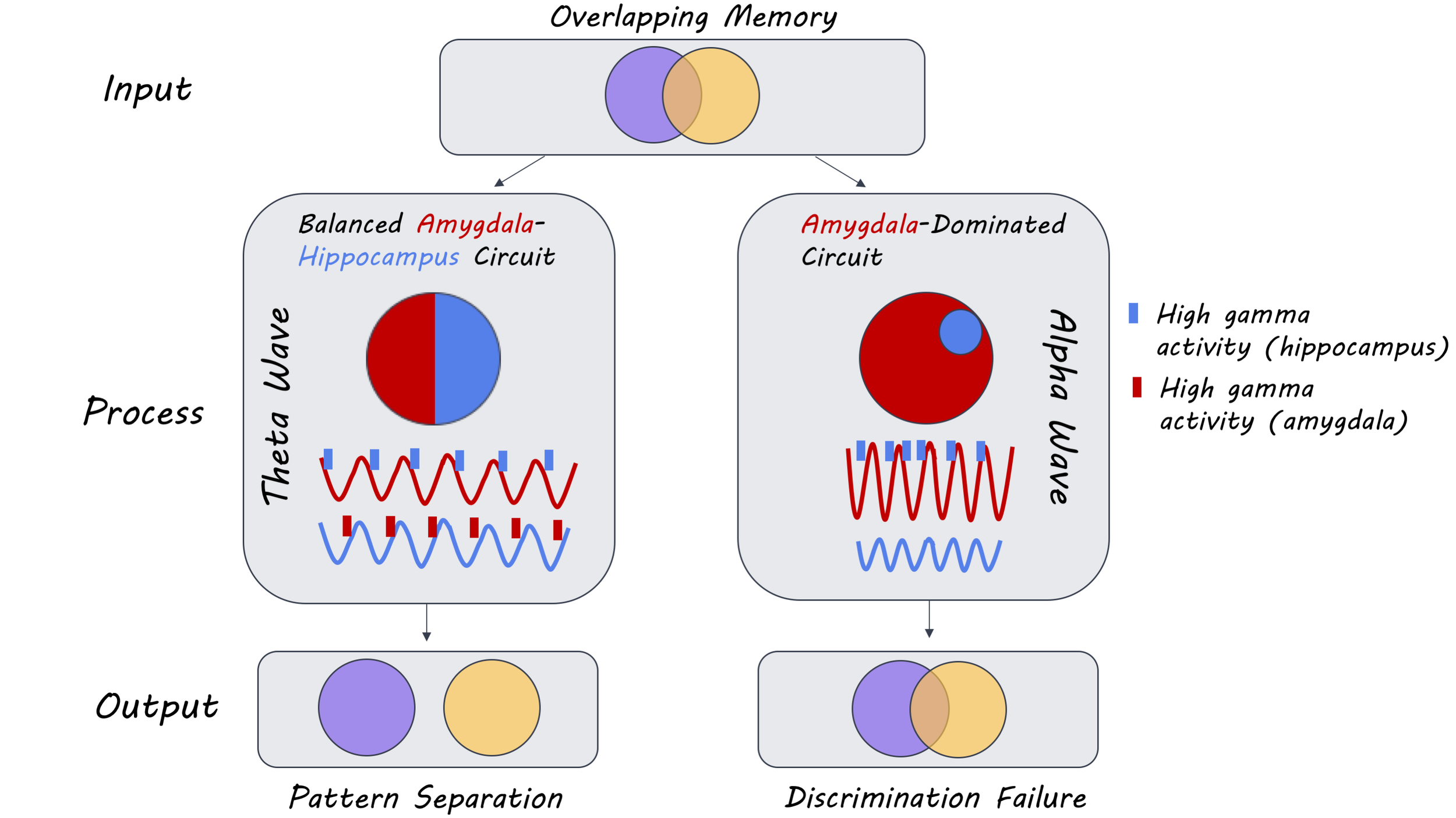
Bidirectional communication between the hippocampus and the amygdala in the theta band was observed on correctly identified lure trials, and unidirectional communication was observed in the alpha band from the amygdala to the hippocampus on lure false alarm trials. Similarly, there was greater phase-amplitude coupling between theta oscillations in the amygdala and HFA in the hippocampus — as well as the reverse — on correctly identified lure trials. There was also a modulation of hippocampal HFA by alpha oscillations in the amygdala on lure false alarm trials. The trough of the theta cycle was associated with HFA in the amygdala (descending slope) and the hippocampus (ascending slope) on correctly identified lure trials. Such theta phase-dependent modulation of HFA was not present on lure false alarm trials. Moreover, the influence of amygdala theta oscillations on the hippocampus was stronger with emotional stimuli than neutral images, especially for negatively-valenced images. However, this emotional modulation of directional connectivity was not observed in the reverse direction (hippocampus to amygdala) or on lure false alarm trials. Thus, bidirectional communication between the amygdala and the hippocampus in the theta band results in the correct discrimination of lures from targets, whereas a disproportionately amygdala-driven interaction in the alpha band results in poor discrimination.

What's the impact?

This study is the first to show that bidirectional theta oscillations between the hippocampus and the amygdala at memory retrieval are necessary for the pattern separation of emotional events. These findings are consistent with the literature demonstrating the role of low-frequency oscillations in memory formation and provide further insight into the mechanism that allows for the resolution of interference between highly similar emotional events at retrieval.

Zheng_quote_April16.jpg

Zheng et al. Multiplexing of theta and alpha rhythms in the amygdala-hippocampal circuit supports pattern separation of emotional information. Neuron (2019). Access the original scientific publication here.

New Gene Expression Technologies Improve Understanding of ALS

Post by Flora Moujaes

What's the science?

Amyotrophic lateral sclerosis (ALS) is a type of motor neuron disease. Motor neuron diseases are caused by the gradual deterioration of motor neurons found in the brain and spinal cord. As the motor neurons die, they can no longer control voluntary movement and the muscles begin to atrophy. The cause of the disease and the mechanisms that underlie ALS are still poorly understood, though previous work has indicated genetic factors play a role. This week in Science, Maniatis and colleagues combined new technologies for mapping gene expression that take into account spatial location (where the genes are expressed), and computational modelling, to provide new insights into the mechanisms that contribute to ALS.

How did they do it?

Researchers can determine whether a gene is activated or ‘expressed’ in a cell by examining messenger RNA (mRNA). mRNA is essentially a template for DNA: it is a set of molecules copied from the DNA code that tells the cell how to behave. There are thousands of RNA molecules per cell, and together they make up the mRNA, or ‘transcriptome’. Thus, the transcriptome reflects the genes that are being actively expressed in a cell at any given time. Standard methods to examine mRNA involve extracting all the RNA molecules from a tissue biopsy, and analysing them together. This results in an average representation of all the genes expressed in that tissue, however, spatial information of where the particular genes are expressed within the tissue is lost. Spatial transcriptomics enables you to capture the mRNA in the tissue while retaining the information about where in the tissue this mRNA is expressed. The method involves placing tissue samples on a glass slide covered in tiny spots. These tiny spots contain DNA strands with built in address labels that 1) capture and copy the mRNA and 2) encode a unique ‘barcode’ in the copy that corresponds to the spatial location of the spot on the slide.

The researchers used spatial transcriptomics to examine gene expression in 1) 56 ALS mice, 2) 11 control mice, and 3) post-mortem spinal cord tissue from seven deceased ALS patients. They collected over 76,000 spatial gene expression measurements (or ‘spots’) from 1,165 mouse tissue sections, and over 60,000 spatial gene expression measurements from 80 human tissue sections. The mice were examined at three disease time points: onset, symptomatic, and end-stage, which enabled the researchers to track gene expression over time. The human samples were collected from either end of the spine, which allowed the researchers to examine how ALS pathology spreads, as ALS symptoms usually appear in one part of the body before going on to cause widespread paralysis. The researchers then used computational methods to combine the spatial location and gene expression information.

What did they find?

The researchers produced a multidimensional gene expression atlas which they made available through an interactive data exploration portal at https://als-st.nygenome.org/ .

One of the main hypotheses explored in this study was that even though motor neurons are the most vulnerable in ALS, neighbouring cells such as microglial cells play a key role in the disease. Microglial cells are of particular interest to ALS as they are involved in removing damaged neurons. This study indicated that microglia dysfunction occurs well before the onset of ALS symptoms. This is important as it helps us understand how mutations may disrupt the function of both neuronal and non-neuronal cells, and how the impaired interactions between the different cell types in the nervous system may then lead to motor neuron loss in ALS. To further explore the spatiotemporal dynamics of microglial activation, the researchers examined two genes that had previously been indicated in ALS: TREM2 and TYROBP. They found that TREM2 and TYROBP were both expressed at higher levels by microglial cells; in particular spinal cord regions of mice with ALS symptoms. Thus, they were able to refine our understanding of such gene expressions, showing TREM2- and TYROBP-mediated signalling is an early step in disease-relevant changes in microglial gene expression.

flora.png

What's the impact?

This is the largest study to examine spatial transcriptomics in mice and humans with ALS. This study highlights the value of examining gene expression in individual cell types in relation to their wider spatial context. The vast dataset generated by this study may aid with the development of therapeutic interventions for ALS and earlier diagnostic markers. This is especially important given that ALS currently affects over 200,000 patients worldwide and results in a life expectancy of up to 5 years following diagnosis.  In addition, the methods used in this study could be extended to examine other neurodegenerative diseases including Alzheimer’s, Parkinson’s, and Huntington’s disease.

Science_quote_april9.jpg

Maniatis et al. Spatiotemporal dynamics of molecular pathology in amyotrophic lateral sclerosis. Science (2019). Access the original scientific publication here.

Deep Brain Stimulation of the Internal Capsule Improves Prefrontal Cortex Function

Post by Leigh Christopher

What's the science?

Gold standard treatments for mood and anxiety disorders are often ineffective. Deep brain stimulation (DBS), where electrical current is applied to brain circuits, effectively relieves symptoms in some cases, however, the results are inconsistent. One theory is that this inconsistency is due to the lack of a biomarker to indicate the ‘effective dose’ of electrical stimulation. The ventral internal capsule is affected in both depression and obsessive-compulsive disorder (OCD) and has shown promise as a target for DBS therapy. This region is involved in cognitive control – it regulates theta oscillations in the prefrontal cortex that could act as a biomarker of DBS efficacy. Recently in Nature Communications, Widge and colleagues use DBS to stimulate the ventral internal capsule to assess whether its stimulation alters brain oscillations in the prefrontal cortex and improves cognitive control in patients with depression and OCD.

How did they do it?

Fourteen patients — 12 with major depressive disorder and 2 with OCD participated in the study — these patients previously had a DBS electrode implanted in the ventral internal capsule/ventral striatum. Patients performed the Multi-Source Interference Task – a cognitive control task that includes emotional distractors. During this task, participants had to identify which of a set of three numbers was different than its neighbours, using a key press. In the conflict trail, the target number is out of position (e.g. a number two is not in the second key position). An emotionally distracting image was displayed during certain trials. The participants then played the Effort Expenditure for Rewards task, in which they had to press a button to fill a bar on the screen - they had to choose quickly between easy and hard options in an attempt to receive as high a payoff as possible. The authors used EEG to record the patients’ brain oscillatory activity throughout these tasks while their DBS was turned either on or off. They analyzed response times and how they were affected by cognitive conflict (interference), emotional distraction and DBS treatment using a linear mixed effects model. They used sliding multivariate regression to assess whether theta activity (as recorded by EEG) was associated with cognitive control.

What did they find?

DBS enhanced cognitive control for both interference and control trials of the Multi-Source Interference task – reaction times were 34 seconds faster on average compared to DBS being turned off. The power of theta oscillations (non-phase-locked, or task-evoked) was higher throughout the prefrontal cortex while participants exercised cognitive control during the decision-making portion of the task. DBS stimulation increased this effect for almost the entire decision-making period. In the Effort Expenditure for Rewards task, the response times were slower while DBS was on, suggesting that the DBS effects were specific to the cognitive control tested in the Multi-Source interference task. Button presses were not different when DBS was on vs. off in this task, suggesting that DBS does not impact movement speed. Change in the theta power in the inferior frontal gyrus during the interference task while DBS was on was associated with a reduction in depressive symptoms. The inferior frontal gyrus showed the most drastic change in theta power of any prefrontal region. These changes were specific to the theta frequency, with no other frequency band showing changes during the task.

Leigh_april9_img.jpg

What's the impact?

This work supports the theory that DBS exerts its therapeutic effects by regulating brain activity in the cortex. Thes study demonstrates that DBS improves cognitive control and that this is related to an increase in theta oscillatory power in the prefrontal cortex. These findings have important clinical implications – clinicians could use change in theta power as a biomarker to assess whether the proper stimulations parameters have been applied during DBS, improving efficacy of the treatment. Further, this study suggests that augmenting cognitive control in general may be an effective treatment strategy for psychiatric illness.

Widge et al. Title. Nature Communications (2019). Deep brain stimulation of the internal capsule enhances human cognitive control and prefrontal cortex function. Access the original scientific publication here.