Decreased Short-Range Brain Connections Linked to Social Symptoms of Autism Spectrum Disorder

Post by Deborah Joye

What's the science?

Autism spectrum disorder (ASD) is a neurodivergent condition that results in a range of symptoms including impaired social communication and restricted, repetitive behaviors. The brain can be broken down into local connections within brain regions, and long-range connections between more distant regions. Researchers have hypothesized that brain regions in individuals with ASD are more highly connected to themselves than with other more distant brain regions. Recent studies, however, have found that individuals with ASD can display reduced connectivity, overconnectivity, or a combination. Just under the surface of the cortex there are U-shaped connections between adjacent cortical gyri (the bumps of the brain). These superficial connections are the last to be myelinated in development, which makes them more adaptable to change. Whether these short-range connections are altered in ASD and how they may change throughout the lifespan remains unclear. This week in Brain, d’Albis and colleagues used brain imaging to investigate whether white matter tract connectivity in adults with high-functioning ASD correlates with social and language symptoms.

How did they do it?

To study how the brain is changed in individuals with ASD, the authors recruited a total of 58 adult subjects, 27 with ASD and 31 controls participants. To investigate how brain changes correlate with clinical measures, the authors tested a subset of 39 participants – 26 with ASD and 13 controls. All participants underwent MRI and completed clinical and cognitive evaluations. The authors analyzed data using whole-brain deterministic tractography, which allows identification of anatomical connections within the brain. Using a newly-developed tractography-based atlas of superficial white matter fibers, the authors were able to study the integrity (via a measure called fractional anisotropy) of 63 superficial white matter bundles. Using principal component analysis  (PCA), they sorted white matter bundles into three groups and then analyzed how connectivity within those groups correlated with scores on clinical/cognitive questionnaires.

What did they find?

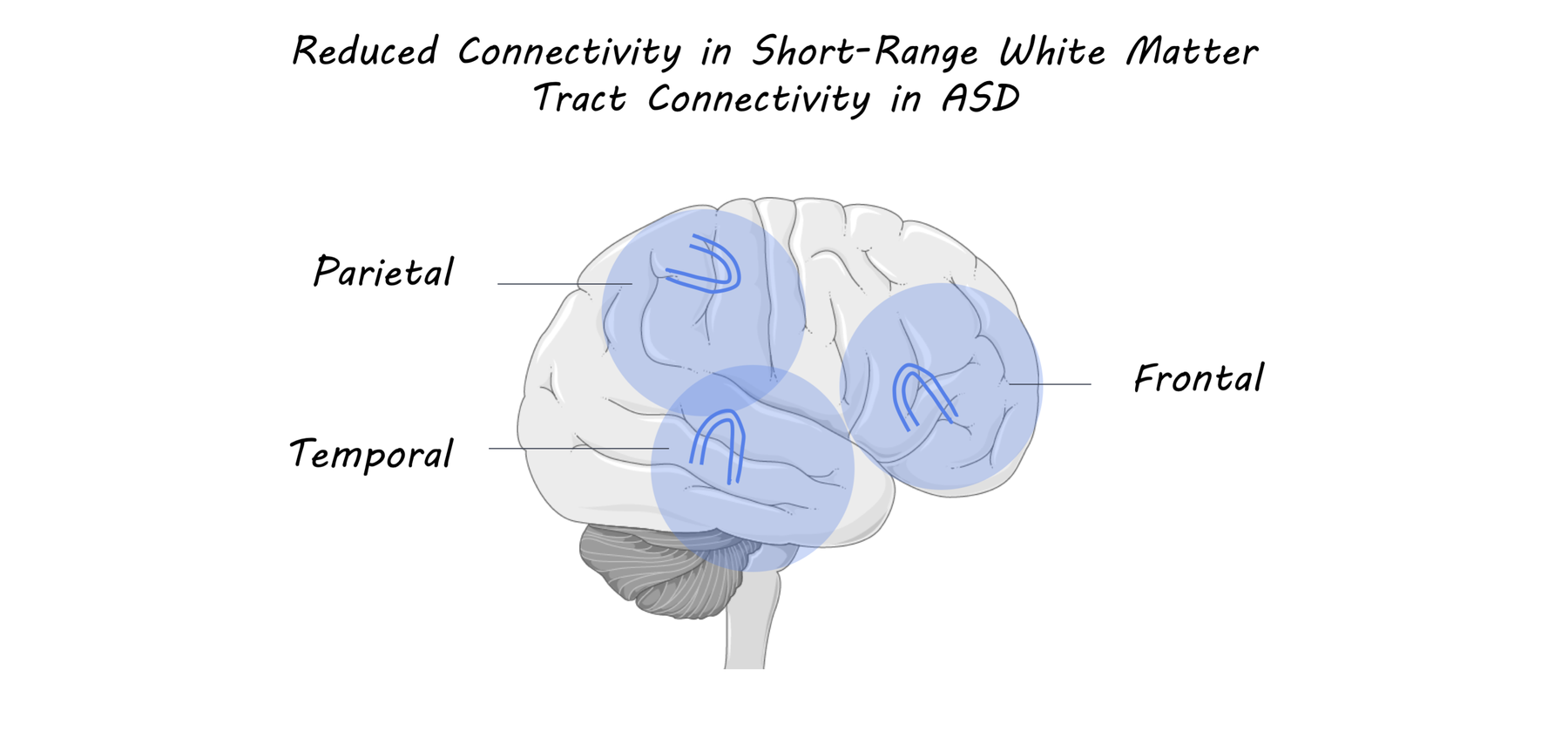
The authors found that, compared to their control population, individuals with ASD exhibited reduced connectivity in temporal, frontal, and parietal superficial white matter bundles. In addition, individuals with ASD exhibited worse performance in clinical measures including 4 social measures (social awareness, cognition, communication, and motivation), 3 adult communication measures (language structure, pragmatic skills, and social engagements), and a measure of empathy. They then tested correlations between white matter tract connectivity in specific brain regions and clinical measures. The authors found that reduced connectivity in the left inferior temporal-middle temporal region was correlated with decreased empathy scores. Additionally, reduced connectivity of the right supramarginal insula was correlated with decreased language structure and social awareness scores.

ASD_image.png

What's the impact?

This study is the first to demonstrate that individuals with ASD exhibit decreased short-range connectivity in specific brain regions associated with social cognition. Since ASD is typically studied in children, this study is also the first to investigate such connectivity in an adult ASD population and relate imaging findings to clinical measures. These findings suggest that highly plastic short-range white matter tracts may underlie some of the key social deficits observed in ASD. Since these short-range white matter tracts may remain malleable until people are in their twenties, these findings suggest a new and exciting possibility for intervention.

autism_quote.png

d’Albis et al., Local structural connectivity is associated with social cognition in autism spectrum disorder, Brain (2018), Access the original scientific publication here.

The Genetic Relationship Between Cardiovascular Health and Alzheimer’s Disease

Post by Anastasia Sares

What's the science?

Many studies have shown a relationship between lifestyle factors and cardiovascular risk factors— like high cholesterol or triglycerides— and Alzheimer’s disease. This connection may be, in part, a result of genetic predisposition. There is already one genetic variant in a gene called APOE (the strongest known risk variant for Alzheimer’s disease) which encodes a protein involved in cholesterol metabolism. This variant is known to be linked to both cardiovascular health and to Alzheimer’s disease. However, it is unclear whether there are other genetic variants like APOE that are linked to both cardiovascular risk factors and the development of Alzheimer’s disease. This week in Acta Neuropathologica, Broce and colleagues set out to identify pleiotropic genes, jointly associated with both cardiovascular health and Alzheimer’s disease risk, and specifically which cardiovascular risk factors are associated with the development of Alzheimer’s disease.

How did they do it?

The authors made use of large publicly available genome-wide association data from studies reporting both Alzheimer’s disease risk and cardiovascular outcomes. They first assessed ‘pleiotropic enrichment’ by analyzing whether Alzheimer’s disease risk increased as a function of cardiovascular risk factors. Pleiotropy is when one gene is associated with two or more distinct phenotypes. The cardiovascular risk factors included triglyceride count, coronary artery disease, body mass index, type 2 diabetes, waist hip ratio, total cholesterol and high- and low-density lipoproteins. Then, they performed a genome-wide meta-analysis using a large dataset with both genetic data and clinical information (Alzheimer’s disease and cardiovascular health) to identify significant pleiotropic genetic variants (i.e. single-nucleotide polymorphisms (SNPs)) that were significantly associated with both Alzheimer’s status and cardiovascular risk factors. They examined these SNPs across two other datasets, a replication dataset and another proxy dataset for Alzheimer’s disease (where parental status of Alzheimer’s disease was provided) to ensure that the association was consistent, both in confirmed cases of Alzheimer’s and people at risk for Alzheimer’s. Finally, they checked whether these pleiotropic SNPs actually altered gene expression in tissue samples (brain and blood), and whether the related gene was more highly expressed in brains of individuals with Alzheimer’s than in normally aging adults.

What did they find?

The authors first assessed ‘pleiotropic enrichment’ to determine whether genetic variants associated with Alzheimer’s disease risk would increase or be enriched as a function of cardiovascular risk factors. The authors found that the strongest pleiotropic enrichment for Alzheimer’s disease risk occurred as a function of triglycerides, high and low density lipoprotein as well as total cholesterol, suggesting a strong genetic overlap between plasma lipids and Alzheimer’s disease. Other cardiovascular health risks, like body mass index and type 2 diabetes, showed little to no overlap with Alzheimer’s disease risk. The authors identified 90 SNPs in the first stage of their genome-wide meta-analysis that jointly conferred risk for both cardiovascular outcomes and Alzheimer’s disease. Of these 90 SNPs, the authors were able to replicate three of the novel pleiotropic SNP associations across all datasets (these were variants within MINK1, MBLAC1, and DDB2 genes). Further, they found that these genes were differentially expressed in both Alzheimer’s disease and healthy control participants. One possible explanation for the relationship between plasma blood lipids and Alzheimer’s disease is that cholesterol is processed differently in the body and the brain. The blood-brain barrier normally keeps these processes separate, however high cholesterol may damage this barrier resulting in a predisposition to Alzheimer’s disease.

AD_image.png

What's the impact?

This study found novel genetic contributors to both Alzheimer’s disease and cardiovascular health. These findings may help to explain other clinical and epidemiological studies, which have already shown that Alzheimer’s disease and cardiovascular risk factors overlap. The identification of these genetic variants could help clinicians to better predict who is at risk for Alzheimer’s, and could also provide targets for new therapies.

Broce et al. Dissecting the genetic relationship between cardiovascular risk factors and Alzheimer’s disease. Acta Neuropathologica (2018). Access the original scientific publication here.

Spinal Cord Stimulation Enabling Movement in Humans with Spinal Cord Injury

Post by Amanda McFarlan

What's the science?

Epidural electrical stimulation is a therapeutic treatment for individuals with spinal cord injuries that involves applying continuous electrical current to the lower part of the spinal cord. This technique has been shown to restore movement in animal models of spinal cord injury, but has been less effective in treating humans. It is hypothesized that action potentials induced by epidural electrical stimulation may collide with naturally-occurring action potentials conveying proprioceptive information (information about where one’s body is in space), disrupting the flow of information traveling to the brain. This may be a larger issue for humans compared to smaller mammals. This week in Nature Neuroscience, Formento and colleagues investigated why treatment of spinal cord injury with epidural electrical stimulation is less effective in humans compared to other mammals.

How did they do it?

The authors tested whether epidural electrical stimulation produces action potentials that travel in the opposite direction (i.e. towards the periphery, away from the brain) from that of sensory afferents (nerve fibers). To do this, they inserted subcutaneous needle electrodes in 2 patients with chronic spinal cord injury and recorded from their sural nerve, the proximal and distal branches of their tibial nerve, and their soleus muscle while applying epidural electrical stimulation. They also developed a computational model of proprioceptive afferents in rats and in humans to determine the probability of having a collision between naturally occurring action potentials and action potentials induced by epidural electrical stimulation. Next, they aimed to determine whether epidural electrical stimulation disrupts proprioception in humans. They had 2 participants with spinal cord injuries sit in a robotic system that passively moved their leg and asked the participants to indicate the direction of movement of their leg as they perceived it (measuring proprioception). They performed this experiment with and without epidural electrical stimulation. In subsequent experiments, they developed computational models to investigate the underlying mechanisms responsible for the disruption of proprioception in humans treated with epidural electrical stimulation. They used these models to investigate the impact of epidural electrical stimulation on proprioceptive feedback circuits during movement in rats and humans. Lastly, they examined how targeting a smaller pool of afferents with high-frequency, low amplitude bursts (rather than targeting all sensory afferents with continuous electrical stimulation) may resolve the issue of disrupted proprioception.

spinalcord_image.png

What did they find?

The authors found that epidural electrical stimulation elicited responses in the proximal and distal branches of the tibial nerve as well as the sural nerve. These responses occurred before motor responses in the soleus muscle, suggesting that these action potentials were traveling in the opposite direction of sensory afferents (i.e. towards the periphery). Using a computational model, the authors determined that the probability of having a collision between a naturally occurring action potential and an action potential induced by epidural electrical stimulation was much higher in humans compared to rats. They determined that patients were able to correctly identify the direction of movement 100% of the time in the absence of epidural electrical stimulation. In contrast, when electrical stimulation was delivered at 1.5 times stronger than the muscle response threshold, patients were not able to identify the direction of movement as they lacked awareness of their leg position and motion. These findings suggest that action potentials traveling in the opposite direction of naturally occurring action potentials in sensory afferents may be responsible for blocking proprioceptive information from reaching the brain in humans, but not rats. Furthermore, when assessing proprioceptive feedback circuits during movement, the authors showed that epidural electrical stimulation blocked proprioception in humans, but not rats, and that this interfered with the recruitment of alternating antagonist motor neurons required to produce movement. Finally, a computational model revealed that high-frequency bursts of epidural electrical stimulation targeting smaller population of sensory afferents greatly reduced the amount of proprioceptive information that was blocked with continuous electrical stimulation. These findings suggest that a high-frequency, low-amplitude stimulation protocol may be key for treating human patients with spinal cord injuries with epidural electrical stimulation. Note: In a companion study published in Nature at the same time, the authors implemented their spatially selective stimulation approach in spinal cord injury patients and found it to be beneficial. 

What's the impact?

This is the first study to show that treatment with continuous epidural electrical stimulation is less efficient in humans compared to other small mammals because it disrupts proprioception. Further studies focused on improving electrical stimulation protocols will provide insight into how proprioception can be better preserved to make this technique useful for treating humans with spinal cord injuries. Improving this technique could be instrumental in helping individuals suffering from spinal cord injuries regain mobility.   

spinalcord_quote.png

Formento et al. Electrical spinal cord stimulation must preserve proprioception to enable locomotion in humans with spinal cord injury. Nature Neuroscience (2018). Access to the original scientific publication here.