Nested Theta Sequences Contribute to Formation of Long-Term Spatial Memory

Post by Kayla Simanek

What's the science?

Spatial exploration causes the sequential activation of specific neurons in the hippocampus (i.e. 'place cells') to track the ongoing location of the animal. The same neuronal sequences of activity are replayed at a faster rate during sleep for long-term memory commitment. How are these sequences initially memorized during exploration so that they can be replayed during sleep? Sequences are formed at two different time scales: a fast (theta) time scale and a slow, behavioral time scale. Theta sequences are ‘nested’ within slow behavioral sequences. The role of these different time scales in initial spatial memory formation during wakefulness had remained untested until now. This week in Science, Drieu and colleagues investigate whether theta sequences contribute to the initial formation of spatial memories.

How did they do it?

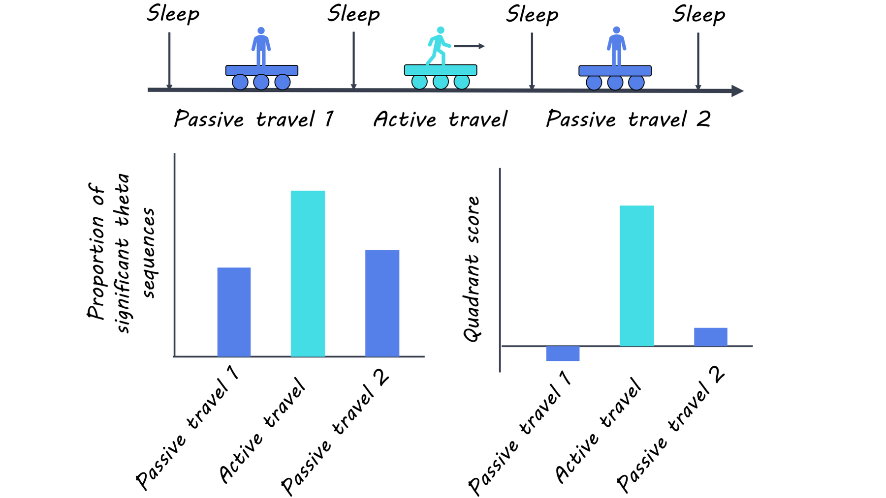
The authors put rats on a moving model train in a novel environment to test their ability to form long-term spatial memories. A treadmill on the model train was either turned off (passive travel) to disrupt theta sequences or turned on (active travel) to leave them intact. Active travel generates intact nested theta sequences while passive transportation is known to disrupt the precise timing of sequential place cell activation. Therefore, passive travel was expected to cause theta sequences to break-down in this study. Rats were tested in three sessions: passively, then actively, and passively again, alternated with periods of sleep, to determine if nested theta sequences were required for accurate replay of spatial memories during periods of sleep. The authors used a Bayesian reconstruction model to statistically analyze time scale patterns. Two quantifications were used: a combined value for trajectory score and slope to assess the quality of memory reconstruction, and a quadrant score to assess the direction of the reconstituted trajectory. To determine if neural sequences formed during initial spatial memorization were committed to long-term memory during sleep, the authors compared sleep sequences to those formed during wakefulness. Additionally, the sleep patterns of pre-active and post-active sessions were compared to confirm that sleep patterns observed were indeed reflective of those patterns formed during wakefulness and not from unrelated, pre-existing connectivity.

What did they find?

As hypothesized, the authors found that slow time scales were identical in all sessions and that genuine theta sequences were present only in active travel sessions. Active travel produced higher valued pairs of slopes and trajectory scores, consistent with greater quadrant scores, compared to passive travel, which confirmed that theta sequences were degraded in passive travel sessions and not active sessions. Neural sequences were found to be intact in sleep sessions after active travel (when theta sequences were previously formed in wakefulness) but not passive travel. This indicates a failure to commit short-term spatial memory to long-term after passive travel. Interestingly, theta sequences were perturbed to a lesser degree in the sleep session that followed the second round of passive travel compared to the first. This rules out the possibility that the emergence of theta sequences during active travel merely resulted from repeated experience. Finally, it shows that previously consolidated memory from active behavior can be undone by perturbation of theta sequences during passive travel.

theta_image.png

What's the impact?

This study is the first to show that nested theta sequences are essential to store initial memory traces which are later consolidated during sleep. Ultimately, this study sheds light on the conversion of short-term spatial memory to long-term memory. These findings advance our understanding of spatial memory consolidation and may have implications for other types of memory consolidation.

theta_quote.png

Drieu et al. Nested sequences of hippocampal assemblies during behavior support subsequent sleep replay. Neuroscience (2018). Access the original scientific paper here.

Abnormal Organization of Brain Networks Predicts Psychosis in At-Risk Youth

Post by Shireen Parimoo

What's the science?

Schizophrenia is a psychiatric disorder that manifests when a psychotic episode occurs, typically in adolescence and young adulthood when the brain is still developing. A prodromal stage of schizophrenia usually occurs prior to the onset of a psychotic episode, where cognitive function or changes in behaviour occur. Previous studies have found that the brain networks are organized differently in individuals with schizophrenia, however it is not known if organization of functional brain networks during the prodromal stage is related to the occurrence of a psychotic episode. This week in Molecular Psychiatry, Collin and colleagues used functional magnetic resonance imaging (fMRI) and graph theory to investigate the organization of brain networks in individuals who were at risk of developing psychosis.

How did they do it?

Participants included 158 adolescents and young adults who were deemed to be at-risk for developing psychosis through the Shanghai At Risk for Psychosis program. They were matched to 93 healthy participants of the same age, sex, and education level. The participants’ brain activity was recorded in an fMRI session at the beginning of the study. Their at-risk status was determined at two time points (roughly a year apart) using the Structured Interview for Prodromal Symptoms, which provides a measure of whether participants are in the prodromal stage. By the second interview, 23 participants had developed psychosis. The authors used graph theory to create functional connectomes of brain networks. Brain regions are said to be functionally connected when their activity is correlated, and a group of functionally connected regions represents a functional network. Functional connectomes were constructed for each participant as well as for each group of participants: these were healthy participants, at-risk participants, and at-risk participants who later developed psychosis. Functional connectomes of those at-risk and at-risk who later developed psychosis were compared to that of the healthy group, which allowed the authors to examine if brain networks are organized differently between groups.

What did they find?

The authors found that functional networks across all groups were organized into five modules consisting of the central-executive, sensorimotor, visual, paralimbic, and default-mode networks, each with different functional roles. An additional cingulo-opercular network was identified in at-risk individuals who later developed psychosis, but not in healthy or at-risk individuals. The functional connectomes of the healthy and at-risk individuals were more similar to each other than to to participants at-risk who later developed psychosis. Moreover, several brain regions that are affected early in schizophrenia were found to be part of different functional networks in the different participant groups. For example, the superior temporal gyrus was part of the sensorimotor network in healthy and at-risk individuals, but this brain region was functionally connected to regions of the paralimbic network in individuals who went on to develop psychosis. Thus, functional networks were organized differently in individuals who later developed psychosis, and this abnormal organization was associated with a three-fold risk of developing psychosis.

psychosis (1).png

What's the impact?

This study found that at-risk adolescents and young adults who exhibit abnormal organization of brain networks early on eventually develop psychosis, whereas at-risk youth with similar functional networks and healthy individuals do not. This novel insight into the predictive value of brain network organization for the onset of psychosis can be used to identify at-risk individuals and potentially establish preventative measures to mitigate the occurrence and severity of psychosis.

connectome_quote.png

Dr. Collin would like to acknowledge support of a Marie Curie Global Fellowship.

Collin et al. Functional connectome organization predicts conversion to psychosis in clinical high-risk youth from the SHARP program. Molecular Psychiatry (2018). Access the original scientific publication here.

How Pain Prediction Affects Perception

Post by Elisa Guma

What's the science?

Our past experiences often shape our expectations of the future, which in turn can influence how we perceive future events. Previous work has shown that our expectation of how painful a stimulus will be can influence our perception of that stimulus and our response to it. Moreover, expectations about pain (e.g., following placebo treatments) can be surprisingly resistant to change, acting much like self-fulfilling prophecies. The brain and behavioural mechanisms underlying these phenomena are largely unknown. This week in Nature: Human Behavior Jepma and colleagues used behavioural assessments and functional magnetic resonance imaging (fMRI) to investigate how expectations about pain affect pain experience, and why expectations of high or low pain sometimes persist despite evidence to the contrary.

How did they do it?

The authors designed two experiments in which they independently manipulated predictive pain cues (to investigate pain expectation), and the intensity of a pain stimulus (to investigate pain perception). In both studies, participants first went through a learning phase where they learned to associate abstract visual cues with either low or high temperatures (displayed on thermometers). In the subsequent test phase participants were presented with both sets of cues followed by a painful heat stimulus to the inner forearm in the first study, and to the lower leg in the second study. Unbeknownst to the participants, the cues were no longer predictive of heat intensity. Participants were instructed to rate how much pain they expected following each cue, and how much pain they experienced following each heat stimulus. In the second study, fMRI activity was also recorded. The authors tested responses in a measure called the Neurologic Pain Signature (NPS); a measure of activity across brain areas validated to be sensitive and specific to pain in tests performed to date. They used these brain areas to guide their investigation of brain signatures of pain perception and expectation in this study. Finally, the authors used computational models to quantify their findings, using both a reinforcement learning model  and a Bayesian model.

elisa (1).png

What did they find?

The authors found evidence that cue based expectations influence pain perception; higher pain expectation led to larger subjective pain rating, and to higher brain activity in the neurologic pain network. In addition, larger pain rating and higher activity in the NPS predicted higher pain expectation in subsequent trials with the same cue. The authors also observed a confirmation bias in learning about pain intensity, with stronger learning from new pain stimuli that confirmed one's initial pain expectation than from new pain stimuli that disconfirmed expectations. The computational models allowed the authors to determine that participants' pain expectations influenced both perceived pain and participants' learning rates. Finally, participants with stronger confirmation biases in their learning rate also showed a greater confirmation bias in the updating of pain-anticipatory brain activity in regions important for threat, anxiety, and value-based decision-making.

What's the impact?

This study is among the first to identify brain and behavioural processes underlying self-reinforcing expectations. This may have implications for chronic pain, as self-reinforcing expectations may be part of what makes people transition from acute to chronic pain. The findings may help in our understanding of how perceptual learning is applied to pain, and can be applied to individuals at risk for chronic pain. More broadly, it may help us understand how beliefs are sometimes so resistant to new evidence.

pain_quote.png

Jepma et al. Behavioural and neural evidence for self-reinforcing expectancy effects on pain. Nature: Human Behavior (2018). Access the original scientific publication here.