Whole-Brain Model Incorporating Serotonin Receptor Density Explains Functional Effects of LSD

Post by Thomas Brown

What's the science?

Lysergic acid diethylamine (LSD) is a potent hallucinogen. Though LSD is most commonly known for its use as a recreational drug, there are studies which suggest that in small doses, it may have therapeutic effects on some conditions such as depression. LSD binds to, and activates serotonergic (5HT-2A) receptors in the brain (which typically bind the endogenous neurotransmitter serotonin). Neuromodulators like serotonin have important effects on brain activity and function, however, the ways in which neurotransmitters like serotonin influence brain activity remain unclear. Understanding how brain structure and activity are modulated by serotonin will provide a greater understanding of the framework that produces effects like those seen with LSD. This week in Current Biology, Deco and colleagues used a whole-brain model (integrating multiple neuroimaging modalities) to understand the contribution of serotonin receptor stimulation in modulating brain activity dynamics while taking LSD.

How did they do it?

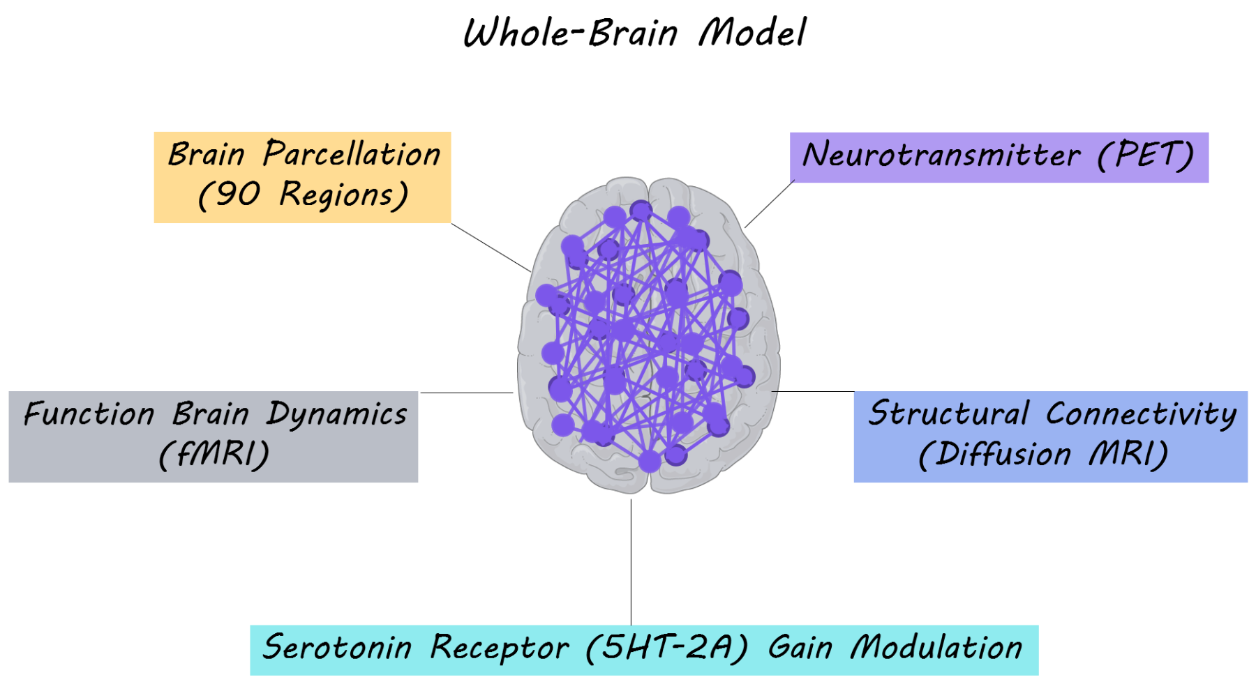
The authors’ primary aim was to understand how structure and function in the brain are modulated by the serotonergic system. In order to study this, they performed a whole-brain multi-modal brain imaging analysis, combining three imaging modalities: resting-state functional MRI (measuring synchrony between fluctuations in brain activity at rest), diffusion MRI (measuring structural white matter tracts/connections in the brain) and PET imaging of serotonergic receptor density in the brain. The whole brain was parcellated into 90 brain regions overlaid on a structural connectivity map and serotonin receptor density maps. To create their whole-brain model, the authors used dynamic mean field modeling which simulates neuronal activity at rest (based on proportions of excitatory and inhibitory neurons). The model was then fitted to the spatiotemporal dynamics of the resting state fMRI activity for each participant in the placebo condition. Subsequently, for the LSD condition they changed the excitatory ‘neuronal gain’ of each region based on the density of serotonin receptors in that region. Gain modulation is a nonlinear way in which neurons combine different inputs to produce an output  — for example, a neuron may amplify input from other neurons to produce a much larger output. By changing the ‘neuronal gain’ for each brain region based on the serotonergic receptor density in their model, they were able to test computationally whether these receptors played a role in modulating neuronal activity. 15 participants with no previous history with LSD use (mean age = 30.5) were administered a placebo and LSD on two separate days. These participants then underwent a resting-state fMRI scan (a brain scan while resting) either while listening or not listening to music (which is known to amplify some experiences related to LSD). Resting state brain activity was extracted and dynamic functional connectivity (the change in synchrony in brain activity between brain regions over time) was measured between each pair of brain regions. They then assessed how well the whole-brain model fit dynamic functional connectivity, in order to understand how neurotransmitter receptor density and structural connectivity modeled brain function.

What did they find?

The authors found that resting state brain activity profiles for the LSD condition were dependent on the serotonergic receptor density map; specifically on the regional distribution and density of these receptors. The whole-brain model, which accounted for the density of the serotonin 2A receptor by adjusting neuronal gain, explained dynamic functional connectivity for participants taking LSD. These results suggest that the whole-brain model, was able to explain the functional effects of LSD. To check that receptor distribution made a critical contribution to the model, the authors randomly shuffled receptor density values associated with each brain region and tested other serotonergic receptors (5HT-1A, 5HT-1B and 5HT-4). The model was significantly worse after these manipulations, demonstrating that the 5HT-2A receptor distribution is crucial in explaining the neural dynamics occurring during serotonergic receptor stimulation by LSD. Overall, the authors were able to demonstrate, using their model, that brain structure (obtained with diffusion MRI), interacts in a non-linear manner with serotonergic receptors (5HT-2A) density to produce changes in dynamic functional connectivity (brain function) associated with LSD.

thomas1800.png

What's the impact?

This study demonstrates that the use of a ‘whole-brain model’, combining several neuroimaging modalities, can be used to better understand the causal influence of neuromodulator systems on brain function. The whole-brain model was able to explain the contribution of serotonin receptor stimulation to brain activity while taking LSD. These findings emphasize the importance of considering the role of neurotransmitter modulation on brain activity at rest. Implementing these methods could be informative for understanding the role of neurotransmitters (e.g. dopamine, acetylcholine or serotonin) on brain dynamics in healthy individuals, during drug use or in neuropsychiatric disorders in which neurotransmitter imbalance occurs. Furthermore, the methods suggest a novel way of making rational drug discovery.

Deco et al. Whole-Brain Multimodal Neuroimaging Model Using Serotonin Receptor Maps Explains Non-linear Functional Effects of LSD. Current Biology (2018). Access the original scientific publication here.

Mutant Protein Aggregates from Humans Aid the Spread of ALS

Post by Sarah Hill

What's the science?

Amyotrophic lateral sclerosis (ALS) is a neurodegenerative disease in which motor neurons progressively die, causing muscles to atrophy. During ALS onset the protein superoxide dismutase 1 (SOD1) misfolds and begins to aggregate, forming a 'seed' within the motor neuron, which subsequently activates SOD1 aggregation in neighboring motor neurons. One theory is that SOD1 spreads in a prion-like manner (replication of the aggregates based on the initial seed). Previously, Bergh and colleagues found that two structurally distinct strains of SOD1 aggregates, termed A and B, form in the spinal cord of mice expressing mutant SOD1, inducing accumulation of aggregates and ALS-like disease. Many genetic mutations contributing to SOD1 misfolding have been identified in ALS, however, It is still unknown which SOD1 mutations result in the formation of aggregates and how these aggregates may differ for various mutations. This week in Acta Neuropathologica, Ekhtiari Bidhendi and colleagues report that seeds prepared from a human ALS patient carrying the SOD1 G127X mutation, a mutation which promotes misfolding, triggers prion-like SOD1 misfolding and aggregation in the spinal cord of transgenic mice.

How did they do it?

Human ALS patients carrying the G127X mutation express low amounts of aggregated SOD1, making it difficult to study the structure of the protein. To get around this, the authors engineered mice to express the human G127X mutation. They then prepared aggregate seeds from spinal cord tissue from the G127X mice, as well as from an ALS patient carrying the G127X mutation. Control seeds were similarly prepared from control mice (without mutation) and a human control patient. Next, the authors implanted either the G127X seeds or the control seeds into the spinal cord of mice expressing mutant human SOD1 and monitored the animals for progression of ALS-like symptoms. Once the animals reached the terminal disease stage, spinal cord and brain tissue were harvested and either used for immunohistochemistry or prepared into extracts for Western blotting. Finally, to determine the structure of the induced SOD1 aggregates, the authors carried out binary epitope-mapping on the isolated tissue extracts, a technique they previously developed to infer aggregate structure based on how well SOD1 binds to specific antibodies in various folding states.

What did they find?

Mice inoculated with mouse- and human-derived G127X aggregates experienced early onset of an ALS-like motor neuron disease, with symptoms including shortened lifespan, weight loss, and decreased number of motor neurons. Interestingly, the onset of G127X-induced motor neuron disease preceded the normal disease onset observed in control-inoculated and non-inoculated mice. Immunohistochemistry confirmed these results, with SOD1 aggregation appearing in G127X-inoculated animals significantly sooner than in control- and non-inoculated groups. The distribution of G127X aggregates was relatively even throughout the spinal cord segments, while control seeds resulted in an uneven aggregate distribution. The varying seed types also resulted in differences in location of symptom onset, with a majority of G127X-inoculated mice first exhibiting hind leg paralysis and control-inoculated mice exhibiting paralysis of hind legs, for legs, or both. Based on the binary epitope-mapping assay, the authors concluded that the G127X mutation led to formation of A-strain SOD1 aggregates in the spinal cord of inoculated animals: the aggregates from mice had a structure similar to that of A-strain seeds. The structure of the induced SOD1 aggregates replicated that of the original implanted mutant seed for all types of seeds, suggesting that transmission of SOD1 occurred in a prion-like manner. Taken together, these results suggest that motor neuron disease induced by the G127X mutation occurs through the prion-like spread of A-strain SOD1 aggregates.     

sarah1800 (1).png

What's the impact?

A current challenge in ALS research is understanding whether different strains of protein aggregates exist and how each aggregate strain is transmitted from cell to cell. We now know that the ALS-inducing SOD1 mutation (G127X) results in the prion-like spread of SOD1 aggregates. The binary epitope-mapping protocol developed previously by the authors is widely applicable to other neurodegenerative diseases, such as Alzheimer's and Parkinson's, and provides a framework for pinpointing differences in the structure and transmission of aggregate strains.  

Ekhtiari Bidhendi et al., Mutant superoxide dismutase aggregates from human spinal cord transmit amyotrophic lateral sclerosis. Acta Neuropathologica (2018).Access the original scientific publication here.

Heritability of Sleep-Specific Oscillations in Adolescents

Post by Shireen Parimoo

What’s the science?

Sleep electroencephalogram (EEG) measures, such as spindle and slow wave activity (SWA), are associated with cognitive and behavioral outcomes. For instance, sleep spindles are related to synaptic plasticity underlying learning and memory, and slow wave activity during sleep is thought to be restorative. Deficits in spindle activity and slow wave activity are associated with neuropsychiatric disorders like schizophrenia and depression. Sleep EEG recordings in adults are heritable (i.e. genetically inherited), and differ from those in adolescents. This is because the brain is still developing during adolescence and undergoes structural changes that influence oscillatory activity. The role of genes in sleep EEG in adolescents is currently unknown. This week in the Journal of Neuroscience, Rusterholz and colleagues used high-density EEG to examine sleep-specific oscillations and their heritability across various brain regions in adolescent twins.

How did they do it?

Eighteen pairs of monozygotic (MZ) and 12 pairs of dizygotic (DZ) adolescent twins between the ages of 11 and 14 years took part in this study. Their sleep was monitored for at least five nights to ensure that they were getting adequate sleep each night, after which high-density sleep EEG was recorded for two consecutive nights. The first night served as adaptation to the sleep EEG equipment. EEG recorded on the second (baseline) night was used in subsequent analyses, except when data quality was poor, in which case EEG data from the adaptation night was used instead. The measures of interest included sleep EEG power (strength of the oscillations) in various frequency bands, ranging from slow oscillations  between 0.6 – 1.2 Hz to gamma oscillations (at a higher frequency) in the 24 – 44 Hz frequency band. The authors also examined spindle characteristics like amplitude, duration, density, and integrated spindle activity, which is the integrated spindle amplitude over time (i.e. intensity of spindles). Structural equation modeling (SEM) was used to estimate the contribution of genetics and environmental factors to the sleep EEG measures, while differentiating between shared and unique environmental factors that the twins were exposed to during development.

What did they find?

Genes strongly contributed to slow wave activity in many cortical regions, accounting for 60 – 93% of variance in slow wave activity, whereas unique environmental factors explained 30 – 44% of variance in slow wave activity in frontal regions. Shared environmental factors did not significantly contribute to slow wave activity. Sleep spindles in posterior regions of the brain were influenced strongly by genetic factors, whereas shared environmental factors contributed to spindles in more anterior regions. Shared environmental factors contributed to spindle amplitude and integrated spindle activity in anterior brain regions for slow (10 – 12 Hz) and fast (12 – 16 Hz) spindles. Moreover, both shared and unique environmental factors contributed to the density and duration of sleep spindles. Specifically, shared environmental factors contributed to the density and duration of fast spindles in fronto-central brain regions, and unique environmental factors influenced the density and duration of fast spindles in posterior brain regions.

eeg1800.png

Slow oscillations in frontal and temporal regions were influenced by unique environmental factors, whereas genes strongly contributed to slow oscillations over widespread cortical regions. For EEG power in alpha, beta, theta, and gamma frequency bands, genetic contribution ranged from 70 – 96%, indicating that sleep EEG power in these frequency bands is primarily determined by genetic factors. Genes and shared environmental factors contributed equally to theta power in lateral fronto-parietal brain regions.

What’s the impact?

This is the first study to show the contribution of genetic and environmental factors to sleep oscillations in adolescent twins. In particular, EEG power (strength of neural oscillations) is highly heritable and determined by genetics, whereas genetic and environmental factors differentially contribute to slow oscillations, slow-wave activity, and spindle activity across brain regions. The onset of many neuropsychiatric disorders occurs during adolescence, therefore, the characteristics and heritability of adolescent sleep EEG could provide neurophysiological biomarkers for these disorders.

EEG_1800_quote.png

Rusterholz et al. Nature and Nurture: Brain region specific inheritance of sleep neurophysiology in adolescence. The Journal of Neuroscience (2018). Access the original scientific publication here.