Shared Narratives are Associated with Similar Neural Responses

Post by Shireen Parimoo

What’s the science?

People can understand narratives presented in a wide variety of formats, ranging from a series of gestures and speech, to entire movies, and simple animations of geometric shapes. When people view the same narrative, such as a movie, their brain activation patterns are correlated. This neural similarity is particularly evident in regions of the default mode network (DMN), a brain network involved in processing complex narratives, among other things. However, it is unclear whether this neural similarity would be observed when a narrative has multiple interpretations. This week in NeuroImage, Nguyen and colleagues used functional magnetic resonance imaging (fMRI) to examine neural similarity across individuals, particularly in DMN regions, when they were presented with an ambiguous narrative.

How did they do it?

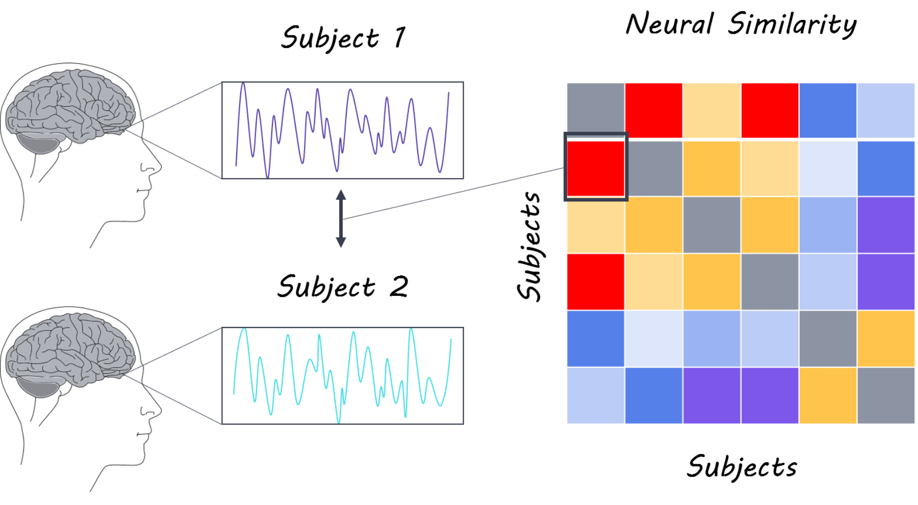
Two groups of participants were presented with either a movie or audio narrative while undergoing fMRI scanning. In one group, 36 adults watched a movie clip of geometric shapes with an ambiguous narrative that contained music to set the mood but no speech. In the other group, 18 adults listened to an audio clip that dictated the narrative corresponding to the movie clip. All participants recalled their respective narratives in detail. In the first analysis, the authors examined whether neural activity was correlated (related) across different participants based on the modality and interpretation of the narrative. Two analyses were performed: 1) Latent Semantic Analysis (LSA) was used to measure similarity of recall among participants within the movie and audio groups, and between the two groups. Participants in the movie group were further split into ‘high’ and ‘low’ recall similarity sub-groups based on their LSA similarity score. Participants in the movie group were also split into ‘high’ and ‘low’ interpretation similarity sub-groups based how correlated their LSA score was to that of participants in the audio group. The neural activity of participants within each of the recall similarity groups was correlated with each other to yield between-participant correlations; likewise, neural activity of participants in the two interpretation similarity groups was correlated with that of participants in the audio group to yield cross-modality between-participant correlations. 2) the authors used between-participant representational similarity analysis (RSA) to identify brain regions where more similar interpretations of the narrative elicited more similar patterns of activity, both within and across the two modalities.

What did they find?

Participants whose recall of the movie was similar (high recall similarity group) had high between-participant correlations in visual and auditory brain areas, as well as in regions involved in complex cognitive processing like the angular gyrus. Importantly, neural similarity in the primary visual cortex and most DMN regions such as the posterior medial cortex and angular gyrus was greater in the high recall similarity than in the low recall similarity group. This suggests that when recall of the movie was very similar across participants, the neural responses in the DMN were more correlated across participants.

When neural responses were compared across modalities, participants who interpreted the movie in a similar way to participants in the audio group (high interpretation similarity group) showed neural similarity in the inferior temporal gyrus. On the other hand, neural responses of participants whose interpretation of the movie was different from that of the audio group (low interpretation similarity group) were correlated in the right angular gyrus. Neural similarity in the posterior medial cortex, angular gyrus, and left medial temporal gyrus was greater in the high interpretation similarity group compared to the low interpretation similarity group, indicating that activity in these regions is more correlated when the people derive similar meaning from a narrative. Finally, the similarity of participants’ interpretation of the narrative was correlated with neural similarity in the right dorsolateral prefrontal cortex, posterior medial cortex, and right angular gyrus.  

shireen1800.png

What’s the impact?

This is the first study to show that when people’s spontaneous understanding of a narrative is similar, so is their brain activity— particularly in the default mode network. Importantly, this neural similarity was modality-invariant, suggesting that the meaning of the narrative, rather than the form in which it was presented, activated those brain regions. This study provides further insight into how social narratives are processed in the brain.

shireenquote1800.png

Nguyen et al. Shared understanding of narratives is correlated with shared neural responses. NeuroImage (2018). Access the original scientific publication here.

Glial Fibrillary Acidic Protein as a Marker for Mild Traumatic Brain Injury

What's the science?

Millions of cases of mild traumatic brain injury occur each year. Computed tomography (CT) scans are used to detect mild traumatic brain injury, and MRI can be used to detect subtle changes in the brain like neuron axonal injury, however these are costly and time-consuming. There is a need for a blood-based biomarker that can detect milder forms of brain injury to ensure proper treatment for these patients. This week in Neurology, Ori and colleagues test whether blood-based biomarkers are associated with neuroimaging changes (on CT and MRI scans) and can successfully detect mild traumatic brain injury. 

How did they do it?

Four blood-based biomarkers have previously been associated with brain changes that follow traumatic brain injury of varying severities: Tau (a neuronal injury marker), Glial Fibrillary Acidic protein, ubiquitin carboxyl-terminal hydrolase L1 (UCH-L1) and neurofilament light. The authors aimed to assess whether any of these biomarkers would be elevated in mild traumatic brain injury and whether they were also associated with subtle structural changes shown on an MRI scan (using diffusion tensor imaging). A group of 277 patients seeking care for a mild brain injury were enrolled in the study. Blood was drawn to measure plasma concentrations of biomarkers and CT and MRI scans were performed within 48 hours of the injury. A control group of 49 healthy participants (with well-matched demographics) was included for comparison.

What did they find?

Glial Fibrillary Acidic protein, Tau and Neurofilament light were all higher in patients with mild traumatic brain injury compared to controls. Glial Fibrillary Acidic protein was the best predictor of mild traumatic brain injury (diagnosis). When patients with mild brain injury were stratified into those with and without changes on their CT scans, Glial Fibrillary Acidic protein, Tau and neurofilament light concentrations were all higher in patients with detectable changes. However, Glial Fibrillary Acidic protein concentration was the only biomarker that significantly predicted trauma-related CT scan changes. Glial Fibrillary Acidic protein, Tau and Neurofilament light all predicted structural MRI changes, however Glial Fibrillary Acidic protein was the strongest predictor of structural MRI changes related to mile traumatic brain injury.

leigh1800.png

What's the impact?

This is the first study to examine whether blood-based biomarkers can be used to detect mild traumatic brain injury. Glial Fibrillary Acidic protein concentration is a sensitive predictor of mild traumatic brain injury and is also closely associated with neuroimaging changes. CT and MRI scans are expensive and time-consuming, so having methods to detect the presence and severity of brain injury early on is important for proper and effective treatment.

leighquote1800.png


Gill et al., Glial fibrillary acidic protein elevations relate to neuroimaging abnormalities acutely following a mild traumatic brain injury. Neurology (2018). Access the original scientific publication here.

Mediated Learning is Dependent on Type 1 Cannabinoid Receptors

Post by Sarah Hill

What's the science?

CB1 receptors bind cannabinoids (a class of chemical compounds) produced within the body (endocannabinoids) and are known to be involved in learning and memory. Binding of CB1R to cannabinoids originating outside of the body, for example during consumption of marijuana, can cause cognitive impairment. In contrast, endocannabinoid binding to CB1R within the hippocampus is involved in direct associative learning (a simple form of learning where an association is made between a certain stimulus and an outcome, such as the pairing of a sound with a subsequent food reward). Whether CB1R and the endocannabinoid system in general are involved in other higher-order forms of learning, such as ‘mediated learning’, is not known. Mediated learning is when a stimulus is paired with an outcome indirectly through association with another stimulus (also known as an incidental association). This week in Neuron, Busquets-Garcia and colleagues demonstrate that hippocampal CB1Rs are specifically required for mediated learning.

How did they do it?

The authors carried out a sensory preconditioning task in mice with a number of transgenic and pharmacological interventions. The sensory preconditioning procedure consisted of three phases: 1) Two low-salience stimuli (such as odor and taste) were presented to male mice simultaneously to promote formation of an incidental association between the two (pre-conditioning phase) 2) One of the original stimuli was directly paired with either an aversive or a reward reinforcer (conditioning phase), increasing the salience of the outcome-associated stimulus and indirectly pairing the other stimulus with the same outcome 3) Mice were presented with either the directly-paired or the indirectly-paired stimuli in order to evaluate direct associative learning or mediated learning respectively (test phase). In the first variation of sensory preconditioning, the authors paired odor and taste with an aversive outcome (gastric malaise). To ensure that their results were not restricted to odor and taste stimuli, nor to an aversive outcome, the authors carried out an additional sensory preconditioning task by pairing two new stimuli, a light and a sound, with a food pellet reward outcome.  

sarah1800.png

As a basic test of CB1R activity in mediated learning, sensory preconditioning was first carried out using mice who had CB1R deleted or 'knocked-out' on a brain-wide level. Next, to determine the timeframe of CB1R activity during mediated learning, the authors administered a CB1R antagonist (i.e. blocker) at different time points throughout the task, either before the preconditioning phase or the test phase. To assess which brain region CB1R-dependent mediated learning occurs within, they repeated the experimental procedure after deleting CB1R within the hippocampus. Finally, to identify the neurons involved in CB1R-dependent mediated learning, the authors carried out the sensory preconditioning task in mice lacking CB1R specifically in forebrain GABAergic inhibitory interneurons. Because CB1R helps to suppress GABAergic inhibitory neurotransmission, the authors hypothesized that CB1R inhibition of GABA neuron firing may be critical for incidental associations and mediated learning to occur. To test this, they infused a viral vector expressing inhibitory designer receptors exclusively activated by designer drugs (DREADD) to either inhibit or excite GABAergic neurons in the hippocampi of mice lacking CBR1 before preconditioning. This allowed them to observe the impact of activating or inhibiting GABAergic neurons on mediated learning.

What did they find?

Wild-type (control) mice that underwent sensory preconditioning consumed reduced amounts of tastes or odors that were indirectly or directly paired with gastric malaise, suggesting that mediated and associative (direct) aversion learning had occurred. Likewise, increased reward-seeking behavior was observed in response to stimuli when two associated stimuli were directly- and indirectly-paired with a reward outcome. However, the results did not hold for indirect-pairing of a stimulus-outcome duo when CB1R knock-out mice or CB1R antagonist-dosed mice underwent sensory preconditioning (direct-pairing was unaffected). This suggests that CB1 receptors are required for higher-order/mediated learning to occur. Administration of a CBR1 antagonist (blocker) at different timepoints revealed that CB1Rs are specifically activated during the formation of incidental associations, but not during expression of mediated aversive or reward-seeking behaviors. Additional transgenic and viral approaches further confirmed that CB1 receptors involved in this mode of learning are uniquely expressed by GABAergic interneurons within the hippocampus.

Suppression of hippocampal GABAergic signaling (accomplished using inhibitory DREADDs) in mice lacking CBR1 in the hippocampus, was sufficient to recover mediated learning deficits (normally observed in animals lacking CB1R inhibitory function). Conversely, excitatory DREADDs used to activate inhibitory GABAergic transmission, effectively overriding suppression by CB1R, resulted in reduced consumption of the indirectly-paired taste stimulus but not the directly-paired stimulus. Taken together, these findings indicate that excess inhibitory signaling during preconditioning disrupts the formation of incidental associations, blocking mediated learning. Therefore, CB1R are necessary to regulate the activity of GABAergic interneurons. The authors performed a follow-up experiment to show that the GABAergic neurons involved are not parvalbumin positive interneurons (approximately half of GABAergic neurons in the hippocampus). This suggests that the remaining half, presumably interneurons containing cholecystokinin (CCK), are the neuronal subpopulation responsible for driving mediated learning.       

What's the impact?

This study identified a unique role for Type 1 cannabinoid receptors in a form of higher-order learning called mediated learning. Specifically, CB1R expressed by non-parvalbumin GABAergic interneurons within the hippocampus contribute to this learning process and are activated during formation of incidental associations. As CB1R has been implicated in a number of psychiatric and neurological disorders, this is an important finding to consider when designing CB1R-targeting therapeutic strategies.

sarahquote1800 (1).png

Busquets-Garcia et al., Hippocampal CB1 Receptors Control Incidental Associations. Neuron (2018). Access the original scientific publication here.