Antiepileptic Drugs Induce Mislocalization of Neurons in the Hippocampus

What's the science?

Valproic acid is an anti-epileptic drug prescribed to some pregnant women who have epilepsy. Use of the drug during pregnancy is associated with autism and Attention Deficit Hyperactivity Disorder in offspring, and these disorders are associated with a higher risk for seizures. High seizure risk has also been linked to mislocalized neural stem cells/progenitor cells in the hippocampus (which will become neurons in the hippocampus; a brain region involved in learning and memory). This week in PNAS, Sakai and colleagues explored whether exposure to valproic acid increased seizure susceptibility through hippocampal mechanisms in mice.

How did they do it?

To test whether exposure to valproic acid could cause seizures, they gave kainic acid (activates glutamate receptors and can promote seizure activity) to mice (at 12 weeks old) who had or had not been previously exposed to valproic acid prenatally. They used immunohistochemistry to locate progenitor cells in these mice. Next they used RNA-sequencing of neural stem cells/progenitor cells in the hippocampus at embryonic day 15, postnatal day 5, and 12 weeks old to identify differentially expressed genes whose expression levels varied due to prenatal valproic acid exposure at embryonic day 12, 13, and 14 (3 times). They then matched abnormal gene expression to known gene function (Gene Ontology analysis). Finally, they examined whether exercise (voluntary running) might mitigate the effects of valproic acid on seizure activity, due to its known role in neurogenesis (production of new neurons).

What did they find?

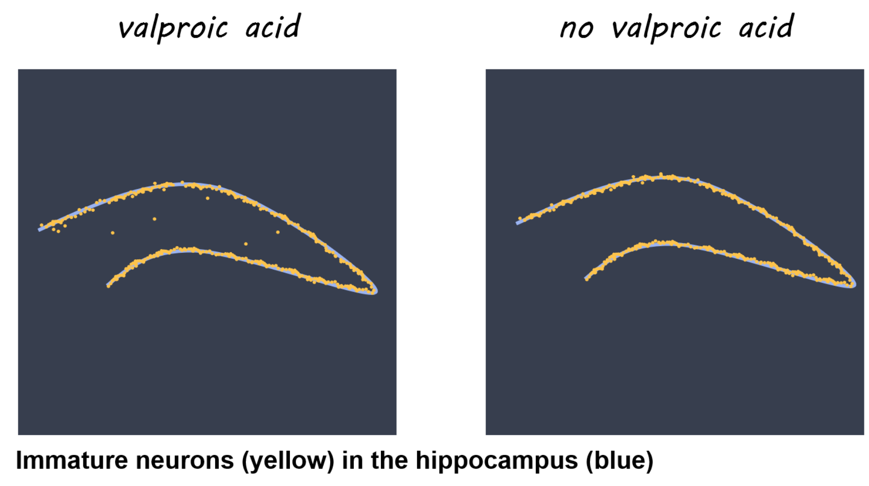
Mice exposed to valproic acid experienced increased susceptibility to seizures at 12 weeks of age, and increased mislocalization of newly generated neurons from stem cells/progenitor cells within the dentate gyrus (decreased in the granule cell layer, increased at the hilus). Several differentially expressed genes were present at different developmental stages in mice exposed to valproic acid, indicating that valproic acid affects gene expression in stem cells/progenitor cells the hippocampus. Using Gene Ontology, they identified several genes involved in cell and neuronal migration, including Cxcr4. When mice exposed to valproic acid voluntarily exercised (ran) for eight weeks, their susceptibility to seizures decreased and their Cxcr4 expression normalized, indicating that exercise may mitigate the effects of valproic acid on the hippocampus through Cxcr4.

Artistic rendering of Figure 1c - Immature neurons in the hippocampus

What's the impact?

This is the first study to link valproic acid with mislocalization of hippocampal neurons and seizure susceptibility in the offspring of pregnant mice. We now have a better understanding of the mechanisms underlying the harmful effects of valproic acid. Exercise may be a particular avenue for focus, as it may mitigate the effects of improper hippocampal neuron placement on susceptibility to seizures.

naka.png

A. Sakai et al., Ectopic neurogenesis induced by prenatal antiepileptic drug exposure augments seizure susceptibility in adult mice. PNAS (2018). Access the original scientific publication here.

The Role of T Cell Populations in Multiple Sclerosis Disease Activity

What's the science?

In multiple sclerosis (MS), effector T cells (immune cells) can travel from the periphery to the central nervous system and mediate symptoms by causing inflammation and neurodegeneration. Some therapies use antibodies to target T cells, but different T cell populations function differently and may release different inflammatory factors, resulting in different effects in MS. This week in Brain, Langelaar and colleagues assessed blood and cerebral spinal fluid in patients with MS to characterize the function of different T cells in MS.

How did they do it?

Healthy controls and patients with MS participated. MS patients included those who had experienced the first presentation of MS (clinically isolated syndrome) who either went on to develop MS <1 year later or did not develop MS for 5 years, and those experiencing relapsing remitting MS who were on antibody treatment (natalizumab) or not. Blood draws and lumbar punctures were obtained. Flow cytometry was used to locate cells based on their antibodies. To confirm their findings, they also obtained blood and spinal fluid and brain tissue samples from 5 patients after death who had late-stage MS.

What did they find?

In patients with the clinically isolated syndrome (i.e. had experienced the first presentation of MS) who developed MS soon after (versus those who did not), a lower proportion of CD4+ cells in the blood were Th1-like Th17 (a type of T helper cell). A lower proportion of Th1-like Th17 cells in the blood was also found in treatment-naive relapsing remitting patients compared to healthy controls, indicating that this finding may be specific to MS. At the same time, the proportion of Th1-like Th17 cells was higher in the spinal fluid of MS patients (and co-produced high levels of inflammatory factors). Therefore, it appears that these cells are being activated and recruited (from the blood) to the central nervous system (lower in the blood, but higher in the cerebral spinal fluid). After death, there was a high proportion of Th1-like Th17 in brain tissues of MS patients but not in controlsproviding further supporting evidence for recruitment of these cells to the central nervous system.  An adhesion molecule called VLA-4, which could help T cells migrate from the periphery to the central nervous system, was reduced following treatment with an MS drug, natalizumab (versus pre-treatment), indicating it was targeted by this antibody.

T cells in Multiple Sclerosis

What's the impact?

This is the first study to demonstrate that a specific subset of T cells (Th1-like Th17 cells) is involved in disease activity in MS. This subset is the main population of inflammatory cells within the cerebral spinal fluid of the central nervous system. The findings suggest that migration of these cells from the periphery could underlie MS disease activity, and that an MS treatment (natalizumab) can target key inflammatory factors involved. This work could help to design more targeted therapies for MS, and demonstrates the usefulness of natalizumab early on in the disease.

J. van Langelaar et al., Characterizing the role of T cell populations in MS disease and treatment. Brain (2018). Access the original scientific publication here.

Ripples During Slow-Wave Sleep Re-balance Neurons

What's the science?

Synapses in the brain are strengthened while awake and synaptic depression (weakening of the synapses) occurs during slow-wave sleep to rebalance synapses. This synaptic depression may help to selectively preserve memories, however, how it occurs during sleep is unclear. During slow-wave sleep, the hippocampus emits high frequency oscillations called “sharp-wave ripples” which reactivate the neurons involved in recent memories. Ripples could be required for synaptic depression during sleep, making “room” for new memories to form. This week in Science, Norimoto and colleagues test whether ripples are required for synaptic depression and subsequent memory formation in mice.

How did they do it?

They silenced sharp-wave ripple events during slow-wave sleep using optogenetic-feedback (i.e. every time a ripple event was detected, they inhibited the hippocampal neurons to reduce firing) in a group of test mice. They then recorded excitatory postsynaptic potentials (activity in the postsynaptic neurons) during slow-wave sleep and compared the test mice to control mice to determine whether synaptic depression during sleep was affected in the test mice. Mice then underwent a spatial object-recognition task. First, they explored an area containing two new objects, and then returned to the same location 2 hours later where one of the objects had moved. The authors compared the memory performance between test and control mice to determine whether disrupting the ripples had an effect on new memory acquisition.

What did they find?

The authors were able to successfully reduce almost all of the ripple events in the test mice. Control mice experienced depressed postsynaptic activity representing the expected synaptic depression that occurs during slow-wave sleep, while mice with the impaired ripple events did not show synaptic depression. Control mice showed normal memory performance, while mice with impaired ripple events were unable to identify a moved object in the spatial object recognition task, suggesting that they could not form new memories. The authors were also able to replicate the lack of synaptic depression by testing the effects of disrupting ripples in hippocampal tissue slices. They used in vitro and in vivo experiments to show that synaptic depression occurring during slow-wave sleep is mediated by NMDA receptors.

Optogenetic ripple silencing

What's the impact?

This is the first study to link sharp-wave ripples during slow-wave sleep with synaptic depression and memory performance. Before this study, we did not understand the mechanism through which synaptic depression occurred during slow-wave sleep. We now know that ripples during slow-wave sleep are critical for balancing of synapses and for new memory formation.

Yuji_quote_week11_small.png

H. Norimoto et al., Hippocampal ripples down-regulate synapses. Science (2018). Access the original scientific publication here.