How Deep Brain Stimulation Affects Decision-Making in Parkinson’s disease

What's the science?

How much time should we give ourselves to make a decision? For example, when faced with a difficult decision, we might give ourselves more time to garner more evidence before we reach the ‘decision threshold’ and decide. One brain region involved in adjusting our decision threshold (meaning we take more or less time before the decision) is the subthalamic nucleus (STN). Deep brain stimulation (DBS) of the STN is often performed to reduce motor symptoms in Parkinson’s disease, however, a negative side effect can be impairment in adjusting the decision threshold, leading to impulsive responses. This week in Current Biology, Herz and colleagues conducted a study in patients with DBS electrodes for Parkinson’s placed in the STN, in order to assess how stimulation of this site affects the decision threshold.

How did they do it?

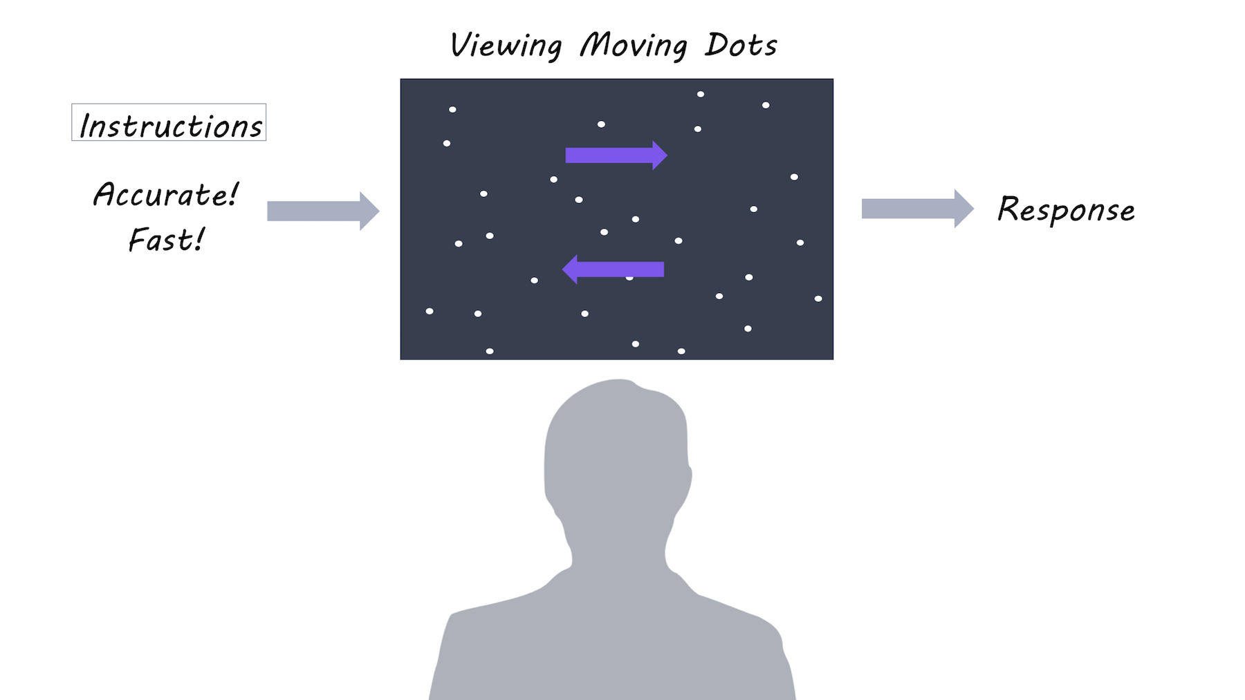
Ten patients with Parkinson’s participated in the study after undergoing surgery to implant electrodes for DBS in the STN. Each patient performed a decision-making task: 1) when DBS was off 2) with DBS on continuously and 3) with ‘adaptive DBS’ where DBS only turns on when necessary. The decision-making task involved looking at dots moving on a screen, and deciding whether the majority of dots were moving to the left or to the right. There were two task conditions and two forms of instruction: In the easy condition, 50% of the dots moved in the same direction, while in the difficult condition only 8% of dots moved in the same direction. Participants were also instructed to focus on either speed or accuracy of their decision.

Dot motion perception

What did they find?

When DBS was off, participants responded more slowly during the difficult task and when instructed to focus on accuracy (versus speed). However, when DBS was on, slowing during a difficult task was diminished, but slowing due to focus on accuracy remained the same. During adaptive DBS, stimulation came on at different times across trials (when beta activity happened to be high). When the DBS stimulation came on during a 400-500 ms time window after the moving dots appeared on the screen, the time required to make a decision (usually increased during the difficult task) was most diminished, suggesting that the effect of stimulation is confined to a short time window. Using ‘drift diffusion modelling’, they found that stimulation affected the decision threshold time specifically, as opposed to, for example, the motor response time. While DBS was off, beta activity increased after presentation of the dots during the difficult condition, and was related to the decision threshold, but these effects were abolished during stimulation. This indicates that DBS may be lowering the decision threshold by changing the relationship between STN activity and threshold adjustments.

What's the impact?

These results are the first to show that the STN may be directly involved in decision thresholds (how much evidence we need before we reach a decision). During a narrow time window, the STN adjusts decision thresholds based on the anticipated difficulty of the decision. This may be a mechanism by which decision-making is impaired in people with Parkinson’s who have DBS.

brown_quote_week11_small.png

P. Brown et al., Mechanisms Underlying Decision-Making as Revealed by Deep-Brain Stimulation in Patients with Parkinson’s Disease. Current Biology (2018). Access the original scientific publication here.


Different Profiles of Microglial Activation in Alzheimer's disease

What's the science?

Microglia, the immune cells of the brain, may contribute to Alzheimer’s disease by becoming activated in response to brain pathology (also known as neuroinflammation). Currently, whether neuroinflammation is associated with Alzheimer’s progression (harms the brain) or whether it may be protective (helps to “eat” plaques in the brain) is still a matter of debate. This week in BrainHamelin and colleagues used PET imaging to examine how microglial activation in the brain is related to Alzheimer’s disease progression.

How did they do it?

PET imaging was used to measure the uptake of a radiotracer (18F-DPA-714) in the brain binding to activated microglia. A large group of patients with Alzheimer’s disease were scanned twice for activated microglia, once at baseline and once two years later. They were then followed up annually and scanned with MRI to measure brain volume (measure of Alzheimer’s progression) and given annual cognitive tests to assess dementia severity and cognitive function. Based on this, patients were split into “fast and slow decliner” categories. The microglial activation levels over time were also analyzed compared to a control group of healthy participants.

What did they find?

Having a high level of microglial activation at baseline was predictive of being a slow decliner. In patients with a high baseline neuroinflammation, cognitive performance was better and brain volume was more preserved, suggesting that more microglial activation at baseline is protective. At two years follow-up, microglial activation was higher in Alzheimer’s participants but not controls as would be expected. Increased microglial activation over time was related to worsening cognitive scores and brain atrophy, suggesting that it is harmful. However, when they examined neuroinflammation over time at an individual level, they found that those with the highest baseline microglial activation had the lowest increase in microglial activation over time. They concluded that there is a dynamic relationship, whereby neuroinflammation may affect patients differently, depending on their original level of microglial activity. Microglial activation appears to be protective initially, but exacerbates Alzheimer’s disease over time; to a greater extent in those who had low levels of microglial activation to begin with.

Microglial activation in Alzheimer’s disease

What's the impact?

This is the first study to show that neuroinflammation may affect individuals with Alzheimer’s disease differently depending on their baseline level of microglial activity. It shows us that microglial activation may be helpful or harmful depending on the individual and how far their disease has progressed. Understanding the role microglial activation play in Alzheimer’s disease is an essential part of understanding how the disease progresses.


L. Hamelin et al., Distinct dynamic profiles of microglial activation are associated with progression of Alzheimer's disease. Brain (2018). Access the original scientific publication here.

Functional Connections in the Brain are Stronger in Females Resilient to Depression

What's the science?

One third of females will be diagnosed with depression (major depressive disorder) during their adolescence. Resilience refers to the ability to adapt well in response to stress and bounce back from challenging life experiences. Currently, we don’t know the brain mechanisms that underlie resilience in adolescents who are at risk for depression. This week in JAMA Psychiatry, Fischer and colleagues test whether brain functional connectivity can be a biomarker for resilience in adolescent females at risk for depression (i.e. depression runs in their family).

How did they do it?

65 adolescent females were recruited: 25 low risk control participants who did not develop depression (control), 20 whose parents had a history of depression and developed depression themselves (i.e. converted) and 20 whose parents had a history of major depressive disorder but did not develop depression (i.e. resilient). The brains of all participants were scanned several times using  resting-state fMRI (which measures brain function at rest) over several years. They compared functional connectivity (synchronous brain activity) between resilient and converted females and between resilient and control females. They assessed the functional connectivity profiles of three brain regions known to be involved in depression: the amygdala (emotion), the anterior insula (attention/cognition) and the dorsolateral prefrontal cortex (planning). They measured the relationship between functional brain connections and life events.

What did they find?

Females who were resilient to depression showed stronger functional connections in the brain between the amygdala (involved in fear and emotion) and the orbitofrontal cortex (involved in impulse control and modulating emotions). A stronger connection between these regions was associated with more positive life events. Resilient individuals also showed stronger connections between the dorsolateral prefrontal cortex (involved in planning and executive function) and the frontotemporal cortex (involved in cognitive control). Both resilient and converted groups had stronger functional connectivity within the salience network (a network of regions involved in attention and cognition) compared to the control group

Functional brain connectivity between orbitofrontal cortex and amygdala

What's the impact?

This is the first study to show that functional connections in the brain can be markers for resilience to depression in adolescent females at high risk for depression. Stronger functional connections could represent adaptation in the brain in response to positive life experience. It is crucial to understand how adolescents can develop resilience to depression in order to better prevent and treat depression.

inkquote4.png

A. Fischer et al., Neural Markers of Resilience in Adolescent Females at Familial Risk for Major Depressive Disorder. JAMA Psychiatry (2018). Access the original scientific publication here.