The Role of T Cell Populations in Multiple Sclerosis Disease Activity

What's the science?

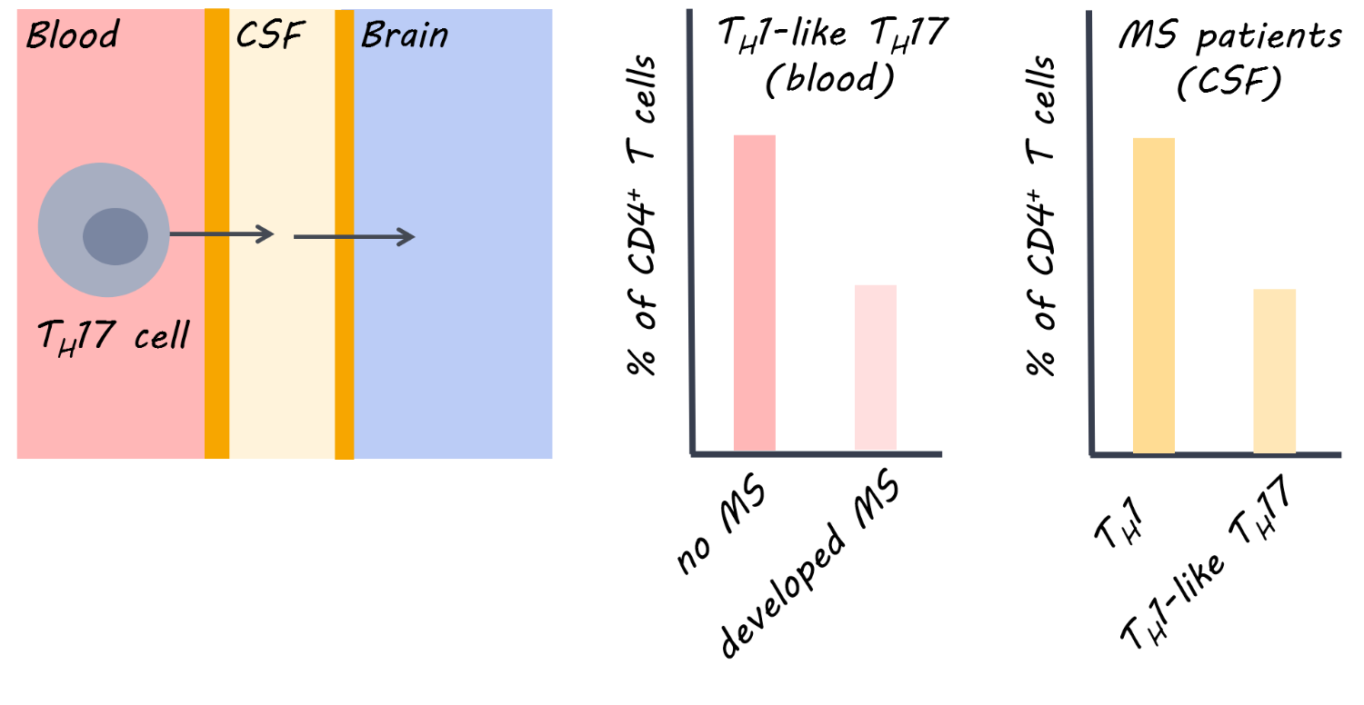
In multiple sclerosis (MS), effector T cells (immune cells) can travel from the periphery to the central nervous system and mediate symptoms by causing inflammation and neurodegeneration. Some therapies use antibodies to target T cells, but different T cell populations function differently and may release different inflammatory factors, resulting in different effects in MS. This week in Brain, Langelaar and colleagues assessed blood and cerebral spinal fluid in patients with MS to characterize the function of different T cells in MS.

How did they do it?

Healthy controls and patients with MS participated. MS patients included those who had experienced the first presentation of MS (clinically isolated syndrome) who either went on to develop MS <1 year later or did not develop MS for 5 years, and those experiencing relapsing remitting MS who were on antibody treatment (natalizumab) or not. Blood draws and lumbar punctures were obtained. Flow cytometry was used to locate cells based on their antibodies. To confirm their findings, they also obtained blood and spinal fluid and brain tissue samples from 5 patients after death who had late-stage MS.

What did they find?

In patients with the clinically isolated syndrome (i.e. had experienced the first presentation of MS) who developed MS soon after (versus those who did not), a lower proportion of CD4+ cells in the blood were Th1-like Th17 (a type of T helper cell). A lower proportion of Th1-like Th17 cells in the blood was also found in treatment-naive relapsing remitting patients compared to healthy controls, indicating that this finding may be specific to MS. At the same time, the proportion of Th1-like Th17 cells was higher in the spinal fluid of MS patients (and co-produced high levels of inflammatory factors). Therefore, it appears that these cells are being activated and recruited (from the blood) to the central nervous system (lower in the blood, but higher in the cerebral spinal fluid). After death, there was a high proportion of Th1-like Th17 in brain tissues of MS patients but not in controlsproviding further supporting evidence for recruitment of these cells to the central nervous system.  An adhesion molecule called VLA-4, which could help T cells migrate from the periphery to the central nervous system, was reduced following treatment with an MS drug, natalizumab (versus pre-treatment), indicating it was targeted by this antibody.

T cells in Multiple Sclerosis

What's the impact?

This is the first study to demonstrate that a specific subset of T cells (Th1-like Th17 cells) is involved in disease activity in MS. This subset is the main population of inflammatory cells within the cerebral spinal fluid of the central nervous system. The findings suggest that migration of these cells from the periphery could underlie MS disease activity, and that an MS treatment (natalizumab) can target key inflammatory factors involved. This work could help to design more targeted therapies for MS, and demonstrates the usefulness of natalizumab early on in the disease.

J. van Langelaar et al., Characterizing the role of T cell populations in MS disease and treatment. Brain (2018). Access the original scientific publication here.

Ripples During Slow-Wave Sleep Re-balance Neurons

What's the science?

Synapses in the brain are strengthened while awake and synaptic depression (weakening of the synapses) occurs during slow-wave sleep to rebalance synapses. This synaptic depression may help to selectively preserve memories, however, how it occurs during sleep is unclear. During slow-wave sleep, the hippocampus emits high frequency oscillations called “sharp-wave ripples” which reactivate the neurons involved in recent memories. Ripples could be required for synaptic depression during sleep, making “room” for new memories to form. This week in Science, Norimoto and colleagues test whether ripples are required for synaptic depression and subsequent memory formation in mice.

How did they do it?

They silenced sharp-wave ripple events during slow-wave sleep using optogenetic-feedback (i.e. every time a ripple event was detected, they inhibited the hippocampal neurons to reduce firing) in a group of test mice. They then recorded excitatory postsynaptic potentials (activity in the postsynaptic neurons) during slow-wave sleep and compared the test mice to control mice to determine whether synaptic depression during sleep was affected in the test mice. Mice then underwent a spatial object-recognition task. First, they explored an area containing two new objects, and then returned to the same location 2 hours later where one of the objects had moved. The authors compared the memory performance between test and control mice to determine whether disrupting the ripples had an effect on new memory acquisition.

What did they find?

The authors were able to successfully reduce almost all of the ripple events in the test mice. Control mice experienced depressed postsynaptic activity representing the expected synaptic depression that occurs during slow-wave sleep, while mice with the impaired ripple events did not show synaptic depression. Control mice showed normal memory performance, while mice with impaired ripple events were unable to identify a moved object in the spatial object recognition task, suggesting that they could not form new memories. The authors were also able to replicate the lack of synaptic depression by testing the effects of disrupting ripples in hippocampal tissue slices. They used in vitro and in vivo experiments to show that synaptic depression occurring during slow-wave sleep is mediated by NMDA receptors.

Optogenetic ripple silencing

What's the impact?

This is the first study to link sharp-wave ripples during slow-wave sleep with synaptic depression and memory performance. Before this study, we did not understand the mechanism through which synaptic depression occurred during slow-wave sleep. We now know that ripples during slow-wave sleep are critical for balancing of synapses and for new memory formation.

Yuji_quote_week11_small.png

H. Norimoto et al., Hippocampal ripples down-regulate synapses. Science (2018). Access the original scientific publication here.


How Deep Brain Stimulation Affects Decision-Making in Parkinson’s disease

What's the science?

How much time should we give ourselves to make a decision? For example, when faced with a difficult decision, we might give ourselves more time to garner more evidence before we reach the ‘decision threshold’ and decide. One brain region involved in adjusting our decision threshold (meaning we take more or less time before the decision) is the subthalamic nucleus (STN). Deep brain stimulation (DBS) of the STN is often performed to reduce motor symptoms in Parkinson’s disease, however, a negative side effect can be impairment in adjusting the decision threshold, leading to impulsive responses. This week in Current Biology, Herz and colleagues conducted a study in patients with DBS electrodes for Parkinson’s placed in the STN, in order to assess how stimulation of this site affects the decision threshold.

How did they do it?

Ten patients with Parkinson’s participated in the study after undergoing surgery to implant electrodes for DBS in the STN. Each patient performed a decision-making task: 1) when DBS was off 2) with DBS on continuously and 3) with ‘adaptive DBS’ where DBS only turns on when necessary. The decision-making task involved looking at dots moving on a screen, and deciding whether the majority of dots were moving to the left or to the right. There were two task conditions and two forms of instruction: In the easy condition, 50% of the dots moved in the same direction, while in the difficult condition only 8% of dots moved in the same direction. Participants were also instructed to focus on either speed or accuracy of their decision.

Dot motion perception

What did they find?

When DBS was off, participants responded more slowly during the difficult task and when instructed to focus on accuracy (versus speed). However, when DBS was on, slowing during a difficult task was diminished, but slowing due to focus on accuracy remained the same. During adaptive DBS, stimulation came on at different times across trials (when beta activity happened to be high). When the DBS stimulation came on during a 400-500 ms time window after the moving dots appeared on the screen, the time required to make a decision (usually increased during the difficult task) was most diminished, suggesting that the effect of stimulation is confined to a short time window. Using ‘drift diffusion modelling’, they found that stimulation affected the decision threshold time specifically, as opposed to, for example, the motor response time. While DBS was off, beta activity increased after presentation of the dots during the difficult condition, and was related to the decision threshold, but these effects were abolished during stimulation. This indicates that DBS may be lowering the decision threshold by changing the relationship between STN activity and threshold adjustments.

What's the impact?

These results are the first to show that the STN may be directly involved in decision thresholds (how much evidence we need before we reach a decision). During a narrow time window, the STN adjusts decision thresholds based on the anticipated difficulty of the decision. This may be a mechanism by which decision-making is impaired in people with Parkinson’s who have DBS.

brown_quote_week11_small.png

P. Brown et al., Mechanisms Underlying Decision-Making as Revealed by Deep-Brain Stimulation in Patients with Parkinson’s Disease. Current Biology (2018). Access the original scientific publication here.