Immune Activation During Development Affects Brain Circuitry Involved in Social Interaction

What's the science?

The immune system could contribute to developmental disorders including Autism Spectrum Disorder. Evidence suggests that immune system activation during pregnancy and shortly after birth can lead to symptoms of autism in mice, however, the brain circuits involved are not known. This week in The Journal of Neuroscience, Li and colleagues test how maternal and postnatal immune activation in mice affect brain circuits implicated in autism.

How did they do it?

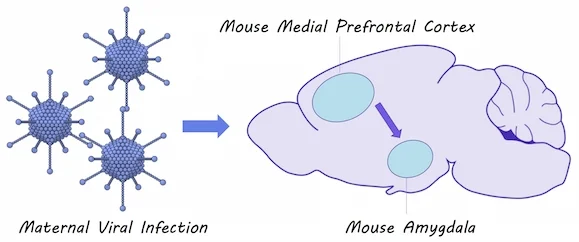
They simulated a viral infection in pregnant mice to induce immune system activation. They also simulated an infection in newborn mice to induce immune system activation shortly after birth. They measured the amount of time that mice spent socially interacting and whether they had more anxiety-like behavior. Using optogenetics and whole cell patch clamp recordings, they tested how early immune system activation would affect the flow of neural signals between the medial prefrontal cortex and the amygdala, a brain circuit known to be involved in social interaction and anxiety. They also used immunohistochemistry to measure the number of microglia (i.e. the brain’s immune cells) present in the amygdala of mice after immune responses.

Immune response to infection

What did they find?

 Mice who were affected by maternal immune activation as well as mice that were affected by postnatal immune activation displayed more anxiety-related behaviors and decreased social interaction compared to unaffected mice. There were also increased numbers of microglia found in the amygdala of these mice. They found that neural connections were stronger between the medial prefrontal cortex and the amygdala in mice affected by maternal immune activation and when maternal and postnatal immune activation were combined. They also found that inhibition of the amygdala by inhibitory neurons, activated by projections form the medial prefrontal cortex, was reduced in mice affected by postnatal immune activation (but not maternal immune activation), suggesting two distinct mechanisms for brain circuit changes in response to immune activation.

What's the impact?

This study confirms that immune responses to infections in early development can result in changes to a brain circuit involved in social interaction and anxiety. Brain changes occurring after immune system activation could contribute to the development of disorders like Autism Spectrum Disorder.

asd_quote_small.jpg

Y Li et al., Maternal and Early Postnatal Immune Activation Produce Dissociable Effects on Neurotransmission in mPFC-Amygdala Circuits. The Journal of Neuroscience (2018). Access the original scientific publication here.

The Brain’s Immune Cells Help Motor Neurons Recover in a Mouse Model of ALS

What's the science?

ALS is a devastating disease where motor neurons degenerate. TDP-43 is a toxic protein that builds up in ALS and contributes to this degeneration. It is unclear whether the immune cells of the nervous system, microglia, react to TDP-43 and whether they play a protective role or make neurodegeneration worse. This week in Nature Neuroscience, Spiller and colleagues test whether microglia contribute to neurodegeneration in a mouse model of ALS.

How did they do it?

They used a transgenic mouse model for ALS in which TDP-43 (toxic protein) build-up can be induced or reversed by suppressing or activating TDP-43 gene expression. With this model they can simulate motor neuron degeneration or recovery. The authors measured whether there were any changes in the number of microglia or their activation state (indicated by the size and shape of the cells) at several time points using immunostaining in the spinal cord, both after inducing TDP-43 damage and after reversing this process. Increases in microglia number and activation indicate that the microglia are actively responding to neuron damage. They also measured gene expression changes in the microglia during the degeneration and recovery of motor neurons. They then measured whether TDP-43 could be found inside the microglial cells, indicating that the microglia are “eating” the proteins to clear them. Finally, they blocked microglia cell replication to test how motor neuron function would recover in regenerating motor neurons.

What did they find?

Microglia were not initially reactive to toxic protein buildup. By the time the motor neurons were severely degenerated, there was a slight increase in the number of microglia and a change in the activation state (i.e. the number of microglia reacting). After reversing the TDP-43 pathology, microglia number and activation increased immediately and dramatically, suggesting that the microglia play a more important role in helping motor neurons recover. This activation of microglia was specific to TDP-43 proteins, and not just a response to neuron death in general. The microglia also showed many changes in gene expression during early recovery (and not early disease), indicating that they are involved in the early recovery process. TDP-43 was found inside microglia, showing that these immune cells actually clear TDP-43 during recovery. When microglial cells were not able to multiply, the motor neurons that were damaged by TDP-43 did not fully recover, and the mouse did not regain full motor function, indicating that microglia are required for a full motor recovery.

Microglia, Servier Medical Art, image by BrainPost, CC BY-SA 3.0

Microglia, Servier Medical Art, image by BrainPost, CC BY-SA 3.0

What's the impact?

This is the first study to show that microglia are involved in the recovery of motor neurons in a reversible mouse model of ALS. Previously, we weren’t sure whether these immune cells of the nervous system actually make things worse or protect neurons from damage in ALS. We now know that microglia play an active role in restoring motor function in neurons damaged by TDP-43 protein buildup in this mouse model of ALS. Currently, we do not have therapies to restore motor function in ALS, and this study suggests that we may be able to target microglia.

krista_test_small.png

Reach out to study author Dr. Krista Spiller on Twitter @krista_spiller

K. Spiller et al., Microglia-mediated recovery from ALS-relevant motor neuron degeneration in a mouse model of TDP-43 proteinopathy. Nature Neuroscience (2018). Access the original scientific publication here.

Your Brain Reacting to Social Injustice

What's the science?

How do we perceive injustice? Many neuroimaging studies have looked at how we perceive the violation of social norms by analyzing brain activity while participants play a computer game. For example, participants might have the option to punish one player who is acting unfairly (e.g. stealing) towards another. Further, different hormones, like oxytocin, influence our social behaviour, suggesting they can play a role in our perception of injustice. This week in The Journal of Neuroscience, Stallen and colleagues performed a new set of experiments using brain imaging to analyze the perception of injustice.

How did they do it?

First, oxytocin was administered to half of the participants. Next, all participants underwent an fMRI brain scan, while playing three computer games: 1) Participants played against an opponent, called a ‘taker’. The taker had the opportunity to steal up to 100 chips away from the participant, and the participant could then punish the taker by giving up up to 100 of their own chips. For each chip they gave up 3 would be taken from the taker, (injustice happening to them) 2) Participants received 200 chips and observed a taker stealing chips from another player, and could then punish the taker (using up to 100 chips, 3 taken from the taker for each chip given up), (observing social injustice, punishing as a third party) and 3) Participants observed a taker stealing chips from another player, and could compensate the disadvantaged player using up to 100 chips (the disadvantaged player was given 3 chips per chip given up) (observing social injustice, compensating as a third party). Participants knew they would receive real monetary compensation after the games according to their performance, and all games were anonymous.

Perception of injustice computer game

What did they find?

Participants who received oxytocin were more likely to dole out small punishments, frequently, to a taker who took chips from the participant or another player, versus participants who did not receive oxytocin. When the authors compared trials in which a participant doled out punishment to the taker versus compensating the disadvantaged player, there was greater activity in the ventral striatum -- a brain region involved in processing rewards. The decision to administer punishment was associated with activity in the anterior insula -- a brain region involved in “gut feelings” and decision making involving risk. Activity in the amygdala, a brain region associated with affective arousal, was correlated with the severity of punishment administered but only in experiment #2, when participants observed a taker behaving unfairly towards someone else.

What's the impact?

This is the first study to assess the perception of social justice in situations where an individual is experiencing injustice firsthand compared to observing injustice as a third party. This study suggests two distinct brain mechanisms might be at play during these unjust situations.

A word of caution: Different brain regions are activated in many different situations. Just because a brain region is known to be activated during reward, for example, does not necessarily mean that brain region will always be active during reward processing.

Stallen_comment_new_small.jpg

M. Stallen et al., Neurobiological Mechanisms of Responding to Injustice. Journal of Neuroscience. (2018). Access the original scientific publication here.