Early Life Adversity Has a Long-Term Impact on Reward Learning

Post by Lani Cupo

The takeaway

Reduced maternal interaction during infant nursing, a marker of early-life adversity, is associated with altered neural responses during a reward learning task in brain regions important for psychiatric health in adulthood.  

What's the science?

Early-life adversity (ELA) events during childhood such as abuse and neglect have been shown to affect reward learning and decision making in adolescence, however, the long-term impact in adulthood of ELA before birth and in infancy (e.g. maternal smoking or stressful life events) is not fully understood. This week in Biological Psychiatry, Sacu and colleagues examined the impact of ELA on the neural processes underlying reward learning in adults with functional magnetic resonance imaging (fMRI).

How did they do it?

The authors used data from an ongoing birth cohort study following 384 participants from birth through adulthood. They included 156 participants who had high-quality fMRI collected during a passive avoidance task. In this task, one of four colored shapes was displayed on the screen. Participants had to decide whether or not to respond to that shape. Responding could result in one of the four following outcomes: winning $1, winning $5, losing $1, or losing $5. Each shape was most likely to result in one of the outcomes. If participants did not respond, they did not receive any feedback. As participants learn which shapes are beneficial, they can choose to respond less to the harmful ones.

The authors then used a series of models to extract information from the participants’ behavior about the expected value (EV) of responding (trials where they responded, expecting to receive a reward) and prediction error (PE; where they received an outcome that deviated from their expectations). A dimensionality reduction technique (principal component analysis) was used to identify factors that represent correlated adversity measures. 

The authors used a t-test to identify brain regions involved in EV and PE signaling in the task. Finally, they used regressions to test the association between ELA factors and activity in 8 brain regions of interest and statistically corrected for multiple comparisons.

What did they find?

The authors identified three factors associated with increased ELA. The first mostly consisted of psychosocial adversities (e.g. family adversity and childhood trauma) and prenatal maternal smoking. The second was mostly informed by perinatal adversity (e.g. complications during birth). The third was mostly maternal sensitivities (e.g. maternal stimulation during nursing).

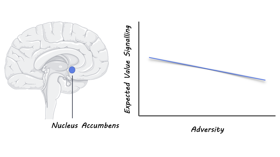
Then, the authors identified that the striatum and medial prefrontal cortex were involved in EV and PE signaling, consistent with previous research. The first and second adversity factors were not significantly associated with neural changes, however, the authors did find lower activity in the nucleus accumbens during EV trials in participants with lower maternal stimulation. Meanwhile, participants with higher maternal stimulation showed increased activity in the striatum and anterior cingulate cortex. Together these results suggest that reduced maternal stimulation alters activity during reward learning. 

What's the impact?

The results of this study suggest even in adulthood, early life adversity associated with psychosocial factors and maternal stimulation in infancy impact neural processes during reward learning. This study suggests interventions targeting the reward system in development may help counteract the effects of ELA. 

 Access the original scientific publication here.

Fluctuations in Heart Rate Influence Brain Activity

Post by Meagan Marks

The takeaway

Heart Rate Variability (HRV), a component of cardiac rhythm, directly affects brain activity important for neural communication.

What's the science?

In neuroscience, it is typically understood that the brain controls the body. But emerging evidence suggests that involuntary functions like heartbeat can influence the brain. Recent research has found that heart rate variability (HRV), the fluctuation in time intervals between adjacent heartbeats, is connected to neural activity. Those with higher HRV – or a heart more adaptable to the environment – are shown to have improved emotional regulation, cognitive function, and well-being. This relationship is especially prominent with high-frequency HRV (HF-HRV, which reflects the variation in heart rate associated with breathing). However, how neural activity and HF-HRV directly affect each other remains unknown. This week in Psychological Science, Sargent and colleagues explored the causal relationship between HF-HRV and neural activity by comparing oscillations from the heart and brain.

How did they do it?

The authors recruited 37 healthy adult participants and asked them to stare at a white cross on a black screen for 5 minutes. During the task, cardiac rhythm was recorded via electrocardiogram (EKG) and brain rhythm via electroencephalogram (EEG). The HF-HRV oscillations were then extracted from EKG recordings and temporally aligned to match EEG data (brain waves), which had been filtered into oscillations occurring at each frequency band (alpha, beta, gamma, delta, theta). The authors then analyzed the oscillations to look for evidence of phase-amplitude coupling, where it was predicted that the phase series (cycles) of HF-HRV oscillations would be coupled with, or associated with, the magnitude of change in brain waves (amplitude). Once phase-amplitude coupling was established, the authors calculated to what extent HF-HRV oscillations successfully predicted brain oscillations and vice versa to establish the direction of the causal relationship (heart-to-brain or brain-to-heart influence). 

What did they find?

Upon analysis, the authors found a strong relationship between HF-HRV and neural activity via phase-amplitude coupling, where the phase series of HF-HRV oscillations modulated the amplitude of the brain waves. It was found that a majority of participants also showed a significant heart-to-brain effect, where HF-HRV oscillations significantly predicted and regulated brain waves. This suggests that cardiac rhythm can influence neural activity. In addition, for all brain wave frequency bands except gamma, the heart-to-brain effect was significantly stronger than the brain-to-heart effect. This was true for EEG signals coming from all areas of the brain, suggesting that the heart was influencing neural communication and activity between multiple regions and multiple brain wave rhythms.

What's the impact?

This study is the first to show that fluctuations in heart rate can modulate neural activity. The findings suggest that improvements in cardiac rhythm may enhance connectivity and communication between neurons in the brain, in turn boosting cognitive functions like emotional regulation, executive functioning, and stress management. Cardiac variables such as HRV could also be a potential therapeutic target for mental health disorders, where methods like HRV biofeedback could help improve well-being. 

How Do Scientists Study Lucid Dreaming?

Post by Laura Maile

What is lucid dreaming?

Lucid dreaming is a state of sleep where the individual becomes aware that they are dreaming. This phenomenon represents a paradoxical situation where the dreamer maintains a level of cognitive awareness while remaining asleep. The concept of lucid dreaming or “sleep awareness” has been written about across the globe for centuries, though it was not studied scientifically until the late 20th century and wasn’t accepted as a legitimate field of study until the 2000s. This phenomenon occurs in about half of individuals, but infrequently. Though lucid dreaming has become a popular field of study among sleep researchers, the small sample sizes in previous research have made the physiology and brain regions underlying this phenomenon difficult to identify.   

How do scientists study it?

In the 1970s, scientists began to study the neuroscience of lucid dreaming using electrooculography (EOG) to detect eye movements during sleep. Eye movements occur during normal dream states, but during lucid dreams, the individual can intentionally move their eyes. This allows the dreamer to communicate with the outside world while still in the dream state, giving experimenters an opportunity to observe and record information about dreaming in real-time. EOG, in combination with fMRI and electroencephalogram (EEG) to record brain activity, allows experimenters to confirm the state of lucid dreaming and collect neuroimaging data on the brain regions underlying this phenomenon. The major challenge with studying lucid dreaming is the low frequency with which it occurs. Some neuroscientists are developing methods to encourage lucid dreams to happen more often. These include exercises that train individuals to reflect on their state of mind before sleeping and during short “inverse naps” during the rapid eye movement (REM) stage of sleep. Others have used virtual reality training or direct electrical or ultrasound stimulation of specific brain regions during sleep.  

What does the future look like?

Scientists in the field of lucid dream research indicate that standard operating procedures are needed to ensure similar methods of cognitive training, experimental procedures, EOG, and EEG to enhance reproducibility and grow the field. Engaging in cross-lab collaborations and open sharing of data will allow scientists to produce more robust, powered studies. Wearable technology is now available to allow study participants to record EEG and EOG during normal sleep at home with simple, user-friendly devices, which will also greatly increase the amount of lucid dream data acquired. Citizen neuroscience also presents unique opportunities for larger-scale data collection and sharing of experiences and resources between community groups of lucid dream enthusiasts and academic researchers. 

Recent advances have allowed for two-way communication between the lucid dreamer and experimenter. Light and sound cues can be used to signal to the dreamer, while the dreamer can communicate with the experimenter using eye movements detected with EOG. Electromyogram technology can also detect subtle facial muscle movements, with the potential for uncovering dream speech. Finally, computational analysis and generative artificial intelligence give scientists new abilities to reconstruct and interpret dream states. Given the surge of interest and the recent technological advances, the future of research of lucid dreaming holds huge potential for understanding the neuroscience of dreaming in general.  

Access the original scientific publication here.