
The Development of Consciousness in Infants

Post by Laura Maile

What is consciousness?

Scientists have long attempted to understand where consciousness resides, whether it involves a network of brain areas, and when in development it emerges. Current theories of human consciousness state that consciousness develops as the brain becomes capable of integrating information, making us aware of ourselves and our environment. There are many different theories, however, on how and where consciousness is represented. Higher-order theories, for example, require that one be able to represent an external experience in the mind, and place importance on the prefrontal cortex. In contrast, integrated information theory places more emphasis on posterior cortical areas, and rests on the ability of the brain to integrate different stimuli to generate information by the whole. In general, it is agreed that consciousness is represented in the brain, likely as an integration of signals across multiple brain areas.  

How do you measure consciousness?

It is of critical importance to develop and agree upon measures of consciousness, specifically for infants, as they are unable to follow directions or communicate verbally. Infants do possess the ability to respond to stimuli such as the sound of their mother’s voice, different facial expressions, and noxious stimuli. Their responses can be measured both behaviorally, through limb withdrawal, facial grimacing, eye movement, vocal and sucking activity, and physiologically, with changes in heart rate and neural activity in brain areas that respond to environmental stimuli. Most current theories of consciousness are based on physical processes that can be measured via electroencephalogram (EEG) recordings and fMRI, and behavioral indicators of consciousness, such as the capacity to respond to environmental stimuli. fMRI studies, which record high-resolution hemodynamic activity in the brain that respresents neural activity, have identified cortical “hubs” and networks of brain regions that are active during different activities and states. By studying brain activity across different states of development, scientists have determined how functional networks change over time. Conceptually similar to EEG, which uses a net of electrodes fitted to the skull to record brain activity, magnetoencephalography (MEG) is a less invasive alternative that can be used to measure fetal brain activity. 

When does consciousness emerge?

There are conflicting theories about when exactly consciousness appears during development. Some theories, for example, require that individuals have a sense of self and an understanding of their own mental state in order to be qualified as “conscious,” which would mean that humans are not conscious until some time after their first birthday. Other recent evidence indicates that consciousness may appear in early infancy or even before birth, as soon as thalamocortical activity appears in the brain at about 24-26 weeks gestation. Rather than base our understanding of the emergence of consciousness on a specific theory or set of conflicting theories, some scientists suggest that the field measure markers of consciousness in adults and observe when they first emerge in infants. 

Activity in the brain shows that the primary cortical areas that process vision, auditory, and sensorimotor information are active in response to external stimuli at birth, indicating that newborn babies can process many sensory inputs. Some of these areas are also present and active prior to birth in the developing fetus. Those brain areas involved in more complex processes such as attention, executive function, and memory appear less complex at birth and develop over the first two years of life. Some specific functional activity networks have been linked to the capacity for or recovery of consciousness after injury in adults. Three of these networks, the default mode network, dorsal activity network, and executive control network, have recently been identified as distinct and functional networks in newborn babies.  

Behavioral data shows that shortly after birth, infants can process auditory and visual inputs that allow them to recognize their mother’s voice, show sensitivity to music, and even show preference for their native language. Visual acuity is low at birth, but brain imaging data shows responses in the visual pathway to distinct visual inputs at two months of age. While most perceived senses expand and develop as the infant ages, there is also data indicating that young infants aged 4-6 months can perceive more distinct sounds and faces than older infants and adults. Additionally, multisensory integration that requires conscious perception of individual stimuli has been demonstrated in 4-5 month old infants. 

What's next?

The study of infant consciousness has become an increasingly important and studied topic in the field of consciousness research. Continued improvement in methods to measure brain activity and other markers of consciousness in fetuses and infants is needed. A more complete understanding of the neural correlates and functional basis of consciousness will also require a tightening of the many theories of consciousness into one universally accepted theory. 

The takeaway

A definitive answer on when human consciousness begins has yet to be identified, but the increase in studies on conscious experience in infants and preterm fetuses is bringing us closer to one. Recent evidence points to an earlier onset of consciousness than was previously described in human infants, indicating some level of consciousness is present at birth, and potentially even in the late stages of gestation when brain activity and behavioral responses to external stimuli can be measured. 

References +

Bayne, T et al., Consciousness in the cradle: on the emergence of infant experience. Trends in Cognitive Sciences. 2023.

Padilla, N et al., Making of the mind. Acta Paediatrica. 2020.

Seth, AK et al., Theories of consciousness. Nature Reviews Neuroscience. 2022.
Alzheimer’s Disease Creates a Positive Feedback Loop of Excitatory Signaling in Brain Networks

Post by Rebecca Hill

The takeaway

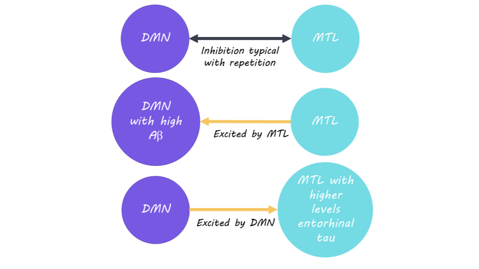
Beta-amyloid and tau are proteins that aggregate in two distinct networks in the brain in Alzheimer’s disease. When there are higher beta-amyloid and tau protein levels, the neuronal networks switch from inhibitory to excitatory signaling, leading to a positive feedback loop of excitation and further protein build-up.

What's the science?

In individuals affected by Alzheimer’s disease, the disruption of normal brain functioning is linked to a build-up of b-amyloid and tau proteins. Early in the progression of Alzheimer’s disease, beta-amyloid proteins build up in the association cortex (the default mode network, also known as the DMN), which is usually active when a person is daydreaming or at rest. Tau proteins on the other hand tend to appear in the entorhinal cortex (in the medial temporal lobe, also known as the MTL), which mainly facilitates memory processing. It’s still unclear why this protein aggregation occurs in two different networks in the brain. This week in Neuron, Giorgio and colleagues investigated this difference in protein aggregation by examining how these two different networks in the brain interact using brain imaging.

How did they do it?

The authors recruited 66 individuals (45 older adults without symptoms of Alzheimer’s and 21 young adults) for this study. A subset of these individuals had evidence of beta-amyloid and tau protein build-up reminiscent of early-stage Alzheimer’s disease. The authors asked participants whether an image presented to them was new or repeated while undergoing brain imaging to measure activity in the association and entorhinal cortices. Typically, neuronal activity is suppressed when viewing an image that has already been seen before. However, previous research has found that Alzheimer’s patients cannot suppress neuronal activity well during these tasks. The authors used functional MRI (fMRI) to measure brain activity and PET scans to measure the amount of beta-amyloid and tau proteins present in the two brain regions.

What did they find?

For participants with low levels of b-amyloid and tau proteins, there was inhibitory signaling between the DMN and the MTL for repeated images, indicating normal brain functioning with an ability to suppress neuronal activity. However, for participants whose protein levels were higher in the two brain regions, there was excitatory signaling instead. This indicates that Alzheimer’s disease alters normal brain functioning by causing neurons to fire excitatory signals from each brain region to the other. As more beta-amyloid builds up in these networks, there is more excitatory signaling to other networks, further increasing the amount of tau building up in the other networks. This creates a positive feedback loop of excitation between these networks, leading to further build-up of beta-amyloid and tau proteins.

What's the impact?

This study is the first to show how Alzheimer’s disease develops in the association and entorhinal cortices by creating a positive feedback loop of excitatory neuronal signaling. This causes abnormal brain functioning in individuals with Alzheimer’s disease by preventing their ability to suppress brain activity in response to new stimuli. Understanding how Alzheimer’s disease develops may help with our ability to diagnose and treat individuals affected by it. 

Access the original scientific publication here.

5 Important Advances in Neuroscience this Year

Post by Meredith McCarty

The takeaway

As the field of neuroscience continues to advance, we highlight 5 notable research advancements from 2023. While this is by no means a comprehensive list, we sought to outline some important advances and exciting future research directions. With these technological and theoretical advances, we step closer to understanding brain function and ways to implement these findings for clinical application.

Mapping the whole brain of the mouse and fly

Mapping the neural diversity and connectivity patterns of the brain has been a research goal of neuroscience for many decades. The idea behind this pursuit is that, through mapping the cells and connections of the brain, we can gain insight into network dynamics, neurodevelopment, and functionality. Within the past year, this has culminated in several major feats: a whole brain atlas of the mouse brain, as well as a whole brain connectome in the fly.

The BICCN (BRAIN Initiative Cell Census Network) has published numerous articles this year outlining their work in developing a whole brain atlas of the mouse brain. They used a variety of methods to measure the connectivity and genetic diversity of both neuronal and non-neuronal cells; a massive effort with broad implications for future work in the field.

The goal of connectome research is to reconstruct each neuron’s connectivity pattern using electron microscopy. As such, this method is comprehensive yet quite methodologically complex and time consuming. The first neuronal wiring diagram of the fly brain has been released this year, outlining the connectivity and diversity of the 130,000+ neurons in the adult fly brain. These efforts to quantify the connectivity patterns and cellular diversity of the brain at many scales have enormous utility for future neuroscientific research, as these resources will help us map out and understand how the brain works.

Advances in decoding brain activity in humans

The goal of neuroprosthesis research is to assist in decoding intended movement, speech, and other faculties in the impaired brain. To implement this process, scientists use neural interfaces to record brain activity and relate these neural signals to behavior, in a process known as decoding. Within the past year, there have been numerous advances in the development and implementation of brain computer interfaces (BCIs) in humans.

In terms of advances in brain recording interfaces, several research groups have successfully used microelectrode devices to record activity from single neurons in the human brain. This scale of recording offers insight into neural dynamics at a level previously inaccessible in human research. Through careful implementation as a part of clinical treatment, these electrode recordings continue to offer invaluable insight into neural dynamics.

A prominent application of BCIs is to decode intended speech from neural data. Within the past year, the algorithms used to interface between neural data and speech output have advanced greatly with the implementation of predictive language models. Experimentally, by recording hundreds of trials’ worth of data from an individual, and labeling the speech sound or word presented in each one, researchers can decode words and syllables from repeated patterns of neural activity. With the use of predictive language models, this process has been advanced, with a higher success rate of predicting intended speech from neural activity within individuals. The continued advances in both BCI hardware and decoding software have exciting implications for clinical and research applications.

Progress in understanding human consciousness 

What exactly makes us human? Although numerous theories of consciousness have gained prominence over the years, there has not been much consensus in the field. In 2018, Melloni and colleagues at the Allen Brain Institute sought to remedy this through an open science adversarial collaboration, also known as COGITATE. The goal of this adversarial collaboration was to design careful experiments to test the predictions of several prominent theories of consciousness. The theories in question are the Global Neuronal Workspace and the Integrated Information theories. The first results from this collaboration have been released as a preprint. Interestingly, there has been much discourse surrounding the predictions, experimental design and analyses, and tentative conclusions ascertained in this collaboration.

The pursuit to experimentally quantify the neural correlates of consciousness (meaning the minimal neuronal mechanisms jointly sufficient for any specific experience) in the brain remains complex and controversial. However, the implementation of adversarial collaborations, both in consciousness research and other research areas, seems a profound and interesting way to collaborate across theories and disciplines. 

New ways of treating mental health disorders like depression and PTSD

In the past year, there has been much research into psychedelic therapy for the treatment of various psychiatric disorders. This area of research primarily implements non-invasive neuroimaging, such as functional magnetic resonance imaging, to study neural changes involved in psychedelic treatment. For example, recent work from Dai and colleagues revealed that nitrous oxide, ketamine, and LSD all led to changes in brain network connectivity, specifically increased between-network and decreased within-network connectivity. Additionally, recent research revealed that MDMA in combination with psychotherapy has promising therapeutic benefits for people with posttraumatic stress disorder (PTSD) and alcohol use disorder. To more clearly understand the mechanism of action of psychedelic therapy in the brain, Moliner and colleagues quantified changes in receptor binding in mice given psychedelics. They found evidence that psychedelics directly bind to a specific brain-derived neurotrophic factor receptor, and promote antidepressant-like effects.

These research directions have interesting implications for understanding the mechanism of action of psychedelic treatment in the brain. Of note, there is some dissent regarding whether psychedelics should be directly compared with psychotherapy and other treatment methodologies in a clinical setting. As such, both classical and psychedelic research offers continued insight and progress in the treatment of various psychiatric disorders.

Predicting language patterns using AI 

This year has seen a lot of excitement around AI systems helping us to analyze and predict language. AI is also being used to help advance our understanding of the human brain. Large language models (LLMs) are trained on enormous datasets, comprising trillions of words and more data than a human could process in their lifetime. While these LLMs have been successfully implemented in many applications, from data analysis to text generation, whether LLMs can tell us anything about how the brain performs language is another matter entirely. An interesting research project called the BabyLM Challenge, sought to explore these unanswered questions more directly. In this challenge, researchers train language models on linguistic data similar to what a human is exposed to while developing language. The results of research efforts like the BabyLM challenge have implications for neuroscientific, linguistic, and machine learning fields. For example, LLMs could be trained on large clinical data sets to identify patterns or associations that would be difficult for humans to detect manually.

The results from the BabyLM Challenge were just published, outlining the 31 submissions from research groups around the world. The submission that performed the best came quite close to human performance (88%) on tested language skills, with a training set of 100 million words. There is some debate as to the metrics used to quantify model performance, but these results have interesting implications. While LLMs are trained to capture the statistics of natural language and predict text generation, whether LLMs are learning any underlying structure or rules of language (and how this compares with human language development) remains an underexplored topic of research.

References +

(1) Krzysztofiak J, 2023. BICCN: The first complete cell census and atlas of a mammalian brain. Nature.

(2) Dorkenwald S. et al., 2023. Neuronal wiring diagram of an adult brain. bioRxiv.

(3) Schlegel et al., 2023. Whole-brain annotation and multi-connectome cell typing quantifies circuit stereotypy in Drosophila. bioRxiv.

(4) The year of brain-computer interfaces. Nat Electron 6, 643 (2023). https://doi.org/10.1038/s41928-023-01041-8

(5) Willett F.R. et al., 2023. A high-performance speech neuroprosthesis. Nature.

(6) Metzger S.L. et al., 2023. A high-performance neuroprosthesis for speech decoding and avatar control. Nature.

(7) Warstadt A., et al., 2023. Proceedings of the BabyLM Challenge at the 27th Conference on Computational Natural Language Learning. Association for Computational Linguistics.

(8) Charpentier L.G.G. & Samuel D., 2023. Not all layers are equally as important: Every Layer Counts BERT. Proceedings of the 27th Conference on Computational Natural Language Learning.

(9) Martinez H.J.V. et al., 2023. Evaluating Neural Language Models as Cognitive Models of Language. Association for Computational Linguistics.

(10) Melloni L., et al., 2023. An adversarial collaboration protocol for testing contrasting predictions of global neuronal workspace and integrated information theory. PLOS One.

(11) Mashour G.A. et al., 2020. Conscious Processing and the Global Neuronal Workspace Hypothesis. Neuron.

(12) Tononi G. et al., 2016. Integrated information theory: from consciousness to its physical substrate. Nature Reviews Neuroscience.

(13) Melloni L., et al., 2023. An adversarial collaboration to critically evaluate theories of consciousness. bioRxiv.

(14) IIT-Concerned et al., 2023. The Integrated Information Theory of Consciousness as Pseudoscience. PsyArXiv Preprints.

(15) Lau H., 2023. What is a Pseudoscience of Consciousness? Lessons from Recent Adversarial Collaborations. PsyArXiV Preprints.

(16) Wall M.B. et al., 2023. Neuroimaging in psychedelic drug development: past, present, and future. Molecular Psychiatry.

(17) Dai R. et al., 2023. Classical and non-classical psychedelic drugs induce common network changes in human cortex. NeuroImage.

(18) Gully B.J. et al., 2023. Treating posttraumatic stress disorder and alcohol use disorder comorbidity: Current pharmacological therapies and the future of MDMA-integrated psychotherapy. Journal of Psychopharmacology.

(19) Hasler G., 2023. Psychotherapy and psychedelic drugs. Lancet Psychiatry.