The Impact of Deep Brain Stimulation on Brain Networks and Treatment Outcomes in Treatment-Resistant Depression

Post by Kulpreet Cheema

The takeaway

Deep brain stimulation of the subcallosal cingulate gyrus in patients with depression induces changes in brain network properties, which is correlated with improvements in depression symptoms.

What's the science?

Treatment-resistant depression (TRD) is a severe form of depression that does not respond to standard treatments. Deep brain stimulation (DBS) is an emerging therapy that involves implanting electrodes in specific brain regions to modulate neural activity and potentially alleviate depressive symptoms. The effectiveness of DBS varies among patients, and personal factors might be the key to understanding this variability. This week in Molecular Psychiatry, Ghaderi and colleagues aimed to understand how DBS affects brain networks in TRD patients using electroencephalography (EEG), a non-invasive technique that measures the brain’s electrical activity.

How did they do it?

The study involved twelve TRD patients who underwent subcallosal cingulate gyrus (SCG) DBS. The researchers collected resting-state EEG data and Hamilton Depression rating scale data from participants at three sessions: before surgery, one to three months after surgery, and six months after surgery. During the second and third sessions, EEG data was collected with DBS turned on and off sequentially.

To analyze brain network properties, the researchers used an analytical method known as graph theoretical analysis to decipher whether there were distinct differences in brain network properties between patients who responded positively to the DBS treatment (responders) and those who did not (non-responders). Using EEG to study oscillatory activity in brain networks, the authors analyzed four different frequency bands — the delta and alpha bands were of particular interest due to their relevance to depression.

What did they find?

The study revealed several important findings. First, the baseline brain network properties in patients — especially in the delta and alpha frequency bands — were associated with the outcomes of DBS treatment. Responders to DBS showed lower levels of network segregation, integration, and synchronization at baseline compared to non-responders. Second, DBS led to changes in brain network properties over time, characterized by increased integration and synchronization of brain regions. These changes were particularly evident in the delta frequency band.

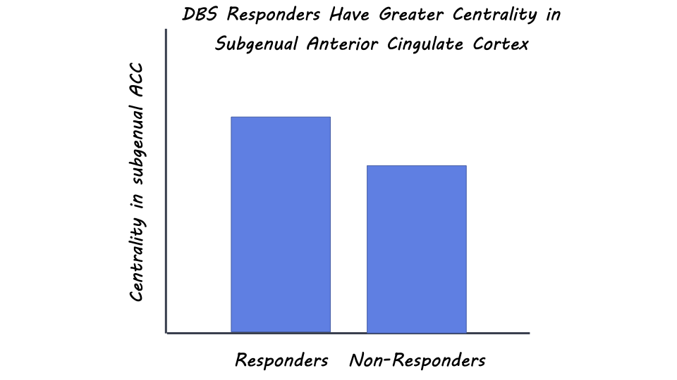
Third, the researchers found that responders had higher centrality - a measure of the importance of a node in a network for information propagation - in the subgenual anterior cingulate cortex (ACC), a brain region associated with depression symptoms. DBS led to a reduction in centrality in this region, which correlated with treatment response. Other brain regions, like the primary somatosensory cortex and parahippocampal gyrus, also showed alterations in centrality, indicating their involvement in TRD and DBS response.

What's the impact?

This study sheds light on the complex effects of DBS on brain networks in treatment-resistant depression. By using EEG and advanced analytical techniques, researchers identified specific brain network features associated with treatment response and demonstrated how DBS induces changes in these networks over time. The findings provide valuable insights into the potential mechanisms underlying the therapeutic effects of DBS in TRD patients. This knowledge could contribute to the development of personalized treatments for individuals with treatment-resistant depression, ultimately offering new hope for improved mental health outcomes. 

Access the original scientific publication here

Myelin Insulation and Risk of Neuron Degeneration in Autoimmune Environments

Post by Meredith McCarty

The takeaway

In the progression of autoimmune diseases such as multiple sclerosis, the degeneration of axons in the central nervous system leads to irreversible damage. Myelin sheaths encapsulating axons increase the risk of axonal degeneration in an autoimmune environment.

What's the science?

Multiple sclerosis (MS) is an inflammatory autoimmune disorder that affects the central nervous system and is characterized by axonal degeneration. Myelination, or the process by which oligodendrocytes encapsulate a neuron’s axons with an insulating sheath of myelin, is largely considered to serve an insulating and predominantly protective role for axons. Based on this assumption, demyelination, or the destruction of the myelin sheath, has been proposed as a likely cause of the axonal degeneration seen in autoimmune disorders. However, recent evidence that axonal damage actually occurs before the onset of demyelination suggests a more complex role for myelination in autoimmune disorders. This week in Nature Neuroscience, Schäffner, and colleagues utilize MS human biopsies and mouse models of demyelinating disease to investigate the role of demyelination and oligodendrocyte function in axonal degeneration.

How did they do it?

The authors first measured the relationship between axonal damage and demyelination in human MS. They performed electron microscopy on 4 MS biopsies in order to quantify the degree of demyelination of axons surrounding the lesions. They compared how the degree of demyelination correlated with the abundance of lesions and accumulation of organelles and condensed axoplasm, as measures of irreversible axonal damage.

In order to study the relationship between myelination, oligodendrocyte function, and acute inflammatory environments, the authors utilized several experimental mouse models and induced experimental autoimmune encephalomyelitis (EAE) to study axon survival.  First, to understand the temporal dynamics of axon degeneration, they induced EAE in mice via immunization. They quantified the proportion of myelinated and demyelinated damaged axons and characterized changes in myelin structure using electron microscopy and immunohistochemistry. To understand changes in gene regulation in EAE, the authors analyzed RNA sequencing datasets.

Next, in order to test whether demyelination could actually improve axon outcome, the authors used a cuprizone mouse model where axon demyelination occurs at an increased rate over time and measured changes in axon organelle accumulation. Lastly, in order to directly test whether the degree of myelination correlates with axonal damage, the authors used a mouse model with decreased expression of myelin basic protein (MBP). These hMbp mice exhibit an increased proportion of unmyelinated axons. They compared the disease progression and degree of axonal damage in hMbp mice relative to controls in an induced EAE environment.

What did they find?

The authors found that in both human and mouse models, irreversible axonal damage was restricted to myelinated axons. These damaged axons displayed increased organelle accumulation and highly condensed axoplasm, which are early biomarkers of axonal degeneration. The physical characteristics of myelin in damaged axons were found to be reflective of the dysfunctional movement of cellular materials to the axon by oligodendrocytes. They found evidence for the downregulation of RNA sequences essential for many cellular processes in EAE. In the cuprizone mouse model of axon demyelination, the authors found a decline in organelle accumulation in demyelinating axons, which suggests that demyelination can improve axonal survival.

When directly comparing axonal damage in hMbp mice in an EAE environment, the authors found significantly less axonal damage, fewer axons with organelle accumulation and condensed axoplasm, and overall better disease outcome relative to the control group. After ruling out differences in immunological response across these experimental groups, they found that axonal damage was limited to myelinated axons, even in a mouse model with an artificially increased proportion of demyelinated axons. Based on these data, the authors hypothesize that the progression to irreversible axonal degeneration is likely due to the accumulation of organelles and condensed cytoplasm, a process that would be prevented with efficient demyelination of the axon. They propose that oligodendrocyte dysfunction and the failure of efficient demyelination leads to this irreversible axonal damage.

What's the impact?

This study untangled the contradictory role of axonal myelination in the axonal damage characteristic of autoimmune diseases such as multiple sclerosis. Through untangling the molecular mechanisms involved in this axonal degeneration, this study puts forth novel theories as to the crucial role of oligodendrocytes and efficient demyelination of axons in an acute inflammatory environment. Future work building off of these findings has enormous implications for identifying new targets for therapeutic intervention to prevent axonal damage early in disease progression.

A New Bird Model for Studying Vocal Production

Post by Rebecca Hill

The takeaway

There has been little focus on female birdsong until recently, leaving many questions unanswered about how the female brain is involved in singing. Male and female red-cheeked cordon bleus that sing at the same rate have similar hormone and brain composition.

What's the science?

Bird song learning can be used as a model for human vocal learning, but up until recently the field has focused on studying species where only male birds sing. Studying bird species where females sing will give us a more complete understanding of how hormones and the brain control vocal production. This week in Journal of Comparative Physiology, Rose and colleagues studied the red-cheeked cordon bleu, a bird species in which both males and females sing, by recording their song behavior, measuring their hormone levels, and analyzing the brain regions involved with vocal production.

How did they do it?

The authors first studied the song behavior of 20 birds (10 males and 10 females) by putting them in sound recording chambers, which cut down on background noise. They recorded each bird for one hour, eight times over a month, and found the recording with the most songs sung. They recorded the birds’ song rate by dividing 20 by the number of minutes birds took to sing 20 songs. They also counted the number of songs the birds sang in the first 30 minutes of singing on the day of tissue collection.

The authors also studied the hormones and brain regions involved in singing by collecting blood and brain tissue. They studied the sex hormones testosterone and progesterone, which are known to be involved in singing rate, by measuring the concentration of them in the blood. They studied three brain regions: 1) Area X of the striatum, which is involved in song learning, 2) RA (the robust nucleus of the arcopallium), which is involved with song production, and 3) HVC (the acronym is the proper name), which connects to both learning and song production pathway and is the central vocal production area in the brain. They measured the volume of these brain regions and the ZENK protein expression, which shows the level of activity in the brain in certain areas.

What did they find?

Male and female birds both sang at high song rates, and their songs were similar in total length and structure. Both testosterone and progesterone hormone blood levels were similar between males and females. This suggests both sexes have similar hormonal mechanisms that drive song behavior. Area X and HVC were larger in males than females, but RA volume was similar between the two sexes. This suggests that song production pathways are similar, but song learning pathways are different between the sexes. Birds that sang had more ZENK expression than birds that were not singing in both sexes, which suggests that there were similar levels of brain activity involved with producing singing.

What's the impact?

This study found that red-cheeked cordon bleus have very few behavioral, hormonal, and brain sex differences compared to other species often studied in the lab. This means they could be a good model species to study mechanisms controlling vocal behavior as a model for human vocal production. Understanding how the brain is involved with speech can help us to diagnose and treat speech and communication disorders.

Access the original scientific publication here