Transcriptomic Changes in Cellular Communities In the Brain Contributes to Alzheimer’s Disease

Post by Soumilee Chaudhuri

The takeaway

Major brain cell types — neurons, oligodendrocytes, endothelial cells, etc. — individually and synergistically contribute towards molecular changes seen in the aging human brain in Alzheimer’s Disease (AD). A high-resolution transcriptomic map in the aging human brain unraveled 1) diverse cell populations associated with AD and 2) how networks of cellular communities coordinate to alter biological pathways, ultimately leading to AD.

What's the science?

Alzheimer’s Disease is an irreversible, neurodegenerative illness, with a complex pathophysiology. Amongst many unknowns in the molecular mechanisms of AD, is the specific contribution of major cell types in the aging brain and how this might trigger AD-related dementia. Even so, knowledge of the contribution of different brain cells in AD pathogenesis has advanced over the last decade due to the advent of high-resolution technologies such as single-cell RNA sequencing. We know that perturbation in transcriptomes (i.e., full range of mRNA molecules produced) of major brain cell types and subtypes like excitatory and inhibitory neurons, oligodendrocytes or microglia for example, synergize to cause events that lead to molecular changes seen in AD. However, we do not know the specific contributions of each cell subtype to this disease due to limited sample size and a lack of robust and sensitive technologies powerful enough to capture interindividual as well as cell-specific diversity in AD brain. This week in Nature Neuroscience, Dr. Cain and colleagues unravel the distinct cellular architecture of the aging brain prefrontal cortex in AD as well as how cells interact, using combined bulk and single cell RNA sequencing analyses and novel bioinformatics pipelines.

How did they do it?

The authors used a powerful approach to get insights about coordinated multicellular communities in the AD brain. They used a) integrative transcriptomics (bulk and single-cell RNA sequencing) as well as b) a robust analytical approach (CellMod: a deconvolution tool that allows estimation and projection of single cellular landscapes from a limited set of individuals to a higher number of individuals). The authors used single-cell data from 24 individuals from the ROSMAP cohort to generate a cellular map of the aging Dorsolateral Prefrontal Cortex (DLPFC) brain region, and used that as input to their novel CellMod deconvolution algorithm to estimate cellular compositions in an independent set of 638 individuals with bulk RNAseq data. Then, network analysis within this model revealed cellular sets and subsets and interacting cellular communities across individuals with AD vs. controls; and advanced statistical modeling associated known AD traits and risk factors to these identified cell subsets and communities across the case (AD) vs control (no AD) groups.

What did they find?

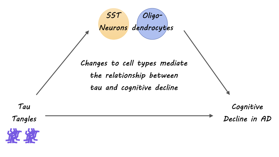
The authors identified specific subpopulations of cells associated with AD pathophysiology. Primarily, the authors discovered oligodendrocyte transcriptional pathways and a downregulation in somatostatin-producing neurons (SST neurons) as perturbed in AD pathogenesis. From the innovative cellular map of the neocortex that the authors constructed, they found interesting insights about oligodendrocyte diversity and interactions, implicating oligodendrocytes  —the myelinating cells of the brain — as a strong cellular contributor to AD. Further, they identified that oligodendrocyte expression from both positive (Oli.4) and negative oligodendrocyte (Oli.1) cells was strongly associated with tau pathology and cognitive decline in AD. Multiple shared pathways within cellular communities were identified and related to known risk factors of AD, as a way of validating that AD indeed, is a pathophysiologic process with multiple interacting cell types. Overall, the findings of this study pinpoint the importance of approaching our understanding of AD through a lens of interacting multicellular communities and networks. 

What's the impact?

This study is the first to show that cell specific as well as coordinated cellular and sub-cellular interactions in the aging brain may contribute to a diseased microenvironment in AD. Additionally, the authors find evidence of the contribution of communities of oligodendrocytes to cognitive decline and tau burden in Alzheimer’s Disease. The result of this study extends research on cellular and subcellular heterogeneity in the diseased aging brain and helps to inform therapeutic targets for AD and dementia.

Access the original scientific publication here

Understanding How Decisions Are Terminated

Post by Kulpreet Cheema

The takeaway

Little is known about how decisions are terminated and translated into actions or plans. This study provides evidence that the superior colliculus (SC), a midbrain structure involved in eye movements and orienting behaviors, plays a crucial role in terminating decisions.  

What's the science?

Previous research has shed light on how the brain accumulates evidence before reaching a decision. This process can be modeled as a stochastic drift-diffusion process or bounded random walk. Neurons in the lateral intraparietal area (LIP) have been shown to accumulate noisy evidence during decisions. However, how this process is terminated and translated into a specific action is still unknown. The SC is known for its role in generating eye movements, and is directly coupled to the LIP; the LIP projects to the SC and the SC projects back to LIP via the thalamus. In a study published in Neuron, researchers investigated the role of SC in applying a decision threshold to the accumulation of evidence represented in the LIP.

How did they do it?

The researchers recorded simultaneous neural activity in the LIP and SC while monkeys performed a motion-discrimination task. In the motion discrimination task, the monkeys were trained to make eye movements based on the direction of a moving stimulus. Researchers used high-density multi-channel electrodes to capture the firing rates of functionally similar neurons in both areas during the decision process. To directly test the involvement of the SC in decision termination, researchers performed focal inactivation by temporarily inactivating the SC using small muscimol injections. They analyzed behavioral measures and neural recordings from the LIP during SC inactivation.

What did they find?

Researchers found evidence that the LIP and SC exhibited different dynamics during decision-making, and that the SC implements the decision threshold. The researchers observed that bursts of activity in the SC terminated the decision process, as opposed to the accumulation signals in LIP. The bursts in the SC were triggered by upticks in excitatory input and were associated with the termination of the decision. When the SC was inactivated, the termination mechanism was impaired, leading to slower, biased decisions and prolonged evidence accumulation in LIP. These findings suggest that, while evidence may accumulate in the LIP during decision-making, decision termination occurs in an area responsible for action selection; in this case, the SC, as the decision is about moving the eyes.

What's the impact?

This study sheds light on how the brain makes decisions and transforms evidence into actions. Understanding how decisions are terminated is crucial for comprehending the entire decision-making process. By identifying the role of the SC in decision termination, the study highlights the importance of a region known for action selection in decision termination. The findings have implications for understanding decision-making processes in humans and other primates. 

Connectivity Between the Amygdala and Frontal Cortex Predicts Youth Depression Treatment Response

Post by Baldomero B. Ramirez Cantu

The takeaway

Connections between the frontal cortex and the amygdala in the brain have shown potential in identifying depression in young individuals, and their responsiveness to standard behavioral and pharmacological treatments for depression.

What's the science?

Youth depression is commonly characterized by difficulties in emotional regulation and a decline in interest in activities. Although there are numerous pharmacological and behavioral interventions available, only approximately 70% of youths exhibit positive responses to treatment, and a substantial portion (40-60%) do not achieve remission even after treatment. The neurobiology and brain systems underlying youth depression are still not comprehensively understood. In a recent study published in Biological Psychiatry, Kung et al. explore whether the dynamics of the frontoamygdalar pathway during cognitive reappraisal (i.e. recognizing and reinterpreting negative thought patterns) can help predict the effectiveness of first-line depression treatments.

How did they do it?

The authors used functional magnetic resonance imaging (fMRI) and dynamic causal modeling to map frontoamygdalar effective connectivity during a cognitive reappraisal task and assess its association with depression diagnosis and treatment response.

They recruited a cohort of 107 young individuals diagnosed with mild to severe depression and 94 healthy individuals in a control group. The participants underwent fMRI scans while they performed cognitive reappraisal tasks to examine the neural connections between the frontal cortex and the amygdala. In these tasks, images that could elicit negative emotions were shown, and participants were asked to use reappraisal strategies to reinterpret the images. The fMRI data were then analyzed to measure the strength and dynamics of the frontoamygdalar pathway during these tasks. The researchers also collected information on the participants' engagement with a clinical trial, including the type of interventions received and their treatment response. Statistical analyses were performed to investigate the relationship between frontoamygdalar effective connectivity, treatment response, and depression diagnosis in youth.

What did they find?

The authors’ results indicate that frontoamygdalar effective connectivity can serve as a predictive factor for youth depression and treatment response. Those in the control group more successfully used reappraisal strategies versus those with depression. Participants with stronger inhibitory connections between the frontal cortex and the amygdala demonstrated a lower likelihood of having a depression diagnosis. Weaker excitatory frontoamygdalar connectivity was associated with positive responses to standard depression treatments. These findings highlight the importance of understanding and targeting the neural circuits involved in regulating negative emotion for optimizing treatment outcomes in youth with depression.

What's the impact?

This study enhances our understanding of youth depression by investigating frontoamygdalar effective connectivity as a potential biomarker for depression and treatment response, offering the possibility of personalized and more effective interventions for young individuals. Ultimately, it holds promise for improving outcomes and quality of life for youth with depression.

Access the original scientific publication here.