Anxiety is Induced by Activating Microglia, the Immune Cells of the Brain

Post by Rebecca Hill

The takeaway

Hoxb8 microglia, the support cells of the brain created by the Hoxb8 gene, play a role in regulating anxiety. When these microglia are activated with light using optogenetics in certain areas of the brain, mice display anxious grooming and freezing behaviors.

What's the science?

Hoxb8 is a gene involved in creating certain microglia, the immune support cells of the brain, but the function of both have yet to be fully elucidated. When the Hoxb8 gene is mutated, or these microglia are removed in mice, they show chronic anxious behaviors and excessive grooming. Recently, in Molecular Psychiatry, Nagarajan and colleagues investigated whether activating these microglia in certain areas of the brain using light has an effect on anxious behaviors in mice.

How did they do it?

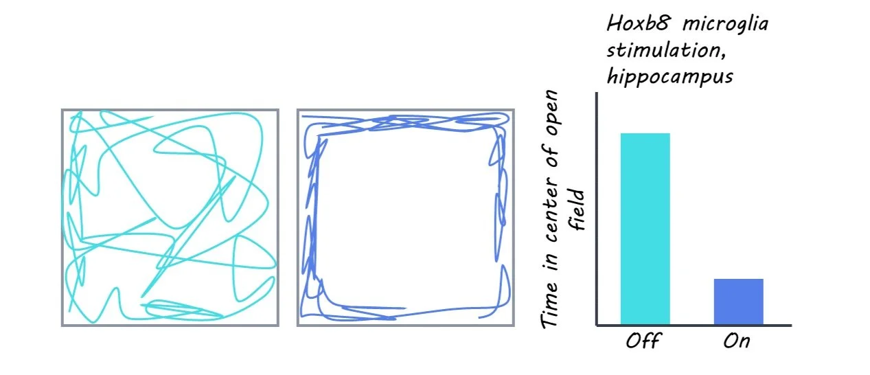
To activate the Hoxb8 microglia, the authors used optogenetic stimulation — using light to control the activity of certain cells in the brain. They activated Hoxb8 microglia in specific areas of the brain such as the dorsomedial striatum, the medial prefrontal cortex, the amygdala, and the hippocampus, which have previously been shown to control anxiety in mice. While stimulating these areas of the brain, they measured the behavioral effects; changes in grooming and other anxiety behaviors in different situations. They ran mice through several behavioral tests, measuring the anxiety-behaviors 2 minutes before stimulation, during the 2 minutes of stimulation, then the 2 minutes after stimulation. To measure anxiety levels, they used both a maze and an open field area to test how much time mice would spend in the fear-inducing open areas of the chambers as opposed to comfortable enclosed areas.

What did they find?

Mice groomed themselves when the dorsomedial striatum and the medial prefrontal cortex were stimulated and demonstrated higher levels of anxiety when areas in the amygdala were stimulated. This suggests that grooming is controlled by the former two areas, while anxiety is controlled by the latter area. When the microglia in the hippocampus were stimulated, mice showed both grooming and anxiety behaviors, in addition to increased freezing, which suggests the hippocampus is involved in controlling all three behaviors related to anxiety. Interestingly, when both Hoxb8 microglia and microglia not created by Hoxb8 (non-Hoxb8 microglia) were stimulated at the same time, mice did not display any anxiety behaviors at all. This suggests that Hoxb8 and non-Hoxb8 microglia work together with opposing effects, to control anxiety. Hoxb8 microglia turn off anxiety behaviors (like brakes on a car), and non-Hoxb8 microglia turn these behaviors on (like the accelerator).

In order to reconcile previous findings of anxiety increasing when Hoxb8 microglia are removed, with the current finding that activating Hoxb8 microglia also causes anxiety increase, the authors suggest that optogenetic activation of these Hoxb8 microglia might somehow cancel out their inhibitory effects on anxiety behaviors. While these mechanisms are still not fully understood, they likely involve the neighboring neurons that were activated when the Hoxb8 microglia were stimulated. Either way, these microglia are key in regulating anxiety, potentially in both directions.

What's the impact?

This study is the first to show that Hoxb8 microglia can be used to control anxiety behaviors using optogenetic techniques. It also suggests the reason for having both Hoxb8 and non-Hoxb8 microglia is to finely control anxiety behavior. Anxiety and related mental disorders are widespread both among adolescents and adults, so understanding the way it works within the brain is crucial so that we can better treat chronic anxiety. Studies like these could play a huge part in creating treatments that target these specific microglia and areas of the brain for chronic anxiety disorders.

Associating Heart and Brain Health using MRI

Post by Laura Maile

The takeaway

Evidence suggests a relationship between heart and brain health. The authors identified associations between structural and functional traits of the heart and brain that share genetic signatures with cardiac and brain diseases. 

What's the science?

Cardiovascular disease is often clinically associated with brain diseases, but the underlying genetic, structural, and functional connections between the heart and the brain remain unknown. Magnetic resonance imaging (MRI) has been used to identify structural and functional abnormalities that are associated with disease in individual organs, though few studies have analyzed MRI data from both the heart and the brain to find correlations between the two. 

This week in Science, Zhao and colleagues used multiorgan MRI to examine the connections between heart MRI features and structural and functional patterns in the brain. They then used genome-wide association studies (GWAS) to correlate these findings with genetic variants associated with both heart MRI traits and brain diseases. 

How did they do it?

The authors analyzed MRI data from >40,000 participants in the UK Biobank study. They identified 82 cardiac MRI traits that included measurements of the four cardiac chambers, the ascending and descending aortas, and wall thickness of different regions of the heart. A variety of brain MRI traits were also identified using imaging techniques that examine both structure and functional connectivity of different brain regions at rest and during specific tasks. Next the authors used statistical association and correlation analyses to explore associations between the identified traits. 

The authors then performed GWASs to identify specific genetic variations associated with the cardiac MRI traits. They repeated the GWAS on several different datasets to confirm the associations in a wider population. Next, they completed association and colocalization studies on the significant genetic variants to determine whether the cardiac and brain MRI traits shared genetic signatures. Finally, they sought to determine a genetic causal relationship between the heart and brain by applying Mendelian Randomization to the 82 cardiac MRI traits and several brain-related clinical outcome databases. 

What did they find?

The authors found 4193 significant associations between the 82 identified cardiac MRI traits and brain MRI traits such as cortical thickness, white matter microstructure, and volume of specific brain regions. Associations were also observed between cardiac MRI traits and the functional connectivity between certain brain networks. GWASs identified associations of 49 cardiac MRI traits at 80 genomic loci, which were found to be repeated across several datasets, indicating the generalizability of the findings across populations. They identified genetic variants that were shared across the cardiac and brain MRI traits that had been associated with diseases of the heart and brain. Mendelian randomization analysis revealed a causal relationship between genetic signals associated with heart traits and neuropsychiatric disorders. 

What's the impact?

This study found associations between heart and brain MRI traits that shared common genetic signatures. These findings denote a causal relationship between heart and brain health. This suggests that early intervention and treatment of heart conditions may improve brain health outcomes. 

Access the original scientific publication here

A Signature of Cortical Information Processing Underlies Shifts in Global States of Consciousness

Post by Soumilee Chaudhuri

The takeaway

Altered global states of consciousness are based on a top-down information processing signature in the cortex and influenced by a) spontaneous brain activity as well as b) regional brain organization. So, consciousness is determined by a hierarchical brain-region and brain-activity dependent signature.

What's the science?

Classically, consciousness has been understood as a neural manifestation of subjective experiences and linked to several dynamic neural processes in the brain. We  know that breakdowns in consciousness (during sleep, sedation, etc.) elicit complex changes in regional brain coordination and neural processing. However, we do not understand the exact relationship between shifts in global states of consciousness and brain activity in certain regions of the brain. This week in Nature Communications, Dr. Ang Li and colleagues unravel the complexity of shifting states of global consciousness by combining behavioral, neuroimaging, electrophysiological, and transcriptomic experiments.  

How did they do it?

The authors hypothesized that the shift in states of global consciousness might result from differential step-by-step processing of brain activity in different regions of the cortex (the brain’s gray matter-containing outer layer). They combined several functional Magnetic Resonance imaging (fMRI) approaches to capture altered consciousness — from deep sleep to full wakefulness — in recruited volunteers. In the first step, the authors captured the change in cortical activity over space and time in three distinct conditions: a) medication induced sedation, b) normal sleep, and c) awake, resting quietly. After this, they compared the cortical fluctuations between these conditions minute by minute. The authors also performed the exact same protocol for volunteers a) on caffeine or after fasting, b) administered a psychedelic drug and c) with neuropsychiatric disorders. They also used fMRI data from the Human Connectome Project to validate the spatiotemporal signatures obtained from the experiments. Electrocorticography (ECoG) recordings from Macaque monkeys were also used to compare to obtained hierarchical signatures. Additionally, the authors used spatial transcriptomic analyses from the Allen Brain Atlas to comment on specific regional contributions to wakefulness in subjects.

What did they find?

After looking at all of the evidence across different conditions, species, and timescales, the authors found that shifts in global state of consciousness can be attributed to changes in cortical neural variability, over time. This means that the global state of consciousness hierarchically associates with the disparity in neural responses across an experiment. Additionally, these complex shifts in consciousness can be translated to a simplified low-dimensional signature, enabling understanding of changes in consciousness in individual people. The authors also noted significant elevations of this hierarchical signature in abnormal states of consciousness (such as on psychedelics, in neuropsychiatric disorders, etc.). This signature also corresponded with the complex patterns of coordination that happen during wakefulness. The authors also found that the heterogeneity in the obtained hierarchical cortical neural variability across different conditions and species was modulated by a) spontaneous waves of cortical activities and b) the histaminergic system, a system that mediates inflammation.

What's the impact?

This study is the first to show that global states of consciousness rely on top-down hierarchical information processing in the cortex. The results also provide critical preliminary evidence of the association between the histaminergic system and hierarchical cortical processing. Most importantly, the authors find that at a global level, consciousness maps to top-down information processing by the cortex and that this may not be dependent on a specific neuroanatomical location in the brain. These findings provide a holistic understanding of the neural mechanisms of different conscious states such as sleeping, caffeinated or on psychedelics. This information may help to guide  therapeutic and behavioral interventions targeting disorders of consciousness, such as sleep disorders, addictions to psychedelics or psychiatric disorders.

Access the original scientific publication here