Human Brain Organoids Can Integrate into the Adult Rat Visual System

Post by Megan McCullough

The takeaway

Human brain organoids, grown from human stem cells, can integrate both structurally and functionally into adult rat brains.

What's the science?

One promising treatment for restoring brain function after an injury is cell transplantation. Human brain organoids, created from pluripotent stem cells, are an avenue of research currently being studied for therapeutic potential. Previous studies have shown that it is feasible for human brain organoids to integrate into rat brain systems, but there has been limited research into the functional integration of these organoids into the networks of injured mammalian brains. This week in Cell Stem Cell, Jgamadze and colleagues transplanted human brain organoids into damaged cavities in the visual cortex of adult rats to examine potential integration. 

How did they do it?

The authors transplanted brain organoids into the visual cortexes of rats. These tissues were generated from human stem cells and expressed the fluorescent marker GFP - a protein that lights up once exposed to ultraviolet light, allowing the authors to identify the organoids once grafted into the rat brains. The organoids had been growing for around 80 days, longer than in previous studies, to allow for maturation and cell differentiation. The authors then conducted a histological (tissue) analysis on the human grafts to confirm the human origin of the cell tissues, examine potential cell maturation, and observe any immune response from the host tissue. This was done to examine how the cell grafts evolved over time. Viral tracers were also injected into the eyes of the rats which allowed the authors to observe the connections between the human cells and rat cells. Next, the authors looked for functional integration of the transplanted organoids. Extracellular recordings were conducted to test for neural activity in the grafted tissue in response to presenting the rats with visual stimuli.

What did they find?

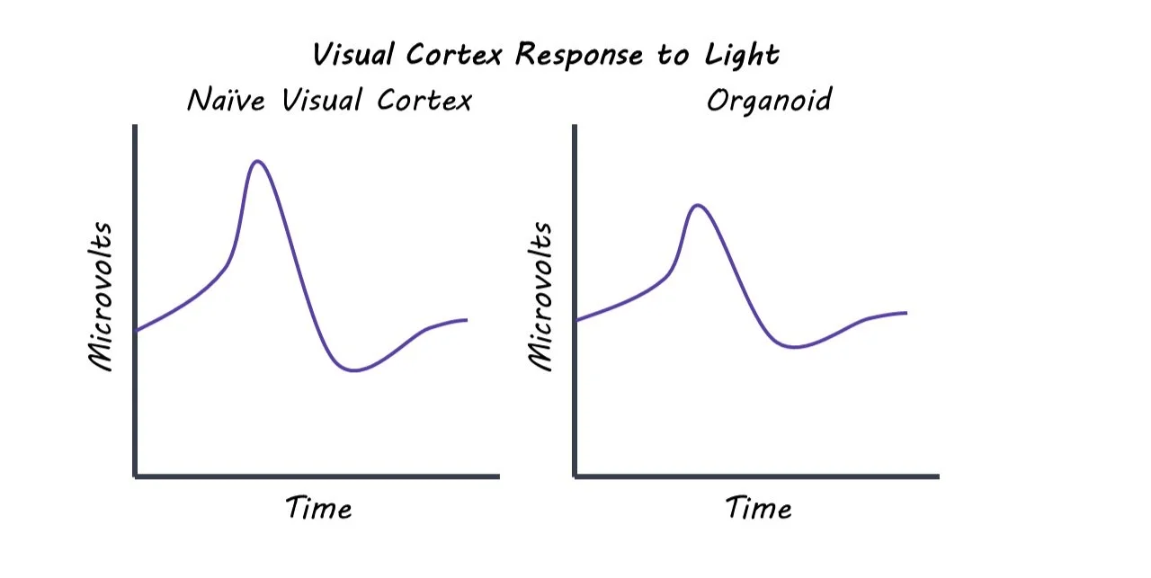
The authors found that in a three-month time period, human brain organoids integrated into the visual system of rat brains. Histological analysis showed that although there was inflammation at the graft site, there was only a mild immune response from the host tissue. This suggests that cell transplant can be a viable option into mammalian brains. This analysis also showed that the human cell tissue continued to differentiate and mature once transplanted. In addition to structural integration, the transplanted organoids also showed functional integration. The grafted human neurons received inputs from the visual system, forming connections via synapses with the host neurons. The brain organoids demonstrated both spontaneous and evoked neural activity, providing further evidence of functional integration. When the rats were exposed to flashing lights, the organoid neurons responded to the stimulation in similar way that the host cells responded.  

What's the impact?

This study found that human brain organoids can both structurally and functionally integrate with the visual system of rats. The human neurons formed connections with the rat neurons and displayed electrical activity in response to visual stimulation. This research shows the possibility of using lab-grown neural tissue to reconstruct brain circuitry after brain injury or stroke. 

CBD’s Anti-Seizure Mechanism of Action

Post by Baldomero B. Ramirez Cantu

The takeaway

Hippocampal hyperactivity is a hallmark of a variety of neurological diseases. Cannabidiol (CBD) is capable of modulating the excitatory-inhibitory balance of hippocampal neurons and its effects are mediated by interactions with the GPR55 receptor.

What's the science?

CBD is known to reduce seizure activity in animal models and patients with treatment-resistant epilepsies. However, the underlying mechanisms that enable CBD to reduce seizure activity and severity remain unknown. This week in Neuron, Rosenberg and colleagues investigate the role of the GPR55 receptor and its endogenous ligand LPI in CBD’s anti-seizure action.

How did they do it?

The authors tested if the GPR55 receptor is the site of action for CBD's anti-seizure effects by using mice with functional or genetically-removed GPR55 receptors. They conducted experiments on hippocampal neurons in-vitro to study how GPR55’s natural ligand, LPI, affects neuronal excitability and how CBD interacts with LPI’s effects. Finally, to investigate changes in GPR55 receptor expression after seizures in control or CBD pre-treatment conditions, the authors used qPCR and immunohistochemistry. Seizures were induced in the mice using the drug pentylenetetrazole or lithium-pilocarpine.

What did they find?

The authors found that seizures were only reduced by CBD if mice expressed GPR55 receptors - there was no improvement if these receptors were removed. This finding suggested a critical role of the GPR55 receptor in enabling CBD to attenuate seizures. They conducted neural recordings from the hippocampus and found that the application of LPI increased neuronal excitability in neurons from normal mice, but not in those from genetically modified mice lacking GPR55 receptors, indicating that LPI drives GPR55-dependent increases in neuronal excitation. LPI was found to disrupt neuronal activity in several ways, such as interfering with the impact of crucial inhibitory interneurons that are essential for maintaining an appropriate excitatory-inhibitory balance in the hippocampus. Importantly, hippocampal neural recordings showed that the presence of CBD counteracts the increased excitability produced by LPI. This provides evidence for the GPR55 receptor as a functional target of CBD’s anti-seizure action. Mice that received CBD pre-treatment showed significantly lower increases in GPR55 immunoreactivity and receptor mRNA expression after induced seizures compared to the control group.

What's the impact?

This is the first study to identify the two-pronged molecular signaling, affecting both excitation and inhibition, underlying CBD’s anti-seizure action. These findings could serve as the foundation for the development of new, targeted anti-epileptic therapies

Mitochondria Set the Tempo for Neuronal Development

Post by Elisa Guma

The takeaway

The development of neurons and mitochondria in the cerebral cortex takes considerably longer in the human brain than in other species, such as mice. Accelerating mitochondrial metabolism seems to accelerate human neuronal maturation, indicating that they are important regulators of the pace at which the brain develops.

What's the science?

There are striking species differences in the amount of time required for brains to develop, wherein the human brain develops over the course of months to years, while the mouse brain develops over the course of weeks. It is unclear what is responsible for these differences, however, given the important role that mitochondria play in driving cell maturation, they may also play a key role in modulating the differences in developmental timelines of cortical neurons. This week in Science, Iwata and colleagues investigate the role that mitochondria have in determining the developmental timelines of cortical neurons across the human and mouse brain.

How did they do it?

To investigate the relationship between mitochondrial metabolism and neuronal maturation, the authors used cultures of human and mouse cortical pyramidal neurons derived from pluripotent stem cells, as well as embryonic mouse brain neurons. For the pluripotent stem cells, the authors devised a method that stages neurons based on their birthdate to allow them to compare neurons in the same stage of development. To birthdate the neurons, the authors tagged the NeuronalDifferentiation1 gene – a gene that is active when the neuron enters a specific maturational stage – with a green fluorescent protein, allowing them to identify and separate it from the other neurons.

Within neurons of the same maturational stage, the authors also tagged mitochondria with an emerald-green fluorescent protein allowing them to visualize the morphology and location of these organelles within the neuron. To monitor mitochondria in developing cortical neurons in the mouse brain, the authors labeled mouse cortical neurons with a fluorescent protein using in utero electroporation in mid-late gestation, or they transplanted fluorescently labeled human neurons in the mouse brain. This allowed them to use light and electron microscopy to examine patterns of mitochondrial development and identify the age at which they reached maximal levels of growth and size.

Following a characterization of mitochondrial development, the authors examined the metabolic activity of mitochondria in both mouse and human cortical neurons at similar times after birth, focusing on mitochondrial oxidative phosphorylation and electron transport chain capacity (two indicators of metabolic function). Finally, to test whether enhanced mitochondrial activity would also accelerate neuronal development, the authors accelerated mitochondrial metabolism by inhibiting specific enzymes that are important in glucose metabolism in human cortical neurons. 

What did they find?

In mouse pluripotent stem cells and newly born neurons (from the embryonic brain), the authors found that mitochondria were initially small and sparse but grew in quantity over the 3-week maturational window of these neurons. In contrast, pluripotent stem cells derived from human cortical neurons, as well as their mitochondria, showed a much slower pattern of maturation, taking several months. These data suggest that mitochondrial morphology and development follow a species-specific timeline that is highly correlated with neuronal maturation.

Consistent with morphological development, mitochondrial metabolism was higher in the mouse neurons than in the human neurons in the early stages of development and continued to increase at a faster rate across development. In human cortical neurons, they also observed lower levels of oxidative stress compared to the mouse neurons, consistent with lower activity of mitochondrial metabolism.

When the authors increased mitochondrial activity in human neurons, they observed an increase in oxidative phosphorylation with no significant alterations to mitochondrial morphology. However, they did observe an increase in the speed at which neurons were maturing both in terms of function (based on synaptic currents and membrane potentials) as well as morphology (larger neuronal size and increased dendritic length and complexity). This indicates the crucial role of these organelles in regulating timelines of neuronal development.

What's the impact?

This study provides evidence for the role of mitochondrial metabolic activity in regulating the species-specific developmental timeline of cortical neurons. When mitochondrial metabolism was enhanced, neurons showed accelerated morphological and functional maturation. This may in part explain why the human brain develops across much longer time courses than other species, such as the mouse. Future work is needed to understand the downstream effects of mitochondrial metabolism on brain function, plasticity, and neurodevelopmental disorders.