On-Demand Gene Therapy for Brain Circuit Disorders

Post by Andrew Vo

The takeaway

Current gene therapies that aim to correct abnormal neuronal activity in different disorders fail to discern between pathological and surrounding healthy neurons. Designing therapies that are activity-dependent allows treatments to be delivered specifically to misfiring neurons in a more targeted manner.

What's the science?

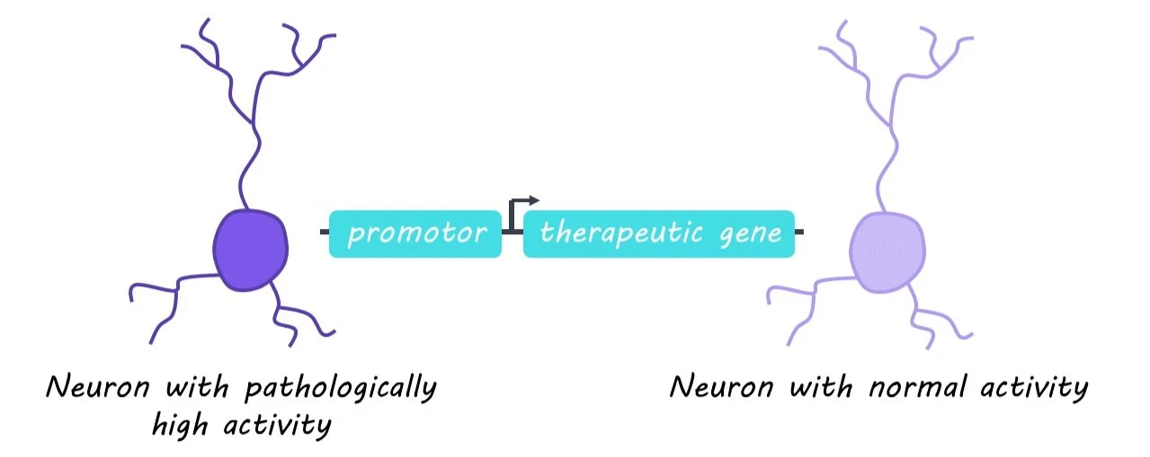
Genetic therapies can augment and treat pathological neuronal activity associated with many neurodevelopmental and neuropsychiatric disorders. However, current options fail to discern between pathological and surrounding healthy neurons, increasing the risk of unwanted side effects. Activity-dependent promoters (gene sequences that only activate following neuronal activity and control the expression of delivered therapeutic genes) could be the key to targeting pathologically overactive neurons. This week in Science, Qiu, et al. test whether an activity-dependent promoter can reduce seizures and seizure-related activity in a model of epilepsy.

How did they do it?

The authors began with cell cultures in which they introduced either (1) a therapeutic gene that encodes potassium channels and decreases neuronal excitability or (2) a control gene that did not affect neuronal activity. The expression of both genes fell under the control of an activity-dependent promoter. They then measured whether their therapeutic gene reduced hyperexcitability. Next, the authors delivered either the therapeutic or control gene into the hippocampus of adult mice. A drug was used to induce generalized seizures and then hippocampal neuronal excitability between the two groups was recorded and compared. In another experiment, mice were injected with a second consecutive dose of the seizure-inducing drug to test whether initial activation of the therapeutic gene had lasting effects. The authors evaluated the mice on different behavioral tests of learning and working memory. Finally, they tested the effectiveness of their activity-dependent therapy in a mouse model of chronic epilepsy with spontaneous seizures, and in neurons derived from human stem cells.

What did they find?

The authors found that cell cultures treated with the therapeutic gene showed reduced neuronal excitability. Closer inspection of the cells revealed that the promoter was selectively activated in excitatory but not inhibitory neurons, which indicates that the therapeutic gene was specific to neurons involved in pathological overexcitability. Similarly, the excitability of activated neurons in response to drug-induced seizures was decreased in the hippocampus of mice treated with the therapeutic gene. When a second dose was injected after a short delay, the authors noted greatly reduced seizures. This suggests that activation of the therapeutic gene following the initial seizure persisted and protected against the second seizure. This benefit was time-limited, however, as a third dose injected after a longer delay when therapeutic gene expression had returned to baseline was no longer attenuated. No significant changes in behavior were observed between the therapeutic and control gene groups. These collective findings were also replicated in a mouse model of chronic epilepsy and human neurons.

What's the impact?

The present study describes a gene therapy for epilepsy that decreases neuronal excitability in an activity-dependent manner. This approach was time-limited to the duration of pathological neuronal activity, effective in mouse models of both discrete and chronic epilepsy, and translatable to human neurons. Activity-dependent gene therapy is an exciting and promising strategy in the targeted treatment of brain disorders.

Patients with Depression Release Less Brain Serotonin

Post by Elisa Guma

The takeaway

Depressive symptoms have been associated with diminished serotonin neurotransmission in numerous brain areas. By directly measuring in vivo serotonin release capacity in the frontal lobe, this study finds that patients experiencing a major depressive episode have reduced serotonin release capacity in their frontal cortices.

What's the science?

Reduced serotonin neurotransmission is thought to play a causal role in the pathology of depression. While depression is one of the most common mental illnesses worldwide, it is even more prevalent in individuals with Parkinson’s disease who experience a loss of serotonin neurons in the raphe nucleus, which provides more evidence for the “serotonin hypothesis”. However, most studies drawing associations between decreased serotonin and depression are based on indirect measurements of this neurotransmitter. This week in Biological Psychiatry Erritzoe and colleagues leverage a novel positron emission tomography (PET) radioligand – serotonin 2A receptor agonist ([11C]Cimbi-36) – to investigate whether individuals with either depressive disorder or a depressive disorder due to Parkinson’s Disease have altered serotonin releasing capacity in their frontal cortex following a d-amphetamine challenge (a common monoamine and serotonin releasing agent).

How did they do it?

The authors recruited seventeen antidepressant-free patients with major depressive disorder (five of whom had the diagnosis due to an underlying Parkinson’s Disease diagnosis) as well as twenty healthy controls. Depressive symptoms were assessed at baseline using the standard Hamilton Depression Rating Scale. Self-rating of effects due to d-amphetamine was recorded, and blood samples were taken, both immediately before and after the PET scan. PET imaging was focused on the frontal cortex, while the cerebellum was used as a reference region (as is customary in many PET studies). The authors measured serotonin release capacity by comparing the change in binding potential (how much radioligand binds to receptors in the brain) in the frontal cortex (relative to the cerebellum) both at baseline and following the dose of d-amphetamine. Thus, an increase in extracellular serotonin (due to d-amphetamine challenge) should result in a decrease in radioligand binding.  

The authors first compared the serotonin release capacity of individuals with depression relative to healthy controls (covarying with age). Additionally, they investigated putative relationships between the severity of depressive symptoms and the effect of baseline binding potential between patients and controls (prior to d-amphetamine challenge).

What did they find?

High (and similar) binding across the frontal cortex for the serotonin 2A radioligand ([11C]Cimbi-36) was observed, confirming the proper function of the radioligand. Age was shown to decrease the binding potential, and thus, accounted for in subsequent analyses. D-amphetamine administration was found to significantly decrease binding potential in healthy individuals, which suggests that they were releasing serotonin extrasynaptically. The effect in patients with depression was less pronounced. This suggests that in response to the d-amphetamine challenge (which elicits serotonin release, among other things), individuals experiencing depression released less serotonin than healthy controls (they had reduced serotonin release capacity). Individuals whose depressive symptoms were a result of Parkinson’s Disease did not appear different from those with major depressive disorder, however, this may be due to a smaller sample size in the Parkinson’s disease group.  Finally, there was no relationship between depressive scores and the serotonin release capacity in either of the groups nor was there a correlation between depressive scores and baseline radioligand binding for the individuals with depression.

What's the impact?

This study provides the first direct measure of serotonin release capacity in the frontal cortex of individuals with depressive symptoms. The authors find a decrease in the serotonin release capacity of individuals with depression relative to healthy controls in response to a d-amphetamine challenge. Increased serotonin catabolism or dysfunction of certain reuptake transporters may contribute to these observed decreases. Future research is required to better understand the mechanisms driving this decrease in serotonin release capacity and whether therapies that increase serotonin release capacity might be effective.

Age-Related Neurodegeneration in Huntington’s Disease is Associated with Impaired Autophagy

Post by Leanna Kalinowski

The takeaway

Age-related neurodegeneration in people with Huntington’s disease is associated with an inhibition of autophagy in neurons, which is when old cells are recycled in order to regenerate new cells. 

What's the science?

Huntington’s disease (HD) is an inherited neurodegenerative disorder that is characterized by uncontrollable movements, emotional and behavioral problems, and cognitive decline. Symptoms of HD progressively get worse over time, however, it is unclear how getting older drives the onset of neurodegeneration in people with HD. This week in Nature Neuroscience, Oh and colleagues examined the underlying mechanisms of age-related neurodegeneration by reprogramming cells from HD patients into medium spiny neurons.

How did they do it?

The researchers used cell reprogramming to convert fibroblasts (non-neural cells that contribute to the formation of connective tissue) into medium spiny neurons (MSNs), which are the primary neurons that degenerate in people with HD. The reprogrammed cells were from three groups of patients: (1) patients with HD who show symptoms (“symptomatic HD MSNs”), (2) patients with HD who do not yet show symptoms (“pre-symptomatic HD MSNs”), and (3) age-matched patients who do not have HD (“controls”).

After reprogramming these cells, they tested the hypothesis that symptomatic and pre-symptomatic MSNs exhibit differences in autophagy, which is the body’s process for recycling damaged cells to regenerate new ones. They measured several markers of autophagy (e.g., the number of autophagosomes) and compared them across each group of MSNs. Then, to directly test the impacts of autophagy on HD symptomology, they treated cells with substances to inhibit or enhance autophagy and measured subsequent levels of MSN degeneration.

They then investigated epigenetic mechanisms that underlie differences in gene expression between pre-symptomatic and symptomatic HD MSNs. First, they conducted chromatin accessibility profiling, which measures how tightly DNA is packed into chromatin and determines how accessible it is for gene transcription (the tighter the DNA is packed, the less accessible it is for transcription). They then probed group differences even further by measuring microRNAs (miRNAs), which inhibit gene expression by preventing translation.

What did they find?

First, they found that, relative to controls and pre-symptomatic HD MSNs, symptomatic HD MSNs have lower levels of autophagy markers. This suggests that autophagy is inhibited in patients with symptomatic HD. Next, they found that inhibiting autophagy in pre-symptomatic HD MSNs increases the degeneration of these cells while enhancing autophagy in symptomatic HD MSNs decreases the degeneration of these cells. This highlights an association between the inhibition of autophagy in patients with HD and subsequent neurodegeneration.  

When evaluating the epigenetic mechanisms of these differences, the researchers found that the genes with reduced chromatin accessibility in symptomatic HD MSNs are genes that are involved in autophagy (e.g., ATG16L1, ATG10). This means that these autophagy genes are more difficult to transcribe in symptomatic HD MSNs, leading to an inhibition of autophagy. These effects are driven by differences in miR-29b-3p, which is an miRNA that inhibits autophagy and promotes the degeneration of MSNs.

What's the impact?

Taken together, these results provide a better understanding of what drives age-related neurodegeneration in HD. Specifically, an increase in miR-29b-3p promotes neurodegeneration by impairing autophagy in patients with symptomatic HD. Results from this study may provide an avenue for the development of therapeutics to slow or even reverse the progression of HD.

Access the original scientific publication here.