Can Combining Brain Imaging Techniques Help Us “See” Our Thoughts?

Post by Christopher Chen

The takeaway

Increasing evidence from brain imaging studies suggests that fingerprint-like patterns of brain activation may reflect information during cognitive processes like emotion and perception. Researchers think that these patterns may represent sparse code at the neuronal level - reproducible patterns of neural firing in a small percentage of neurons.

What underlies perception, cognition and emotion?

Even with the advent of modern brain imaging techniques in the late twentieth century, researchers have still not fully answered a question dating back to ancient Greece and Aristotle’s musings on the mind: what is the basis of human thought?

That said, science has made great progress in painting the big picture regarding the brain’s role in discrete tasks. For example, viewing an image of a face elicits distinct activity patterns in a region of the brain called the fusiform gyrus, and thinking about the connection between two events is related to activity in a brain region called the hippocampus. Showing that specific tasks stimulate specific brain regions, however, is not quite the same as determining how the brain processes information.

Recently in Neuroimage, Jaaskelainen and colleagues offer evidence that combining two techniques – functional magnetic resonance imaging (fMRI) and two-photon calcium imaging (2PCI) – will help researchers demonstrate that the brain processes information through the activation of specific populations of neurons. Combining these two techniques could potentially help neuroscientists take a huge step forward toward uncovering a biological foundation for human thought.

Fingerprint patterns of fMRI brain activity

Since its invention over thirty years ago, fMRI – a medical imaging technique measuring changes in the brain’s blood flow – has been critical in showing how specific stimuli like a human face, for example, activate specific brain regions. Since the early 2000s, a technique called multi-voxel pattern analysis (MVPA) has helped enhance fMRI studies by providing greater resolution in differentiating activity across brain regions, allowing researchers to describe stimulus-specific “fingerprints” in our brains. From these studies, we have determined that these fingerprints most likely represent information gleaned during tasks and presentation of various stimuli, an enormous victory in the quest to decode how our brains process information. Studies pairing fMRI and MVPA have generated a lengthy trail of evidence suggesting fingerprint patterns are linked to information representation in nearly all brain regions and helped establish fingerprints as a reliable biological representation of information in the human brain.   

What is sparse neural code and why does it matter?

Studies in animals have been able to probe even deeper than fMRI studies. While fMRI measures the activity of brain regions, 2PCI can measure the activity of neurons within these brain regions by measuring neuronal calcium levels. These findings also highlight how efficient the brain can be when representing information, with one of the most notable 2PCI experiments showing that activation of only a minuscule (~0.5%) population of visual cortex neurons is required for visual processing in a monkey. This limited activation of specific neuronal populations in response to stimuli is aptly called sparse distributed representation, or sparse code.

Some believe sparse code is another level of detail in the journey to define the biological basis of cognition. Thus, some neuroscientists believe translating 2PCI sparse coding to fMRI fingerprints will help uncover how neuronal populations underlie the basis of thought formation. 

What’s the relationship between fingerprints and sparse neural code?

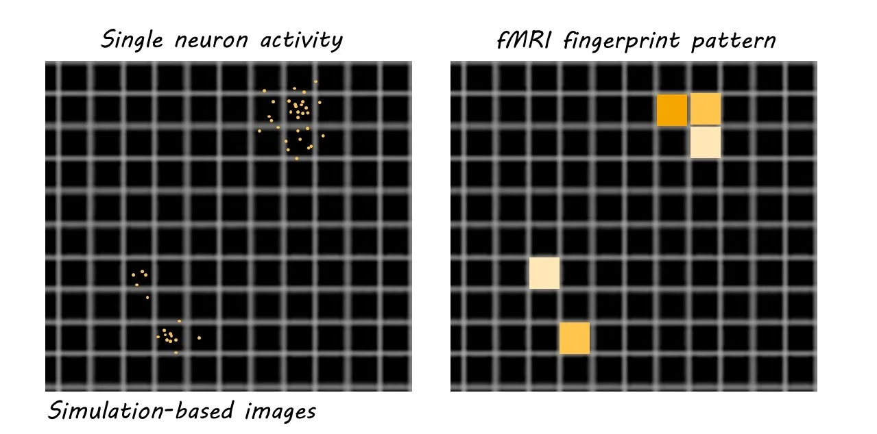
Determining whether 2PCI-generated sparse coding underlies fMRI fingerprints is not an easy task. One of the primary questions is whether fMRI has the spatial resolution to record sparse representation. In other words, can fMRI which usually measures regional activity in millimeters, capture activity happening at the micrometer (1000-fold smaller) level? Evidence does suggest that the primary readout of 2PCI – calcium signaling – scales with the primary readout of fMRI, hemodynamic signaling, suggesting that fMRI can at the very least indirectly capture micrometer-level activity. Additionally, there is evidence from fMRI data suggesting a sparse coding-like distribution activity pattern in visual areas, which corresponds to findings from the visual area in 2PCI studies. And perhaps the most compelling evidence suggesting a functional similarity between sparse coding and fingerprints comes from a simulation experiment the authors performed, that generated amplified 2PCI data resembling an fMRI fingerprint. This evidence is a promising first step in a fuller characterization of just how intimately linked sparse coding and fingerprints are.

What does the future hold?

While still speculative, the idea that 2PCI sparse coding may inform fMRI fingerprints is enormous in scope. If true, it would show that the activation of very distinct, small populations of neurons lie at the core of basic human functions like cognition and emotional processing. A critical step, of course, is to measure the integration of sparse codes from multiple brain regions in higher-order cognitive tasks such as decision-making and learning. Experimentally, using both 2PCI and fMRI imaging in parallel will be instrumental in determining the similarities between sparse coding and fingerprint patterns. Studies could also employ a sparse coding amplification technique like the one just described to predict how well 2PCI data from animals scales with fMRI data in humans. Whether these studies reveal the neural basis of human cognition remains to be seen, but they will undoubtedly teach us more about how the interplay between sparse coding and fingerprints informs human behavior.  

Access the original scientific publication here.

How Diet Influences Your Brain and Mood

Post by Elisa Guma

Brain-body connections that allow food to influence mood

Our moods and emotions govern our lived experiences and are inextricably linked to the function of our brains and bodies. Ingesting certain macro- and micronutrients through our diet can impact the chemicals in our brain that contribute to many emotional states, such as happiness, sleepiness, and sadness. Food choice has been strongly implicated in both mental and overall health and well-being.  Gaining insight into how the nutrients that make up our food affect our brain and behaviour could help us understand how to improve our mental health and overall well-being through our dietary choices.

The role of the vagus nerve

One of the primary pathways through which the brain and body communicate is a bidirectional superhighway known as the vagus nerve. The vagus nerve is the 10th cranial nerve and the longest cranial nerve in the body– starting at the base of your neck, and innervating the brain as well as the organs of the body, including the heart, lungs, stomach, intestines, and liver. Through the vagus nerve, the brain receives information regarding the state of our organs, such as how distended our gut is, how full our intestines are, and how quickly our heart is beating. It can also send motor signals back to the organs, such as telling our heart or our guts to slow their pace. This nerve is one way in which the brain and body are connected and can integrate our emotional states, mood, and well-being. 

Food and neurotransmitters

The production of neurotransmitters such as serotonin, dopamine, and norepinephrine relies on appropriate levels of critical building blocks that we receive from food, such as amino acids, fats, carbohydrates, and minerals. For example, the amino acid L-tyrosine, is a precursor to dopamine and norepinephrine, while both tryptophan (a different amino acid) and carbohydrate-rich foods increase the production of serotonin. Depending on the number of precursors present in the food you eat, you may produce more or less of a certain neurotransmitter. Importantly, imbalances in these neurotransmitters have been associated with numerous neuropsychiatric illnesses, and affect our mood and alertness.

Neurotransmitters are also released in anticipation of and in response to certain foods. For example, the locus coeruleus has been found to release norepinephrine to activate the lateral hypothalamus (the nucleus that regulates food intake) during food preparation. This increases alertness around food, which can lead to feelings of excitement, but also anxiety. Additionally, ingestion of certain foods can induce dopamine release, potentially leading to cravings that cause us to seek out more of that food. For example, certain neurons in the stomach will release dopamine in response to sugar. Interestingly, this is not related to the rewarding sweet flavor of sugary foods, but to specific sugar-sensing neurons in the gut; the same dopamine response occurs even if the sweet taste of sugar is masked. This indicates that there are certain circuits in the body that drive behaviour towards certain types of food based on information that comes directly from our bodies, and not necessarily from our cognitive appraisal of how much we “like” that food. 

Brain-derived neurotrophic factor (BDNF) is an abundant signaling molecule intimately related to both cognitive function and metabolic regulation in the brain. BDNF is involved in reshaping synaptic connections in the context of learning and memory but also plays a role in energy mentalism, appetite suppression, and energy balance in the body. Importantly, low serum levels of BDNF have been identified in individuals with psychiatric diseases, such as schizophrenia and depression, and it is thought to be a key player in mediating the positive effects of antidepressants on the brain. Several studies have investigated the relationship between diet and BDNF and found that diets rich in polyphenols were associated with elevated levels of BDNF. Interestingly, these micronutrients may play a role in disease prevention and longevity as well. 

Omega-3 fatty acids

Omega-3 fatty acids are an integral part of neuronal cell membranes and play a key role in several central nervous system functions including neurotransmission, gene expression, neurogenesis, and neuronal survival. These fatty acids can be found in fatty fish, eggs, flax seeds, hemp seeds, and chia seeds, and can also be ingested as supplements in capsule or liquid form. 

If ingested in the correct ratios (i.e., high omega-3 relative to -6), these fatty acids may have antioxidant and anti-inflammatory properties. In one human study, a low dose (20 mg) of the antidepressant Prozac, a serotonin reuptake inhibitor, and a high dose (1000 mg) of EPA, an essential fatty acid high in omega-3, were found to have similar antidepressant effects. Taken together, the two compounds had synergistic effects, suggesting that the omega-3 fatty acids may amplify the effects of antidepressants. The shift in omega-3 to -6 ratio has been shown to lower inflammation in the body and increase heart rate variability, both of which may improve symptoms of depression and allow antidepressants to do their work.

Micronutrients such as B and D vitamins, zinc, and more

In addition to adequate levels of amino acids, carbohydrates, and fats, our brain, and body also rely on numerous minerals and micronutrients to function optimally. When we are nutritionally deficient in certain micronutrients such as vitamin B12, vitamin B9 (folate), vitamin D, or choline, we may experience symptoms like depression, low mood, fatigue, cognitive decline, and irritability. Similar results have been found for trace minerals such as calcium, zinc, copper, iron, and selenium. Often, diets high in processed food can put individuals at greater risk for nutritional deficiencies, increasing the likelihood of negative impacts on both mood and general cognitive function. Vitamin supplementation has been found to combat cognitive impairment and energy metabolism in both rodent and human studies. 

Conclusions on diet and mental well-being

The nutrients within our food likely have profound effects on our brain and body, affecting both our health and how we feel. Prioritizing and promoting diets that are healthy for our brains may have important benefits for our daily lives. Furthermore, food is a critical component of cultural heritage and can connect us to family and friends, as well as places. Understanding how our mood and well-being are impacted by all these factors can help us to better understand and regulate our emotions and our overall health.

References +

Bremmer JD et al., Diet, Stress and Mental Health. Nutrients, 2020.

Gomez-Pinilla. Brain foods: the effects of nutrients on brain function. Nature Review Neuroscience, 2008.

Gravesteijn E et al., Effects of nutritional interventions on BDNF concentrations in humans: a systematic review. Nutritional Neuroscience, 2022.

Jazayeri S et al., Comparison of Therapeutic Effects of Omega-3 Fatty Acid Eicosapentaenoic Acid and Fluoxetine, Separately and in Combination, in Major Depressive Disorder. Australian & New Zealand Journal of Psychiatry, 2008.

Lachance L & Ramsey D. Food, Mood, and Brain Health: Implications for the Modern Clinician, Missouri Medicine, 2015.

Meeusen R & Decroi L. Nutritional Supplements and the Brain. Int J Sport Nutr Exerc Metab, 2008

The Neural Signature of Mind Blanking

Post by Lani Cupo

The takeaway

Occasionally while we are awake, our minds seem to go “blank”; we cannot say what we were thinking about. Mind blanking is a state that occurs by default, is characterized by a unique behavioral signature, and is linked to an underlying pattern of brain activity.

What's the science?

In recent years, an increasing number of studies have examined the phenomenon of mind blanking, examining the frequency of “zoning out” compared to other mental states, and characterizing the activation of brain regions during mind blanking. However, despite a growing body of research, the physiological and behavioral processes underlying this state remain inconclusive. This week in PNAS, Mortaheb and colleagues used functional magnetic resonance imaging (fMRI) data to characterize the dynamics of brain activity associated with mind blanking.

How did they do it?

Data were previously acquired from 36 healthy adults (27 women, 9 men) who were at rest (resting quietly) in the MRI scanner with their eyes open to ensure they did not fall asleep. A sound played randomly fifty times throughout the scanning period, prompting the participants to respond with a button to report whether their current mental state was one of four options: mind-blanking, perceiving sensory stimuli, thoughts related to external stimuli (stimuli-dependent), or thoughts independent of external stimuli (stimuli-independent). To characterize the behavioral profiles of mind blanking, the authors examined how frequently it was reported, whether the response time (speed to bush button after the auditory cue played) differed based on the reported state, and the probability to re-report mind blanking multiple times in a row.

To characterize brain activity during the mental states, the authors analyzed a 10-second window around each probe (each auditory stimulus prompting a response). First, they measured the amplitude of the global signal - a physiological proxy of arousal. To do this, the mean absolute value of the signal amplitude for each window is calculated across all regions of interest in the brain, giving a gross estimate of brain activity. Then, to determine whether the functional connectivity of the brain during mind blanking differed from other states, the authors trained a machine learning algorithm to classify participants' reports into different categories based on functional connectivity alone.

Finally, the authors used a clustering technique to derive four different patterns of connectivity over the entire resting state period and examined whether one of the four patterns was more strongly associated with the patterns of connectivity during mind blanking states.

What did they find?

First, the authors replicated results from previous studies reporting a low frequency of mind blanking compared to other mental states. Additionally, mind blanking was reported relatively fast compared to stimuli-dependent and independent states, potentially implying that content-less states were easier to recognize and report than states with content. It was extremely rare for participants to re-report mind blanking multiple times subsequently, which may suggest mind blanking reflects a transitional state between other states.

Second, there was a subtle difference between the amplitude of the global signal between states, with a slightly higher average signal amplitude in mind blanking than in stimuli-dependent or independent states. Global signal amplitude has been negatively correlated with alertness before, suggesting global silencing during wakefulness. This is consistent with these results which report a high global signal during an attentional lapse.

Third, the authors found that the trained machine learning classifier could accurately separate mind blanking states from the others based on functional connectivity, suggesting mind blanking is accompanied by a unique pattern of brain activity and functional connectivity.

Finally, the authors derived four distinct patterns of functional connectivity during the resting state. The pattern most closely related to the pattern during mind blanking states was characterized by positive functional connectivity between brain areas across the brain, and the similarity between this pattern and mind blanking states was higher than the similarity between this pattern and any other state. Like an increased global signal, this pattern of positive whole-brain functional connectivity may be associated with decreased cortical arousal.

What's the impact?

The results of this study imply that mind blanking is a unique, possibly default, mental state which might represent a transition between other states. Moments of unreportable thoughts during wakefulness can occur spontaneously. These findings challenge traditional ideas of a constantly-accessible conscious human brain.