Early Intervention in Huntington’s Disease Mutation Carriers Delays Symptom Onset

Post by Andrew Vo

The takeaway

Genetic mutations that cause Huntington’s disease later in life also affect neurodevelopment in early life. Medications that enhance the glutamate neurotransmitter system in mutation carriers during infancy have the potential to prevent the later onset of disease.

What's the science?

Huntington’s disease (HD) is a neurodegenerative disease that is largely inherited through mutations in the huntingtin gene (HTT). HD symptoms do not become apparent until middle or late adulthood, but the underlying mutation has been shown to affect brain development much earlier in life. Although the brain can compensate for this early dysfunction, it is unknown whether intervening during this period can delay or prevent the later development of disease. This week in Science, Braz et al. use a mouse model of HD to demonstrate how treating early brain abnormalities in mutation carriers can alter the disease course.

How did they do it?

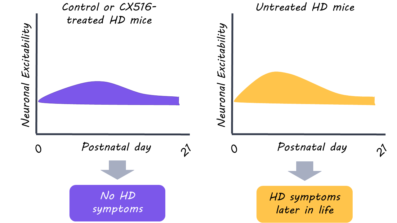
First, the authors recorded activity from neurons of newly born mice expressing the HTT mutation (i.e., HD mice). Similarly, they conducted brain recordings in postnatal mice depleted of HTT (i.e., HTT-depleted mice). Together, these experiments investigated the HTT gene's critical role in HD development. Next, the authors investigated whether enhancing excitatory neurotransmission affected both early brain physiology and the later onset of HD symptoms. Postnatal HD mice and non-mutant control mice were treated with CX516, which increases glutamate neurotransmitter signalling, and the effects on behavioural symptoms were observed during adolescence and adulthood. The effects of HTT mutation and CX516 treatment on brain structure were also measured using magnetic resonance imaging.

What did they find?

During their first week, postnatal HD mice showed evidence of reduced excitatory activity and less complex neurons compared to non-mutant mice. By the second week, however, the brains of the HD mice were able to normalize. Postnatal HTT-depleted mice displayed similar defects in neuronal activity as HD mice, however, these abnormalities failed to normalize. The results suggest that at least partial HTT gene function is critical for the brain to compensate against early HD dysfunction.

Administration of CX516 in postnatal HD mice restored neuron shape and motor function in comparison to untreated HD mice. Astonishingly, this postnatal treatment of excitatory glutamate signalling prevented the later development of HD symptoms in adulthood. Early intervention also appeared to normalize brain structure (as measured with magnetic resonance imaging) in adult HD mice; brain volume was reduced in untreated HD mice but not in those with early intervention. It should be noted, however, that CX516 had negative effects in non-mutant mice. This indicates that there is an optimal range of excitatory neurotransmission for normal brain function.

What's the impact?

This study demonstrated that treatment of early brain dysfunction in HD mutation carriers might delay or prevent the onset of disease. Although this was achieved in a mouse model of HD, it nonetheless represents an important step toward a cure for human patients.

Neuroimaging as a Tool for Diagnosing and Tracking Multiple Sclerosis

Post by Anastasia Sares

The takeaway

Neuroimaging is a common and useful tool for diagnosing multiple sclerosis. Over the last 25 years, imaging technologies have improved to give us clearer pictures of the main features of MS at greater resolution. Further, machine learning algorithms are being used to advance our knowledge of MS. 

MRI: a key tool for diagnosis

Multiple Sclerosis (MS) is a chronic and progressive autoimmune disorder that happens when the body’s immune cells invade the central nervous system and begin to attack myelin: a protective fatty tissue wrapped around the axons of neurons. Symptoms of MS can vary wildly, depending on the region under attack. If the optic nerve is attacked, people can experience loss of vision; if the spinal cord is attacked, shooting nerve pain can be the result. 

Because MS symptoms are so variable, MRI is one of the key tools for diagnosis. MRI is non-invasive and allows a clinician to see lesions: areas of the brain that have been attacked by the body’s immune cells, appearing in the brain or spine even when the patient is not currently experiencing any symptoms. However, identifying the lesions in the first place can be difficult to the untrained eye, and being sure they are caused by MS (and not another disease) is also challenging. Therefore, to better diagnose and treat MS, we need to make it as easy as possible for clinicians to find and examine these lesions.

Using MRI to search for lesions

There are many types of MRI sequences, and each of them can highlight different characteristics of human tissue. MRI uses a strong magnetic field to align hydrogen nuclei (protons) so that they are oriented in the same direction. Then, it hits these protons with a radio frequency pulse that sends them all spinning. When the protons relax back into alignment, they emit radio energy that can be detected by the machine. Protons embedded in different types of tissue will have different resonant properties that affect how long they spin before relaxing. So, it is possible to optimize the radio pulse frequency and the time of detection in such a way that MS lesions will be more visible in the final image.

The current recommended MRI sequence for finding MS lesions is called T2 FLAIR. T2-weighted images use a slow radio pulse frequency and a longer wait time after a pulse for detection. These properties make the sequence excellent for picking up tissues with increased water content, whose hydrogen protons have a longer period of resonance. FLAIR stands for “Fluid Attenuated Inversion Recovery,” which uses an extra radio pulse to suppress signals coming from free-flowing fluids (water, cerebrospinal fluid), making those parts of the image less bright. MS lesions still show up brightly on the image.

Adapted from Bakshi et al. 2001

Similar to FLAIR, other “inversion recovery” sequences have been developed (with names like PSIR and STIR), but these have not overtaken the FLAIR sequence for brain MRIs.

Most early imaging research in MS focused on the brain, but it is now recognized that getting a good spinal cord MRI can be important for a diagnosis. There can be lesions here as well, and they may cause more disabling symptoms. Spinal cord lesions can also help confirm a diagnosis of MS, ruling out other diseases known as MS “mimics” that look similar on brain MRIs. Spinal cord imaging is more prone to interference from bodily processes like heartbeat, breathing, and swallowing, so every advance in this field matters. In the spinal cord, the recommended MRI sequences are different, including STIR or PSIR mentioned above.

Another technique for lesion detection is to use a contrast agent called gadolinium, which is injected into the blood right before an MRI. It is called a contrast agent because it helps to enhance the contrast of the MRI signal wherever it goes, causing a bright glow on a scan. Normally, gadolinium cannot get through the blood-brain barrier (the tight network of cells that separates the central nervous system from the rest of the body). However, if a person is currently having an MS attack, the blood-brain barrier becomes leaky at the site of the lesion. So, the location of “active” lesions will glow brightly on the MRI. However, it is not ideal for the health of the patient to use gadolinium repeatedly since it can accumulate in the central nervous system.

How can artificial intelligence help?

MS is a field ripe for machine learning applications. The basic problem is one of image classification, which is a staple in the machine learning world. Some algorithms for lesion detection have recently been proposed, and these can identify lesions, calculate their volume, and measure overall brain size, These measures can also be tracked over time in patients to get a clearer idea of disease progression.

Many of these algorithms need to be fed a large set of training data—for MS, this means getting a huge number of correctly classified MRIs to learn from. If there are any systematic biases in our diagnosis of MS, these will be replicated by the machine learning program. Finally, someone ultimately must take responsibility for the diagnosis (a doctor, not a machine!), so adding artificial intelligence, while extremely useful, does complicate the accountability landscape.

Moving forward

MS is a disease of the central nervous system, and it has become easier to diagnose as our neuroimaging methods improve in this area of intense research. More advanced MRI sequences are on the horizon, even ones that can detect and quantify the myelin itself (the tissue under attack). Combined with powerful algorithms, these sequences offer clinicians much more information to draw on when making decisions about an MS diagnosis.

References +

Barkhof, F. (1997). Comparison of MRI criteria at first presentation to predict conversion to clinically definite multiple sclerosis. Brain, 120(11), 2059–2069.

Tintore, M., Rovira, A., Martınez, M. J., Rio, J., Dıaz-Villoslada, P., Brieva, L., Borras, C., Grive, E., Capellades, J., & Montalban, X. (2000). Isolated Demyelinating Syndromes: Comparison of Different MR Imaging Criteria to Predict Conversion to Clinically Definite Multiple Sclerosis. 5.

Bakshi, R., Ariyaratana, S., Benedict, R. H. B., & Jacobs, L. (2001). Fluid-Attenuated Inversion Recovery Magnetic Resonance Imaging Detects Cortical and Juxtacortical Multiple Sclerosis Lesions. Archives of Neurology, 58(5), 742.

Dolezal, O., Dwyer, M. G., Horakova, D., Havrdova, E., Minagar, A., Balachandran, S., Bergsland, N., Seidl, Z., Vaneckova, M., Fritz, D., Krasensky, J., & Zivadinov, R. (2007). Detection of Cortical Lesions is Dependent on Choice of Slice Thickness in Patients with Multiple Sclerosis. In International Review of Neurobiology (Vol. 79, pp. 475–489). Elsevier.

Thompson, A. J., Banwell, B. L., Barkhof, F., Carroll, W. M., Coetzee, T., Comi, G., Correale, J., Fazekas, F., Filippi, M., Freedman, M. S., Fujihara, K., Galetta, S. L., Hartung, H. P., Kappos, L., Lublin, F. D., Marrie, R. A., Miller, A. E., Miller, D. H., Montalban, X., Mowry, E. M., … Cohen, J. A. (2018). Diagnosis of multiple sclerosis: 2017 revisions of the McDonald criteria. The Lancet. Neurology, 17(2), 162–173.

Chen, Y., Haacke, E. M., & Bernitsas, E. (2020). Imaging of the Spinal Cord in Multiple Sclerosis: Past, Present, Future. Brain Sciences, 10(11), 857.

Wattjes, M. P., Ciccarelli, O., Reich, D. S., Banwell, B., de Stefano, N., Enzinger, C., Fazekas, F., Filippi, M., Frederiksen, J., Gasperini, C., Hacohen, Y., Kappos, L., Li, D., Mankad, K., Montalban, X., Newsome, S. D., Oh, J., Palace, J., Rocca, M. A., Sastre-Garriga, J., … North American Imaging in Multiple Sclerosis Cooperative MRI guidelines working group (2021). 2021 MAGNIMS-CMSC-NAIMS consensus recommendations on the use of MRI in patients with multiple sclerosis. The Lancet. Neurology, 20(8), 653–670.

Moazami, F., Lefevre-Utile, A., Papaloukas, C., & Soumelis, V. (2021). Machine Learning Approaches in Study of Multiple Sclerosis Disease Through Magnetic Resonance Images. Frontiers in Immunology, 12, 700582. https://doi.org/10.3389/fimmu.2021.700582

Martí-Juan, G., Frías, M., Garcia-Vidal, A., Vidal-Jordana, A., Alberich, M., Calderon, W., Piella, G., Camara, O., Montalban, X., Sastre-Garriga, J., Rovira, À., & Pareto, D. (2022). Detection of lesions in the optic nerve with magnetic resonance imaging using a 3D convolutional neural network. NeuroImage: Clinical, 36, 103187.

Investigating the Link Between Infections and Dementia

Post by Megan McCullough

The takeaway

There was no link found between common infections and cognitive decline later in life in a large cohort of individuals.

What's the science?

Common infections such as sepsis, pneumonia, and urinary tract infections, have previously been linked to dementia. Although this research has shown an association between infections and cognitive decline, there is limited information on the precise relationship. There might also be a link between infections and neuroimaging markers for dementia such as hippocampal atrophy and white matter hyperintensities (WMH), but data is limited. This week in Translational Psychiatry, Muzambia and colleagues aimed to address these gaps in research by exploring the association between common infections, cognitive decline over time, and neuroimaging markers, using data from the UK Biobank study. 

How did they do it?

The authors recruited participants from the UK Biobank study, a large database with medical information for over half a million individuals across the UK. The authors used data from participants within this study that had primary and secondary care data for at least a year and no history of dementia or cognitive problems. The larger study population was divided into two cohorts: a group with baseline data for cognitive function (16,728 participants), and a group with baseline data for neuroimaging (14,712 participants). To measure cognition, participants completed tests that measured reaction time, visual memory, fluid intelligence, and prospective memory. For the neuroimaging measures, the authors looked at hippocampal volume and WMH; markers for preclinical dementia. Participants with and without infections in the five years before baseline tests were included in the study to measure any link between infections and cognitive and neuroimaging markers for dementia. The cognitive assessments were repeated over time to measure any cognitive decline. Linear regression models were then used to match presence of infection to changes in cognitive function and to any appearance of neuroimaging markers associated with dementia. 

What did they find?

The authors found no link between having an infection and cognitive decline over time except for a small association between the presence of an infection and performance on the visual memory test over time. There was also no association found between infection and hippocampal atrophy or WMH. The UK Biobank study provided vast amounts of demographic and lifestyle information for participants which allowed the authors to adjust for many confounding variables in these analyses. 

What's the impact?

This study is the largest longitudinal study thus far to examine the link between contracting common infections and the development of dementia markers. Overall, the data in the study does not support a link between infections and developing dementia. Although this singular study doesn’t rule out the possibility of a link, these data suggest that other factors are likely more important in the development of dementia. 

Access the original scientific publication here