Going Beyond the Diagnosis: What it Means to be “Transdiagnostic”

Post by Anastasia Sares

A diagnosis isn’t the whole story?

“Transdiagnostic research” is a current buzzword in psychiatry. It comes from the prefix “trans,” meaning through/across or beyond, and “diagnostic” meaning a medical category, so transdiagnostic means “across or beyond diagnosis”. The current world of psychiatry is dominated by the DSM (Diagnostic and Statistical Manual of Mental Disorders) and ICD (International Classification of Diseases), which defines the symptom criteria for conditions like depression, anxiety, post-traumatic stress, eating disorders, personality disorders, and psychotic disorders. If you’ve ever taken a psychology class, this is likely the model you’ve been taught.

However, there are some problems with this system. First, there is a high amount of variability between individuals with a given diagnosis - in extreme cases, two individuals with the same diagnosis may only share one or two symptoms. Second, some symptoms overlap between disorders, and comorbidities (when someone has 2 or more diagnoses simultaneously) are more common than we would expect. Third, our categories are based largely on symptoms, not on causes or effective treatments. So, some scientists and clinicians think we need to put the old categories aside and search for a new classification system.

What would transdiagnostic research and treatment look like?

To come up with a new psychiatric classification system, we would have to find a set of traits that span across current diagnostic categories. These new traits would need to do a better job of predicting a patient’s symptoms and/or their response to treatment.

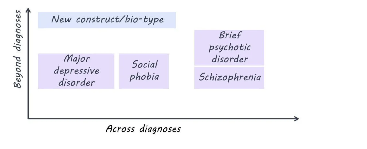
For example, one study that looked at both schizophrenia and bipolar depression found three distinct “biotypes” that were spread across the two diagnoses. One biotype showed marked cognitive impairments and lowered sensory reactivity, another showed some cognitive impairment but heightened sensory reactivity, and a third group had cognitive and sensory features comparable to the non-patient group. Knowing someone’s diagnosis doesn’t help very much in predicting what their biotype is, so these biotypes could be considered transdiagnostic.

What’s new?

Transdiagnostic research is gaining momentum, with large groups of researchers combining their efforts to try and find a new classification system. However, some are critical of the recent flood of research. A systematic review by Fusar-Poli and colleagues found that only three out of the 111 studies they examined were truly transdiagnostic. Less than half of self-titled “transdiagnostic” studies compared their new classifications directly to the current gold standard diagnoses, and when they did, the transdiagnostic approach often performed no differently than the current diagnosis-based approach. Twenty percent of the studies were not transdiagnostic at all. The authors came up with guidelines for future transdiagnostic research to guide the growing field.

What's the bottom line?

It’s obvious that the current diagnostic system in psychiatry is imperfect, but it’s the best we have so far. While there are some good examples of transdiagnostic research out there, the field needs to be rigorous and insightful if it ever hopes to replace the DSM and ICD.

References +

Dalgleish, T., Black, M., Johnston, D., & Bevan, A. (2020). Transdiagnostic approaches to mental health problems: Current status and future directions. Journal of Consulting and Clinical Psychology, 88(3), 179–195. https://doi.org/10.1037/ccp0000482

Fusar‐Poli, P., Solmi, M., Brondino, N., Davies, C., Chae, C., Politi, P., Borgwardt, S., Lawrie, S. M., Parnas, J., & McGuire, P. (2019). Transdiagnostic psychiatry: A systematic review. World Psychiatry, 18(2), 192–207. https://doi.org/10.1002/wps.20631

Clementz, B. A., Parker, D. A., Trotti, R. L., McDowell, J. E., Keedy, S. K., Keshavan, M. S., Pearlson, G. D., Gershon, E. S., Ivleva, E. I., Huang, L.-Y., Hill, S. K., Sweeney, J. A., Thomas, O., Hudgens-Haney, M., Gibbons, R. D., & Tamminga, C. A. (2022). Psychosis Biotypes: Replication and Validation from the B-SNIP Consortium. Schizophrenia Bulletin, 48(1), 56–68. https://doi.org/10.1093/schbul/sbab090


The Role of Dopamine in Forgetting Competing Information

Post by Shannon Kelly

The takeaway

Actively forgetting memories that compete with useful memories is important for our ability to remember information that supports our goals. This study found that the neurotransmitter dopamine in the prefrontal cortex plays a critical role in the active forgetting of competing memories.

What's the science?

Unlike the passive decay of memories that are not used over time, active forgetting is a process by which distracting memories are selectively forgotten to support our ability to retrieve useful memories. Active forgetting can happen when we retrieve certain memories and must inhibit competing memories (i.e., retrieval-induced forgetting). Although the neurotransmitter dopamine is known to play an important role in skills that relate to active forgetting, such as the ability to flexibly change behaviors, its direct involvement in retrieval-induced forgetting is not well understood. This week in The Journal of Neuroscience, Gallo and colleagues found that active forgetting in rats was hindered by reducing the brain’s ability to respond to the presence of dopamine and improved by increasing dopamine-related activity in the prefrontal cortex.

How did they do it?

The authors tested the effects of dopamine-related drugs on rats’ memory. They first showed rats pairs of objects to remember, then injected either harmless salt water or a drug that affects dopamine-related activity in the medial prefrontal cortex (mPFC; similar to the dorsolateral prefrontal cortex in humans). In the retrieval condition, rats then were shown pairs of one previously seen object and one new object so that they used their memory to guide their exploration. In the control conditions, rats either were shown pairs of two new objects or rested for the same amount of time. Finally, they showed the rats a pair of one new and one previously seen object and measured how long the rats examined each object. Since rats are known to prefer new objects, a large difference in time spent exploring the new object compared to the old one indicated that the rats remembered the previously seen object. If the rats preferred the new object in both control conditions but showed no or less preference for the new object in the retrieval condition, this indicated that the rats showed retrieval-induced forgetting of the previously seen object. The authors tested the effects of (1) a drug that decreases reactivity to dopamine by blocking dopamine receptors, (2) a drug that inhibits dopamine release into the mPFC from the ventral tegmental area (VTA), and (3) a drug that activates dopamine receptors on retrieval-induced forgetting.

What did they find?

Rats that were injected with either the drug that blocks dopamine receptors or the drug that suppresses the release of dopamine from VTA into mPFC showed no difference in behavior between the retrieval and control conditions, indicating no retrieval-induced forgetting. These findings show that dopamine-related activity in the mPFC is necessary for active forgetting of competing memories. For rats whose dopamine release from VTA was suppressed, retrieval-induced forgetting was improved back to normal levels when given the drug that activates dopamine receptors in mPFC. Finally, rats given only the dopamine-enhancing drug showed better retrieval-induced forgetting compared to control rats during a more difficult version of the memory test. Together, these findings show that active forgetting can be causally improved or hindered by increasing or decreasing dopamine-related activity in mPFC.

What's the impact?

This study showed that the ability to selectively forget distracting information can be directly influenced by affecting the level of dopamine-related activity in the prefrontal cortex of rats. These findings clarify the mechanisms by which experience helps determine which memories are maintained and which are forgotten. Based on findings that active forgetting involves similar brain mechanisms in humans as in rats, these findings suggest that prefrontal dopamine plays a critical role in our ability to adapt our memories based on the demands of our environments.

Access the original scientific publication here.

Thermal Pain Sensitivity is Driven by Circadian Rhythms

Post by Lincoln Tracy

The takeaway

Keeping patients awake for an extended period in a highly controlled environment reveals thermal pain sensitivity is driven by circadian rhythms, with the peak in pain sensitivity occurring between 3:00-4:30 am.

What's the science?

How intense we perceive a painful sensation to be, changes throughout the day. It’s possible this variation is related to our internal circadian clock (or rhythm), but the exact reason is unknown. This week in Brain, Daguet, and colleagues used a highly-controlled constant-routine protocol to determine whether sensitivity to thermal pain displays rhythmic changes over a 24-hour period and assess the specific contributions of the circadian clock and other sleep-related processes to the potential change in thermal pain sensitivity.

How did they do it?

The authors recruited 12 healthy males (mean age 22.7 years) who maintained a regular sleep/wake schedule for three weeks before entering the laboratory to complete the 56-hour experimental protocol. The participants arrived at about 10 am on the first day and familiarized themselves with the dark laboratory environment which was free of external time cues (e.g., clocks, television, and visitors). Participants slept for eight hours in darkness as per their normal sleeping habits. Once they awoke on the second day, participants were kept awake for the next 34 hours until the end of the experiment (approximately 6 pm on day three). Salivary melatonin levels were assessed hourly during the 34-hour awake period; thermal heat pain sensitivity, body temperature, and heart rate were evaluated every two hours. Specifically, the authors tested participants’ heat detection threshold (the point at which they felt a warm sensation), heat pain threshold (the point at which the stimulus became painful), and sensitivity to 42°C, 44°C, and 46°C stimuli at these intervals. The authors then explored if and how pain sensitivity and the collected physiological measures changed over time.   

What did they find?

First, the authors found that pain sensitivity increased with sleep debt, with the 44°C and 46°C thermal stimuli (but not the 42°C stimuli) being rated as more painful the longer the participant was kept awake. This suggests the known relationship between sleep deprivation and pain sensitivity may not apply to lower levels of pain. Second, they found pain sensitivity is driven primarily by circadian rhythms, rather than by sleep pressure. Instead of simply steadily increasing as sleep deprivation increased over the 34-hour time period, the peak in pain sensitivity occurred between 3:00 am and 4:30 am. Third, they found the peak in pain sensitivity occurred at a similar time to the troughs (low point) in body temperature and heart rate (3:00 and 2:00 am, respectively). In addition, pain sensitivity peaked one and a half hours after the peak in melatonin secretion (2:00 am). These findings imply a phase relationship between the circadian components of pain modulation and other physiological rhythms.

What's the impact?

The results of this study demonstrate that pain sensitivity is driven by our circadian timing system and that sleep (deprivation) has less of an influence on pain sensitivity than previously thought. Further research is required to identify the neural pathways linking circadian rhythms to pain perception. These findings suggest the effectiveness of pain relief could be optimized using circadian medicine, administering treatments based on the patient’s internal time.

Access the original scientific publication here.