Using Brain Lesions to Identify a Common Addiction Brain Circuit

Post by Megan McCullough

The takeaway

Brain lesions brought on by brain damage from events such as strokes have been known to interrupt addiction in some individuals with substance use disorders and even lead to remission. Lesion mapping shows that these regions connect to a specific brain circuit that can be used as a therapeutic target for the treatment of addiction. 

What's the science?

Substance use disorders (SUDs) are a leading cause of death in young populations. Neuromodulation of brain regions implicated in addiction —  a current therapy for addiction — involves delivering electrical stimuli to induce changes in specific neural circuits. However, there is a need to better identify therapeutic targets for neuromodulation. Previous research has shown that brain damage from strokes has led to addiction remission in some individuals with SUD. This week in Nature Medicine, Joutsa and colleagues aimed to link brain lesions that resulted in addiction remission to the human connectome in order to gain a better understanding of the specific brain regions involved in addiction remission.

How did they do it?

The authors examined data from 129 patients who were daily nicotine smokers at the time they acquired a brain lesion, with a subset of these patients entering addiction remission right after the lesion. Lesion locations within the patients entering addiction remission were analyzed to identify the specific brain regions connected with remission. The authors next tested if lesions within the addiction remission cohort mapped to a specific brain circuit rather than a single brain region. Utilizing lesion network mapping, connectivity patterns between lesion locations were then compared across groups: those entering remission versus non-quitters. Finally, the authors looked at which brain voxels [a location in three-dimensional space] have a similar connectivity profile to the profiles of the lesion locations that led to addiction disruption.

What did they find?

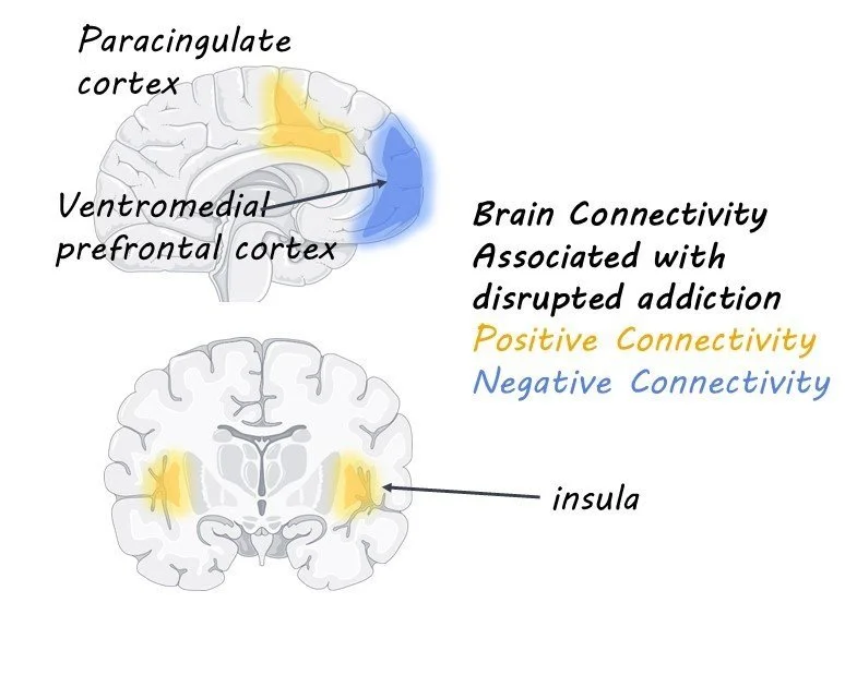
The authors found that lesions that led to a disruption in nicotine addiction occurred in multiple different brain regions. These data suggest that there is no one specific brain region implicated in addiction remission. However, the lesions all had the same pattern of connectivity to brain regions implicated in models of addiction. Brain regions that best matched the connectivity pattern of the lesions included the frontal operculum and paracingulate cortex. This study helps explain why some previous studies found that lesions to the insula are more likely to lead to remission while others have not replicated those results; lesions map to specific brain circuits, not specific regions.

What's the impact?

This study is the first to show that brain lesions that disrupt addiction map to specific brain circuits. This work has therapeutic potential, as neuromodulation techniques such as DBS and TMS may be most effective if they target brain regions with the same connectivity profile as the lesions that disrupted addiction in this study. This study provides targets for therapeutic neuromodulation, which has the potential to induce remission in those with SUDs.

Sleep Health and Emotional Reactivity

Post by Anastasia Sares

The takeaway

In a study of over 26,000 participants, emotional reactivity was associated with sleep duration, a result that adds to the conversation on sleep hygiene and mental health.

What's the science?

Previous research has established a relationship between sleep patterns and mental health issues like depression and anxiety. People with insomnia have shown altered processing of emotional stimuli, and a similar effect can be found in people who are temporarily deprived of sleep in an experimental setting. However, there are a number of ways to measure sleep quantity and quality, which are not applied consistently.

This week in Biological Psychiatry, Schiel and colleagues used a massive dataset to test whether emotional reactivity was related to different aspects of sleep health.

How did they do it?

The authors used data from the UK Biobank, a large study that has been collecting MRI data since 2014. Before accessing the data, they preregistered the study—meaning they submitted a publicly accessible document that outlined their hypotheses and planned analyses. Preregistering is a modern process that is increasingly recommended to counteract publication bias: either publishing results that were not hypothesized as if they had been, or failing to publish hypothesized results because they do not turn out to be true.

The authors were interested in the activity of the amygdala, a small almond-shaped region deep in the brain that responds to fearful or negative stimuli. Biobank participants had done an experiment while in the MRI where they were shown images of negative facial expressions (like anger). The authors isolated the amygdala and measured its reactivity to these negative stimuli, and then tried to see if any aspects of the participants’ sleep health correlated with this reactivity. Measures of sleep health included duration of sleep, insomnia, daytime sleepiness, and chronotype (see a previous brainpost for details on what chronotype is).

What did they find?

In this large sample, only sleep duration over the long term was associated with amygdala reactivity—people with shorter sleep duration had lower reactivity. This result was not intuitive, and it contradicted the authors’ original hypothesis, which was based on previous studies about amygdala reactivity (these other studies found that short-term sleep deprivation results in increased amygdala reactivity). The authors proposed that the decrease in emotional reactivity could be a sort of blunting effect—that is, people who habitually get less than 7 hours of sleep lower their overall amygdala reactivity so that they won’t suffer emotional fatigue. Interestingly, insomnia was not related to amygdala reactivity in this large sample, though previous studies with fewer people had found this association.

What's the impact?

This study confirmed that there is a link between sleep and emotion regulation, however, the exact nature of the relationship was unexpected given the previous literature. This shows why it can be beneficial to preregister research, so we can see when the results of a study diverge from our predictions.

Access the original scientific publication here.

How Sleep Facilitates Relational Associative Memory

Post by Andrew Vo

The takeaway

Sleep plays a critical role in our ability to make connections among indirectly learned but overlapping items in our memories. Building a theoretical neural model that can learn and simulate sleep states revealed specific mechanisms by which sleep may improve associative memory.

What's the science?

Relational memory refers to the ability to form associations between individual items. An important feature of relational memory is transitive inference, the ability to form associations between indirectly learned but overlapping items. For example, indirectly learning that A→C after directly learning that A→B and B→C. Previous research has suggested that sleep is important in forming such memories. The exact mechanisms underlying this process are not entirely clear, however. This week in Journal of Neuroscience, Tadros and Bazheno build a thalamocortical network to test how sleep might strengthen relational memories.

How did they do it?

The authors built a computer model of a thalamocortical network that could learn a relational memory task as well as simulate awake/sleep states. The network contained a cortex, composed of two layers of neurons that represented the primary visual cortex and associative cortex, and thalamus. Excitatory and inhibitory connections among neurons were randomly modelled. Network states were simulated by changing levels of neuromodulators, such as acetylcholine and GABA, until neuron firing rates became characteristic of different sleep cycles.

The relational memory task was comprised of three stages: supervised training, unsupervised training, and sleep. During supervised training, each of six individual items was stimulated in cortical layer 1 followed by layer 2, forming network pathways that represented these items. Next, during unsupervised learning, pairs of items were stimulated at the same time to induce associative learning between individual items. During a sleep phase, neuromodulator levels were changed to simulate slow oscillation neuron firing typical of slow-wave sleep and no stimulation was provided. Finally, the network’s ability to learn and recall direct and indirect relational memories following sleep was tested by stimulating each individual item in layer 1 and measuring responses in layer 2.

What did they find?

The authors found that following supervised training, stimulation of a given item in layer 1 induced corresponding activity for that item measured in layer 2. After unsupervised training, they observed an increase in direct but not indirect relational memories. Stimulation of a given item in layer 1 led to activity in layer 2 that corresponded only to directly associated items. It was only after a sleep phase that the network demonstrated increases in indirect relational memories. Stimulation of a given item in layer 1 now produced activity in layer 2 corresponding to indirectly associated items. This sleep-related improvement in relational memory was modulated by the length of training and the duration of sleep. These findings reveal that sleep is necessary for the formation of indirect relational memories.

To investigate the exact mechanism by which sleep can improve relational memory, the authors looked specifically at neuron spiking events during slow oscillations that were considered replay events. These replay events are related to the reactivation and consolidation of a memory. The authors found that the number of replay events was correlated with the strengthening of neural connections, suggesting that the replay of memory traces during sleep underlies the formation of not only direct but also indirect relational memories.

What's the impact?

The present study highlights the importance of sleep in our ability to form connections between indirectly learned but overlapping memories. It also demonstrates how building models of the brain that simulate different states can allow for controlled testing of specific hypotheses about human cognition.

Access the original scientific publication here.