Mental Disengagement From Navigation Degrades Spatial Codes for the Environment

Post by Lani Cupo

The takeaway

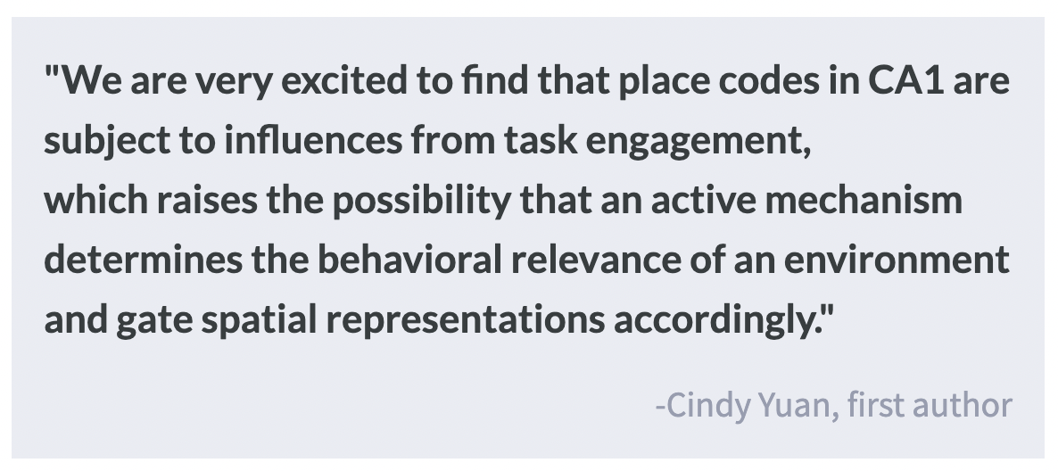
When animals navigate an environment, the hippocampus forms a spatial code based on sensory cues and motion. Mentally disengaging from navigation leads to the degradation of place codes, even when physical motion through the environment is still occurring, suggesting that internal state, not just external information, is critical to forming a spatial code.

What's the science?

Previous research in rodent navigation has established the existence of “place cells” — neurons in the hippocampus that respond to specific positions or directions in the external environment that allow animals to navigate. Specific patterns of activity among these neurons can be viewed as a spatial code, however, it is yet unknown how mental engagement impacts the activation of these spatial codes. This week in Nature Neuroscience, Pettit, Yuan, and colleagues investigated the role of mental engagement in activating spatial codes in mice by examining their behavior and neuronal activity during reward-based navigation tasks in a virtual environment. 

How did they do it?

The authors constructed a virtual environment for male adult mice, with each mouse placed on top of a spherical treadmill. The heads of the mice were restrained to allow for concurrent cellular imaging, however the mice could freely rotate the treadmill. Motion was captured with optical sensors and the information was relayed to a projection on a screen in front of the mouse’s head displaying an environment with visual cues that the mice were trained to recognize. Mice could achieve water rewards in certain “reward zones” of the virtual environment by licking a spout in front of them. Cell imaging was achieved with a method known as two-photon microscopy: lasers are shined on cells, and, because the mouse strains express fluorescent calcium indicators in neurons, light is emitted back upon neuronal activation and can be recorded during waking behavior. This allows the researchers to examine when neurons fire, linking neuronal activation with mouse behavior. In this study, the authors quantified the degree to which mice were engaged or disengaged via lick-based metrics, based on how spatially selective and abundant mouse licks were.

What did they find?

While some sessions included almost only engaged trials, other sessions included larger proportions of disengaged trials, and these usually occurred together at the end of sessions, indicating that mice switched from engaged to disengaged behavior. This could indicate satiety as they received about 1 mL of water. During engaged sessions, neural activity formed specific sequences or place codes. However, the activity of the population of cells differed during disengaged trials. Regardless of whether the trial was engaged or disengaged, the mouse was moving through the environment, indicating that the change in neural activity was not due to placement in the environment, but was instead associated with the mouse’s altered behavior reflecting mental disengagement, even when matching trials on variables such as running speed. Furthermore, when considering the activity of all neurons at a population level, there was no difference between engaged and disengaged trials, suggesting a degradation of the spatial code, not just a general alteration in neural activity. Finally, by examining neuronal activity in streaks of engaged and disengaged trials, the authors found that the shift in activity happened in less than a minute.

What's the impact?

This study found that the hippocampal place codes that are associated with rodent navigation of the environment degrade when mice mentally disengage from a goal-directed task. The authors’ findings suggest that beyond sensory cues and motion information, mental engagement is required to establish hippocampal spatial maps of the surrounding environment. These findings challenge the established idea that spatial maps form automatically in rodent hippocampi and demonstrate that internal state impacts neural encoding of the external environment.

The Anterior Cingulate Cortex Signals the Need to Control Intrusive Thoughts

Post by Leanna Kalinowski

The takeaway

The human brain has evolved mechanisms that enable it to suppress unwanted memories from coming to mind. This study identified a crucial role for the anterior cingulate cortex in detecting and responding to these intrusive thoughts.

What's the science?

Recalling an unwanted memory can be distressing, but luckily, the human brain has adapted to be able to prevent such memories from coming to mind. When such memories intrude, the brain detects a need for control, which engages the prefrontal cortex to inhibit activity in the hippocampus, which then stops retrieval of the unwanted memory. However, it is unknown which brain region is responsible for detecting and coordinating a response to this need for control. During non-memory contexts (e.g., moments of surprise), many scientists believe that the anterior cingulate cortex (ACC) coordinates a need for control, but this region’s role in inhibiting unwanted memories has not yet been examined. This week in the Journal of Neuroscience, Crespo García and colleagues tested the role of the ACC in processing and preventing unwanted memories.

How did they do it?

Twenty-four participants first underwent the “study phase”. Here, they were tasked with studying 64 pairs of words that were shown side-by-side on a computer screen for 5 seconds each. After being shown each word pair, they were given one word from each pair and were asked to recall its associated word. Each participant repeated the study phase until they were able to correctly recall at least 50% of the words, and the words that they recalled were then used in the subsequent phase.

Participants next underwent the “Think, No-Think (TNT) phase”. This phase consisted of six blocks, each containing 80 words. During “Think” trials, a word appeared with a green frame around it, and participants were asked to recall and think about its associated word. During “No-Think” trials, a word appeared with a red frame around it, and participants were asked to pay full attention to the word on the screen but to actively prevent the associated word from entering their memory. During this phase, participants underwent functional magnetic resonance imaging (fMRI) scans and electroencephalography (EEG) recordings to measure brain activity.

Finally, they performed two types of tests: a same probe test, in which they were shown a word and were asked to say out loud its associated word, and an independent probe test, where they were given a category and were asked to say out loud any word from the original list that was within this category (e.g., category = “vehicle”).

What did they find?

From the EEG recordings, the researchers found that theta signals in the ACC play a role in detecting a need for memory control, particularly during two key time points. The first time point was early in the “No-Think” trials, which suggests a proactive control prior to the unwanted memory. This signal was associated with reduced activity (via fMRI) in the hippocampus, ACC, and prefrontal cortex, suggesting that the suppression of memory early on led to a decrease in demand for activity from these brain regions. The second time point was later into the “No-Think” trial, which suggests a reactive response following the successful intrusion of the unwanted memories. This was associated with increased communication from the ACC, to the prefrontal cortex, to the hippocampus, suggesting that these brain regions work together to facilitate forgetting of the unwanted memory.

What's the impact?

This study showed that the ACC not only detects the need for memory control but also responds proactively and reactively to unwanted memories by triggering other brain regions that are necessary for memory processing. During instances where unwanted memories still emerge, the ACC communicates with the prefrontal cortex, and these regions then work together to inhibit the hippocampus and suppress the intrusive thought.

The Effects of Living in High Crime Neighborhoods on Neonatal Brain Function

Post by Elisa Guma

The takeaway

Living in neighborhoods with high rates of crime has an impact on neonatal brain activity at birth, and this is mediated by maternal psychosocial stress. Weaker neonatal brain connectivity in some areas of the brain is directly associated with higher crime rates.

What's the science?

Maternal exposure to stress and adversity during pregnancy has been shown to impact fetal brain development, with lasting effects in postnatal life. Exposure to adversity may increase mothers’ stress and inflammatory factors, which may cross the placenta and interfere with fetal development. Previous work has found that mothers living in high-crime neighborhoods may not only have an increased risk of being victims of crime (a significant risk factor) but also experience higher levels of psychological stress. This week in Biological Psychiatry, Brady and colleagues investigate the relationship between neighborhood crime and neonatal brain function at birth.

How did they do it?

Mother-infant pairs were recruited as part of a larger birth-cohort study at Washington University in St. Louis. Shortly after birth, fed and swaddled neonates, underwent T2-weighted structural magnetic resonance and resting-state functional imaging. Reliable fetal brain networks were identified and connections between limbic regions (amygdala, hippocampus, thalamus) and frontal networks (anterior default mode network, anterior frontal-parietal network) were examined.

Crime data was obtained from a commercial database with data from law enforcement agencies and sorted into two categories: violent (i.e., crimes committed against persons such as murder, rape, robbery, and aggravated assault) or property crimes (i.e., burglary, larceny, and motor vehicle theft). Maternal addresses at birth were used to determine their block level crime exposure.  

The authors also assessed potential protective actors or additive factors of socioeconomic status, referred to as “advantage”. This included family income relative to household size, insurance status, maternal education, area deprivation index (based on census data ranking neighborhood socioeconomic status based on income, education, employment, and housing quality), and maternal nutrition. At each trimester during pregnancy, measures of psychosocial stress were obtained, including measures of maternal depression, lifetime stressor exposure, and racial discrimination.  

Researchers investigated the effects of prenatal exposure to crime on fronto-limbic connectivity as alterations in their connectivity have been previously associated with violent crime exposure, correcting for “advantage”. Potential mediating effects of maternal psychosocial stress were also investigated.

What did they find?

The authors found that disadvantaged mothers tended to live in areas with higher crime rates, but were widely distributed across both safe and dangerous neighborhoods, whereas advantaged mothers almost exclusively lived in areas with lower crime levels. Additionally, the women living in higher crime neighborhoods had higher psychosocial stress during pregnancy. 

Exposure to violent and property crime during pregnancy, when correcting for “advantage”, was related to weaker connectivity between neonates' thalamus and anterior default mode network. Exposure to violent crime was additionally related to weaker amygdala-hippocampus connectivity. Furthermore, maternal psychosocial stress partially mediated the direct associations between the thalamus and anterior default mode network for both violent and property crimes.

What's the impact?

These important findings indicate that living in neighborhoods with objectively higher violent and property crime rates may have specific effects on neonatal brain function above and beyond those due to living in impoverished areas. Additionally, these effects may be mediated by maternal psychosocial stress associated with these circumstances. The work presented here sheds light on the pervasive, intergenerational effects of crime, which disproportionately impact minority communities. Future work should investigate the long-term impact of this prenatal exposure.