Amyloid Versus Tau Proteins in the Path to Alzheimer’s Disease

Post by Anastasia Sares

The takeaway

The two main hallmarks of Alzheimer’s disease in the brain are plaques, made from proteins called amyloid-beta, and neurofibrillary tangles, made from proteins called tau. Brain activity and performance on memory tasks depend on the levels of both of these proteins in the brain, supporting the idea that amyloid is related to disease onset, while tau is related to disease progression.

What's the science?

In recent years, the field of Alzheimer’s research has hit a wall. Both amyloid and tau proteins are correlated with having the disease, but many researchers had guessed that amyloid proteins were the key, and that tau tangles were just a downstream effect. Some animal models of Alzheimer’s were created so that they naturally over-produced amyloid, which seemed to recreate the disease state. From work on these animals, treatments were developed that targeted only amyloid, but when these treatments were tried on humans in clinical trials, they didn’t work as well as people hoped. Now, researchers are re-examining the role of tau proteins in Alzheimer’s. This week in Brain, Düzel and colleagues showed how amyloid and tau status can interact in humans without Alzheimer’s dementia, affecting memory and brain activity. 

How did they do it?

Participants gave samples of cerebrospinal fluid (the fluid that bathes the brain and central nervous system) so that the researchers could determine the amount of amyloid and tau proteins present in each person. Their brain activity was also recorded with MRI while they performed a memory test about recognizing familiar scenes. There were three groups of participants: people with mild cognitive impairment but without Alzheimer’s disease, people who complained of cognitive decline but nevertheless had a good memory, and healthy controls. With this sample, the researchers were able to get a variety of levels of amyloid and tau, both in individuals with cognitive decline and those without.

What did they find?

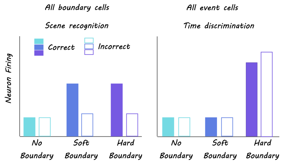
The researchers found that both memory performance and brain activity were predicted by an interaction between amyloid and tau. In “amyloid-negative” groups, tau levels were not related to memory performance, while in “amyloid-positive” groups, tau levels were related to memory performance. This result was also consistent with a paper-and-pencil test of memory recall and held true even with different statistical corrections (for example, accounting for group, age, sex, site of testing, etc.). This same interaction also showed up in brain activity during the tasks, specifically in the hippocampus and surrounding entorhinal cortex, key brain regions for forming new memories and recognizing what is familiar versus what is new. Individuals with a well-known genetic variant (APOE4) related to Alzheimer’s did not significantly differ from those without this variant.

What's the impact?

The findings of this study will help researchers in the field of Alzheimer’s to decide between competing theories of the disease, one of which is an “amyloid x tau” model. Once we have a correct understanding of the cause of Alzheimer’s in humans, we can look for treatments much more effectively.

The Brain Dynamically Changes Size Throughout Life

Post by D. Chloe Chung

The takeaway

Researchers have created a growth chart of the human brain, reflecting how the brain changes size throughout the lifespan. This chart can be used as a reference tool for the neuroimaging and clinical communities.

What's the science?

The advancement of neuroimaging techniques such as magnetic resonance imaging (MRI) has helped many clinicians, patients, and researchers over the past decades. However, unlike how we understand the change of height and weight throughout our lives, there has been no standard reference of what the brain looks like at a certain age. An inclusive map that describes developmental milestones and aging-related changes of the brain will benefit both researchers and clinicians. This week in Nature, Bethlehem and colleagues comprehensively examined how our brain dynamically changes its size throughout our lifespan by analyzing more than 100,000 MRI scans of the brain.

How did they do it?

The authors collected 123,984 MRI scans from 101,457 human participants (with or without medical conditions), aged from 16 weeks after conception to 100 years old. These brain scans were obtained from both primary studies and publicly available open databases. To create the brain size chart over the lifespan, the scans were quantified for structural changes in the brain and how fast these changes occur through aging. For this analysis, the authors adapted a modeling approach recommended by the World Health Organization that can help neutralize differences in measurement derived from diverse techniques and machines used across studies. The final brain chart was made into an interactive tool that can be used to analyze additional MRI datasets generated by tool users in the future.

What did they find?

The authors found that, up to 6 years of age, grey matter rapidly increases in its volume and thickness. The white matter volume also showed strong growth during early childhood but in a more delayed fashion than grey matter, peaking in size at around 30 years of age. The authors noted that these changes early in brain development highlight grey/white matter volume differentiation. After their respective peaks, both grey and white matter volume began to decrease over the rest of the lifespan. In contrast to these early developmental milestones, the authors found that the amount of cerebrospinal fluid in the brain ventricles that maintains its plateau throughout life starts to exponentially increase from around 60 years of age. In addition to defining developmental milestones using the brain chart, the authors demonstrated the utility of their brain chart in studying brain-related conditions. For example, the authors observed a faster decrease of the grey matter volume in Alzheimer’s disease patients, especially those who are biologically female, compared to non-patients of the same age.

What’s the impact?

This study generated a comprehensive growth chart of the human brain by examining the largest collection of MRI brain scans to date covering a 100-year age range. This brain chart will serve as a highly useful, standardized reference for neuroimaging in the future. The authors pointed out that even this brain chart is not inclusive enough as it covers mostly European and North American populations because neuroimaging tools are not as readily available to all global communities. Future studies will hopefully improve demographic and socioeconomic diversity in MRI research.

Neurons Detect Cognitive Boundaries to Separate Memories

Post by Andrew Vo

The takeaway

We experience our lives as a continuous stream that is organized and stored in our memories as discrete events separated by cognitive boundaries. A neural mechanism in the medial temporal lobe (MTL) detects such boundaries as we experience them and allows us to remember the ‘what’ and ‘when’ of our memories.

What's the science?

How is our continuous experience of the world transformed into discrete events separated by boundaries in our memories? Whereas we have a clear understanding of how the brain encodes our spatial environments with physical boundaries, the neural mechanism by which nonspatial memories are shaped by abstract event boundaries remains unknown. This week in Nature Neuroscience, Zheng et al. recorded neuronal activity within the MTL of human epilepsy patients and tested their memories for video clips separated by different types of event boundaries.

How did they do it?

The authors recorded single-neuron activity within different regions of the MTL (including the hippocampus, amygdala, and parahippocampal gyrus) of 20 epilepsy patients as they performed a task. During an encoding phase, individuals watched 90 distinct and novel video clips that contained either no boundaries (i.e., a continuous clip), soft boundaries (i.e., cuts to different scenes within the same clip), or hard boundaries (i.e., cuts to different scenes from different clips). During a scene recognition phase, individuals were presented with single static frames (either previously presented ‘target’ clips or never-before-seen ‘foil’ clips) and asked to identify the frames as either ‘old’ or ‘new’ along with a confidence rating. During a time discrimination phase, individuals were shown two old frames side by side and asked to indicate the order they had previously appeared along with a confidence rating.

What did they find?

Scene recognition accuracy did not differ between boundary types. In contrast, time discrimination accuracy was significantly worse when discerning the order of frames separated by hard boundaries compared to soft boundaries. These findings suggest a tradeoff effect in which hard boundaries improve recognition but impair temporal order memory. The authors identified ‘boundary cells’ as those neurons in the MTL that showed firing rate increases following both soft and hard boundaries whereas ‘event cells’ were those neurons that responded only to hard but not soft boundaries. The level of boundary cell firing rate during encoding predicted later scene recognition accuracy, while the coordination of event cell activity with ongoing oscillations in the brain predicted later time discrimination performance. When examining neural state shifts (i.e., changes in the population activity across boundary-responsive neurons), larger shifts were positively related to improved recognition accuracy but negatively related to time discrimination—revealing a neural mechanism for the tradeoff between recognition and temporal order memories. 

What's the impact?

This study revealed a neural mechanism in the MTL that responded to boundaries separating discrete events and helped to shape the content and temporal order memories for these events. A particular highlight of this paper is the use of single-neuron recordings in human patients, which allows for a more direct study of memory-related brain activity compared to less invasive approaches such as functional MRI or EEG.