Loneliness Distorts Neural Representations of Social Connection

Post by Cody Walters

What’s the science?

Social connection is a key component of well-being. Social isolation and loneliness, on the other hand, are known to pose significant health risks. Despite the important role that social relationships play in one’s overall wellness, it remains unclear how the brain represents relationships between oneself and others and whether those representations are modified by loneliness. This week in The Journal of Neuroscience, Courtney and Meyer show that there are distinct neural representations stratified along social-closeness categories, with lonelier individuals having representational distortions between themselves and others.

How did they do it?

The authors used both univariate (i.e., average activity across voxels) and multivariate (i.e., multi-voxel patterns of activity; a voxel is like a 3D pixel in an image of the brain) functional magnetic resonance imaging (fMRI) analyses. Multivariate analyses typically involve training a classifier (machine learning model) to distinguish between multi-voxel activation patterns that correspond to specific stimuli. The authors used two multivariate methods: representational similarity analysis (RSA) and whole-brain searchlight analysis: RSA is a method for comparing patterns of blood-oxygen-level-dependent (BOLD) brain activity between distinct stimuli to quantify how similar (or dissimilar) they are, whereas whole-brain searchlight analysis is an approach for identifying voxelated neighborhoods that exhibit specific patterns of brain activity.

cody.png

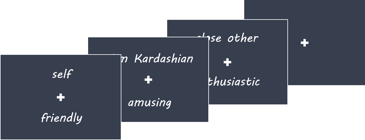
Prior to placing the participants inside the MRI scanner, the authors had participants list out and rank the names of five close others as well as five acquaintances. While in the scanner, participants fixated on a screen that displayed these target names (either their own name, one they supplied or one of five well-known celebrities) along with traits (e.g., polite, amusing, etc.). Participants then had to indicate how well the trait described that person on a scale from 1 (‘not at all’) to 4 (‘very much’).

What did they find?

The medial prefrontal cortex (MPFC) is known to represent information about the self as well as close others, thus the authors examined the activity of a predefined MPFC region of interest. Specifically, they constructed a representational dissimilarity matrix in order to test whether there was any meaningful structure in how self-other relationships are categorized in the MPFC. The authors identified that there were three representational clusters corresponding to self, social network members (i.e., close others and acquaintances combined), and celebrities. The authors then employed a whole-brain searchlight analysis to look for other brain regions that shared a similar clustering profile as the MPFC. They found that regions commonly implicated in social cognition — the posterior cingulate cortex (PCC), precuneus, middle temporal gyrus, and temporal poles — also exhibited a three-cluster structuring of self-other representations. Next, the authors investigated whether ranked closeness to the targets influenced neural responses. Restricting their analysis to the predefined MPFC region of interest, they found that mean MPFC activation linearly increased with perceived closeness to the target. The authors examined the extent to which representations of the self overlapped with representations of others. While self-other overlap did not linearly increase by target category (close others, acquaintances, and celebrities), they did find greater overlap between representations of the self and close others relative to acquaintances and celebrities. They identified the PCC/precuneus, as well as the MPFC as regions where the representations of others, were more similar to the representation corresponding to the self.

cody_image_June23.png

To examine whether loneliness modulated self-other representations, the authors used an established loneliness questionnaire. Between target categories, they found that the MPFC of individuals who reported higher loneliness represented adjacent (e.g., close others and acquaintances) and distal (e.g., close others and celebrities) targets as being more similar to one another. Furthermore, they found that within categories, acquaintances were represented more similarly to one another in both the MPFC and PCC of lonelier individuals. These data suggest that there is a blurring of representational similarity within and between social groups in lonely individuals. The authors also found that loneliness was negatively correlated with self-other similarity across all categories (close others, acquaintances, and celebrities) in the MPFC, whereas loneliness was positively correlated with self-other similarity across all categories in the PCC. These findings suggest that lonelier individuals might perceive others as being dissimilar from themselves owing to a lack of self-other representational similarity.

What’s the impact?

The authors provided evidence indicating how the brain might map out subjective social closeness in terms of representational similarity and how these representations are blurred and skewed in lonelier individuals. Developing a better understanding of how the brain processes interpersonal ties and how that processing is disrupted as a result of social isolation has implications for advancing our scientific understanding of happiness and well-being.

Courtney_quote_Jun23.jpg

Self-other representation in the social brain reflects social connection. The Journal of Neuroscience, (2020). Access the publication here.

Decoding of Natural Sounds in Congenitally Blind Individuals

Post by Stephanie Williams

What's the science?

Previous work has shown that patterns of brain activity measured with functional magnetic resonance imaging (fMRI) data can be used to classify sounds. Typically, these studies are performed with complex sounds (traffic, nature sounds) as the stimuli, and the classifiers (machine learning models) are built to predict groups of sounds. For example, fMRI could be used to predict whether an individual was listening to traffic noise or to a group of people speaking. One region that can be used for this decoding of auditory information is the early “visual” cortex (V1, V2, V3), which suggests that early visual cortex processes non-visual auditory information. Earlier work on auditory decoding in the early visual cortex was performed in sighted individuals only, leaving open the question of whether the same auditory information could be decoded from the visual cortex of blind individuals. This week in Current Biology, Vetter and colleagues show that sound decoding can be performed in both sighted and blind individuals with similar accuracy.

How did they do it?                             

The authors collected fMRI data from 8 congenitally blind individuals while they listened to three different natural scene sounds. The authors compared these data to previously published data (N=10) from sighted individuals, which was collected with similar stimuli and MRI acquisition parameters. The sounds consisted of 1) a bird singing and a stream 2) people talking without any clear semantic information and 3) traffic noise with cars and motorbikes. Participants listened to four rounds (‘runs’) of 18 randomized repetitions of the three sounds. The authors focused their analysis primarily on three visual areas called V1, V2, and V3, and further subdivided these into three eccentricities: foveal, peripheral and far peripheral regions. They also conducted some whole-brain analyses, searching on a voxel-by-voxel basis across the brain, rather than using predefined regions, for voxels that could be used to predict which sounds the subjects were listening to. The authors used multivariate pattern analysis (MVPA) to predict which of the three sounds participants were listening to based on the activity patterns derived from the fMRI data. They trained their classifier on three of the four runs and tested on the left-out fourth run for each subject. They compared their decoding accuracy results from the early visual cortex to the auditory cortex (which acted as a positive control) and motor cortex (negative control). The authors then analyzed how the sounds were represented in the eccentricity pattern across the early visual cortex. 

What did they find?

The authors successfully decoded natural sounds from the early visual cortex of congenitally blind individuals, showing that visual imagery and experience is not a prerequisite for the representation of auditory information in the early visual cortex and that there’s a similar cortical organization for auditory feedback in visual cortex between sighted and congenitally blind individuals. The authors saw both higher decoding accuracy for the early visual cortex and lower decoding accuracy for the auditory cortex in the blind group compared in the sighted group. This result indicates that visual deprivation may cause sound representation to be more distributed across the auditory and visual cortex in congenitally blind individuals. When the authors analyzed how eccentricity affected decoding results, they found that they had higher decoding accuracy for peripheral regions of visual cortex compared to foveal regions. This finding is supported by previous research showing that the peripheral visual cortex is connected to many non-visual brain regions. Interestingly, the authors point out that none of the three sounds induced a statistically significant response in the overall brain activity while listening to sounds compared to at rest in any of the 3 early visual areas. This suggests that their decoding accuracy is driven by small activity differences across voxels in each region of interest

stephanie.png

What's the impact?

The authors extend previous work on auditory decoding in the early visual cortex to include blind individuals, showing that there may be a similar organization of auditory information in the early visual cortex of both sighted and blind individuals. This study provides further evidence that the early visual cortex is involved in functions other than the feedforward processing of visual information in both sighted and blind individuals.  

Vetter_quote_Jun23.jpg

Petter et al. Decoding Natural Sounds in Early “Visual” Cortex of Congenitally Blind Individuals. (2020). Access the original scientific publication here.

Abnormal Circadian Rhythm Can Predict Parkinson’s Disease

Post by D. Chloe Chung

What's the science?

Parkinson’s disease is a debilitating neurodegenerative disease that is characterized by the loss of dopaminergic neurons in the brain in an area called the substantia nigra. In addition to severe motor symptoms, Parkinson’s patients often experience a disrupted sleep-wake cycle, sometimes early on in the disease course. However, no study has actually measured behavioral markers of circadian rhythm to find out whether disruption of the internal biological clock can precede the development of Parkinson’s disease. This week in JAMA Neurology, Leng and colleagues reported that abnormal circadian rhythm in healthy older adults can be regarded as an early sign of developing Parkinson’s disease in the future.

How did they do it?

The authors enrolled almost 3,000 healthy older males (average age of 76.3 years old) for the initial evaluation of circadian rhythm and followed up with them for the following 11 years. The participants were mostly Caucasians and lived in a community setting. At the beginning of the study, participants wore a wristband-like device that can track any movement during sleep. For a minimum of three separate 24-hour periods, the monitoring device recorded various circadian rhythm parameters of wake and rest. Sleep efficiency was determined based on the percentage of time the participants were asleep after “lights off”. Other important factors such as sleep apnea or periodic limb movement have been also taken into account. During the 11-year follow-up, participants were subject to in-person visits or questionnaires five times and reported whether they have been diagnosed with Parkinson’s disease, as well as their medication history. 

What did they find?

While none of the participants had Parkinson’s disease at the beginning of this longitudinal study, 78 out of 2930 study subjects were later diagnosed with Parkinson’s disease over the course of 11 years. After adjusting for variables such as demographics, education level, medication or substance usage, comorbidities, and baseline cognition, the authors found a strong association between the decrease in three out of four circadian rhythm parameters and the development of Parkinson’s disease later in life. Strikingly, participants who showed the most irregular circadian rhythm were three times more likely to develop Parkinson’s disease compared to those with the regular circadian rhythm. These findings indicate that decreased circadian rhythmicity can act as an important early symptom of Parkinson’s disease

PD_image_Jun23.png

What’s the impact?

This study is the first one to analyze a large cohort for a long time and reveal that circadian rhythm abnormalities in healthy adults are associated with their chance of developing Parkinson’s disease as they get older. Therefore, the authors suggest that the detection of abnormal circadian rhythm in healthy adults may help early prediction and diagnosis of Parkinson’s disease, ultimately allowing for early disease intervention. It will be interesting to further investigate whether circadian rhythm might directly contribute to the onset of Parkinson’s disease.


Leng et al. Association of circadian abnormalities in older adults with an increased risk of developing Parkinson disease (2020). Access the original scientific publication here.