Exploring the Long-Term Effects of Psychedelics on the Brain

Post by Flora Moujaes 

What's the science? 

Psilocybin, the psychoactive compound in magic mushrooms, has recently proven an effective treatment for depression, anxiety, tobacco addiction, and alcohol use disorder. Treatment with psilocybin can have long-lasting effects: 1-3 psilocybin sessions can lead to a reduction in clinical symptoms that lasts for up to one year. We still don’t fully understand the psychological and neural mechanisms that underlie psilocybin’s therapeutic effects. Molecular studies have shown that psilocybin is a serotonin 2A/5-HT2A partial agonist, while therapeutic studies have indicated that psilocybin exerts its clinical effects by reducing negative affect and increasing positive affect. The reduction in negative affect may be linked to the amygdala, the brain region responsible for tracking the salience of the stimuli in the environment. Functional magnetic resonance imaging (fMRI) studies have shown that psilocybin reduces amygdala activity when viewing negative stimuli. This week in Scientific Reports, Barrett et al. use fMRI to explore the long-term effects of psilocybin on emotional and brain plasticity in order to better leverage it as a clinical tool. 

How did they do it?

To explore the long-term effects of psilocybin, the researchers administered a single high dose of psilocybin (25mg/70kg) to twelve healthy volunteers in an open-label within-subjects pilot study. To investigate if psilocybin could lead to an enduring increase in positive affect and decrease in negative affect, a battery of self-report state and trait measures was completed one day before, one week after, and one month after psilocybin administration. Responses were then compared between time-points. At each time-point, in order to determine whether psilocybin could lead to an enduring change in neural response to emotional stimuli, participants also took part in an fMRI session in which they completed three emotion-processing tasks. fMRI analysis of the emotion-processing tasks focused on the amygdala as a key region of interest. Finally, to determine whether psilocybin could lead to an enduring change in brain plasticity, participants’ resting-state fMRI data were also collected at each time-point. Functional connectomes were then compared between timepoints.

What did they find?

Long-term behavioural effects of psilocybin on emotions: Behavioural measures indicated that one-week post-psilocybin there was a reduction in negative affect and an increase in positive affect. One month post-psilocybin, the reduction in negative affect returned to baseline levels, while positive affect remained elevated. Ratings of trait anxiety were also reduced one-month post-psilocybin, despite showing no significant reduction one-week post-psilocybin. 

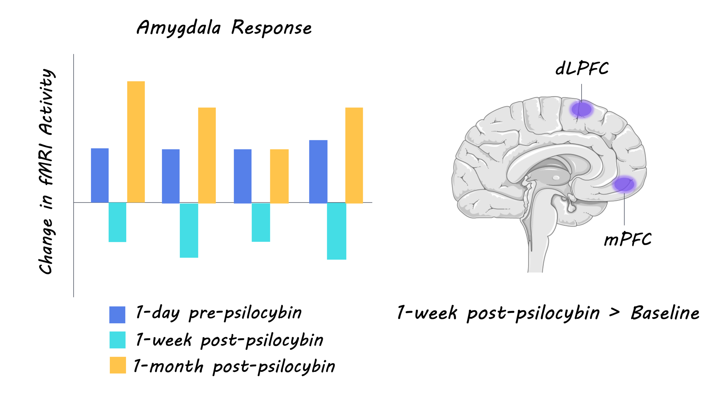
Long-term neural effects of psilocybin on emotions: Analyses of the fMRI data revealed that psilocybin led to reduced amygdala response to facial affect stimuli one-week post-psilocybin, however, this change was not sustained after one month. Overall, these results suggest that acute psilocybin administration leads to shifts in emotional affect, and the neural correlates of affective processing, which may endure one month later. fMRI data also showed that psilocybin resulted in increased dorsal lateral prefrontal and medial orbitofrontal cortex activity in response to emotionally conflicting stimuli after one week, and increased somatosensory cortex and fusiform gyrus activity in response to emotionally conflicting stimuli after one month. This indicates that psilocybin may also increase the top-down control of emotional processes, which may have a modulatory effect on the amygdala.

psilocybin_quote_Feb18.png

Long-term effects of psilocybin on brain plasticity: Global increases in functional connectivity were found both one week and one-month post-psilocybin. The increase in functional connectivity strength that was observed indiscriminately across multiple networks may reflect a domain-general cortical plasticity process that supports the observed changes in affective processing. 

What's the impact? 

Overall this study shows that despite the fact that the half-life of psilocybin is roughly 3 hours, psilocybin induced behavioural and neural changes were seen one-week and one-month post-psilocybin administration. This indicates that acute psilocybin may lead to a dynamic and neuroplastic period that lasts for a number of weeks, during which the neural processing of affective stimuli is altered. These findings also help explain psilocybin’s therapeutic effects: reduction of negative affect may undermine ruminative processes that contribute to depression and explain the antidepressant effects of psilocybin. Studies that utilize a larger sample size and placebo-controlled design are needed to explore this key neuroplastic period following acute psilocybin administration. 

Barrett et al. Emotions and brain function are altered up to one month after a single high dose of psilocybin. Scientific Reports (2020). Access the original scientific publication here.

Individual Variation in Amygdala Connectivity

Post by Deborah Joye

What's the science?

The amygdala is a part of the brain most known for its role in aggression and fear. Amygdala function is affected in many psychiatric illnesses including post-traumatic stress disorder, anxiety, depression, and phobias. Most in-depth studies of the amygdala in humans measure brain signals averaged across a group of individuals to determine the location and connections of the amygdala. While this approach has helped us to better understand the amygdala in general, it has limited our ability to tailor treatment of amygdala dysfunction to individual patients. This week in PNAS, Sylvester and colleagues use extensive functional magnetic resonance imaging (fMRI) of individuals to characterize three functional subdivisions of the amygdala and their specific patterns of connectivity with other networks in the brain.

How did they do it?

The authors analyzed over 5 hours of fMRI data per individual from 10 individuals to determine different amygdala subdivisions based on activity patterns within the amygdala and associated activity in other cortical regions. The authors then used both group-averaged and individualized data to demonstrate that group-averaged analyses can obscure the specific locations of amygdala regions and mask their functional patterns. The authors compared their amygdala subregions from the individualized dataset against a much larger independent dataset to understand whether amygdala subdivisions and their connectivity patterns were roughly consistent across people. Finally, the authors investigated possible differences in the timing of activity across the amygdala and other cortical networks to investigate whether individual differences exist in the timing of amygdala-cortex connectivity.

What did they find?

The authors characterized three subdivisions of the amygdala that are roughly consistent across individuals with some differences in spatial location. The authors also found that each subdivision of the amygdala had its own unique connections to other brain networks and that the magnitude of these connections varied from person to person. One subdivision was anatomically superior and preferentially connected to the default mode network, a widespread neural network important for reflecting on the self and others, as well as thinking of the past and future. This amygdala connection’s role could be to integrate important environmental information with an individual’s past history regarding the emotional significance of that stimuli. Another subdivision was anatomically medial in most people and preferentially connected to the dorsal attention network which is active during attention-demanding tasks. This amygdala connection’s role might be the top-down modulation of attention networks.

amygdala_connectivity_Feb11.jpg

The last subdivision was anatomically ventral and did not show a preferential connection to a specific neural network but had connectivity properties that were shared across the rest of the amygdala. When the authors compared these findings with a larger, publicly available dataset they found similar amygdala subdivisions, but the selectivity of each subdivision for particular neural networks was much weaker compared to individual analyses. Lastly, the authors found that the timing of activity between amygdala subdivisions and other neural networks was consistent across both datasets, suggesting that though location and magnitude of amygdala connections may vary from person to person, the networks themselves are consistent.

What's the impact?

This study is the first to use extensive fMRI from individuals to demonstrate that three distinct subdivisions of the amygdala are roughly consistent across people, but with important individual variation in location and magnitude of connectivity. The study also revealed that subdivisions of the amygdala can have preferential connectivity with specific neural networks, providing a framework for a more detailed understanding of how the amygdala interacts with other brain regions in individual patients. These findings could lead to improvements in personalized psychiatry and potential therapeutics for amygdala dysfunction.

amygdala_quote_Feb11.jpg

Sylvester et al., Individual-specific functional connectivity of the amygdala: A substrate for precision psychiatry, PNAS (2020). Access the original scientific publication here.

Threshold for Odor Detection Adapts Based on Past Experience

Post by Shireen Parimoo

What's the science?

Animals react to sensory input from the environment, but sometimes the input isn’t strong enough to elicit a behavioral response. How much sensory input is needed for organisms to detect it? Several models attempt to explain how external sensory information, like sound, is detected in the brain. For example, the absolute threshold model proposes that a sound will be detected once it reaches a certain intensity (i.e. the threshold). According to the derivative model, the rate at which a sound’s intensity changes will determine when it is detected, whereas the fold change model posits that detection depends on how much the sound changes in proportion to its original intensity. Although these models have been applied to explain sensory detection in various organisms and across different modalities, no study has directly compared them with each other. This week in Neuron, Levy and Bargmann used computational modeling and calcium imaging to develop a unified model for odor detection in Caenorhabdtis elegans (roundworms).

How did they do it?

Roundworms have a simple nervous system that makes it possible to record the activity of specific neurons. The authors measured the sensory activity of an olfactory neuron called AWCON in response to changes in levels of the odorant butanone. Specifically, using a microfluidic setup, AWCON calcium activity was recorded in immobilized animals across a wide range of odor concentrations and timescales. Neuronal activity and navigation decisions were also examined in animals freely moving in odor gradients controlled by a specialized microfluidic device.

The authors rigorously tested many models that predict neuronal activity features (such as neuronal response and latency of response) and navigation behavior, including the absolute threshold, derivative, and fold change models. They also created an adaptive concentration threshold (ACT) model in which sensory activity is initiated when the odor concentration reaches a threshold, however, this threshold is continuously adapting to the odor. The ACT model includes (i) a threshold constant, which changes the neuron sensitivity, and (ii) adaptation time, which determines how long is the neuron memory of the external information. To determine whether the ACT model is generalizable, it was also tested on a separate dataset of neuronal activity in zebrafish in response to visual input. To identify the molecular basis of sensory detection, they examined the role of EGL-4, a protein kinase in the AWCON neuron that is involved in olfactory learning. They compared the effect of butanone concentration in loss-of-function mutants without functional EGL-4, gain-of-function mutants with enhanced EGL-4 activity, and wild-type animals. Finally, they performed theoretical studies to determine which model can allow both accurate and fast sensory responses, two key features for sensory neurons performance. 

What did they find?

Previous models did not adequately predict the observed neuronal responses and latencies, and could only match a subset of the experimental observations. For instance, they found that calcium responses depend on butanone concentration and the rate of concentration change, inconsistent with the absolute threshold and the derivative change models. The ACT model, on the other hand, predicted neuronal responses for both slow and fast changes in butanone concentration. The ACT model also predicted neuronal activity and aversive navigation decisions, like reversals and pauses, in more natural conditions, while animals freely navigated in odor gradients. This indicates that odor sensation and navigation are driven by an adaptive threshold mechanism that allows a comparison of past and current sensory inputs.

Shireen_neuron_pic.png

Interestingly, loss of EGL-4 function elongated the threshold adaptation time relative to wild-type animals and enhancement of EGL-4 function shortened it, suggesting that the protein kinase EGL-4 tunes the adaptation time of the sensory detection threshold. The ACT model also predicted activity in the optic tectum of zebrafish in response to visual input, demonstrating generalizability. Finally, the authors show that in contrast to alternative models, an adaptive-threshold mechanism allows sensory neurons to respond both fast and accurately to external stimuli, highlighting its benefit in reliable environment sensation.  

What's the impact?

Combining computational modeling with quantitative assays, this study is the first to systematically compare previous sensation models and to demonstrate how sensory detection is driven by a combination of current and past sensory inputs from the environment. The ACT model is powerful because it encompasses elements of previous models under different conditions and further generalizes to visual stimuli. These findings pave the way for future research to uncover the neurobiological basis of sensory detection and test the generalizability of the model across organisms and sensory modalities. 

Levy & Bargmann. An adaptive-threshold mechanism for odor sensation and animal navigation. Neuron (2020). Access the original scientific publication here.