Activity in Insular Cortex Reflects Current and Future Physiological States

Post by Stephanie Williams 

What's the science?

Information related to hunger and thirst is thought to be reflected in the ongoing activity of a region called the insular cortex. Interoception — the perception of internal bodily signals such as hunger or thirst — is critical for regulating physiological states, emotion and cognition. Models of interoceptive processing suggest that the insular cortex integrates external sensory information with internal bodily sensations (related to physiological changes like heart rate, thirst or hunger) and generates ‘interoceptive predictions’, however, the neural circuitry underlying this process remains unclear. This week in Neuron, Livneh, Lowell, Andermann and colleagues show that activity patterns in insular cortex represent current and future physiological need states. 

How did they do it?                             

The authors used two-photon calcium imaging, circuit mapping and manipulations in mice to investigate representations of physiological need and predictions in insular cortex. For the behavioral task, the authors trained water-restricted mice to perform a visual discrimination task for water rewards. While the mice performed this task, the authors imaged neurons in layer 2/3 of insular cortex using a method involving a reflective microprism that acts as a kind of periscope. To ensure that their results were not affected by arousal, the authors monitored the pupil diameter of the mice, along with a myriad of other factors (eg. motion), which they accounted for in their analysis. The authors compared activity recorded during the behavioral task (tens of minutes) with activity imaged during rapid thirst quenching (2-5 min of continuous consumption). The authors also performed two-photon imaging of axons (N=257) that extended from the basolateral amygdala into insular cortex to better understand the pathway of cue-related information into insular cortex.

To further investigate ongoing insular cortex activity, the authors analyzed the interval between trials to see if the pattern of activity tracked the hydration state of the animal. The authors then trained a Naïve Bayes classifier — a model trained to predict whether an animal was in a thirsty or a quenched state from the activity patterns in insular cortex. They performed the same classification procedure on temporaly shuffled data and also on identity-shuffled data. They also trained the classifier on data collected in primary visual cortex and postrhinal cortex for comparison. Next, the authors mapped the circuits connecting specific types of hypothalamic neurons (eg. “hunger” or “thirst” neurons) to insular cortex neurons. They used retrograde tracers to trace neurons and then recorded light-evoked current in the labeled neurons to understand the connections between the hypothalamus, thalamus, amygdala, and insular cortex. Then, they manipulated the hypothalamic neurons to induce artificial thirst or to suppress thirst and recorded the corresponding changes in insular cortex activity. Finally, they then performed a loss of function experiment, and inhibited glutamatergic neurons in order to suppress thirst. To confirm their results in a separate dataset, the authors analyzed similar data collected under hunger versus sated state conditions, this time stimulating other hypothalamic neurons to induce artificial hunger. The authors artificial manipulation techniques allowed them to analyze both 1) cue-evoked activity related to water seeking and  2) the ongoing activity patterns related to the animal’s physiological state to artificial induction or suppression of thirst. 

What did they find?

The authors found that individual insular neurons responded to 1) the visual water cue 2) the onset of licking or 3) water delivery during the behavioral task. Combined with their previous work, this result suggests that insular cortex neurons respond to learned water cues. The majority of neurons that the authors recorded from responded to the visual water cue (80%) and/or actual water consumption. They found that most cue-responsive neurons, which responded to the water cue in the thirsty state, were significantly attenuated in the quenched state. They observed this trend in data collected during both rapid thirst quenching, as well as during the behavioral task. When the authors imaged axons projecting from basolateral amygdala to insular cortex, they found that single axons responded to water cues, the onset of licking, and/or water delivery. Most of the axons they recorded from responded to the water cue or water reward, while others responded to visual cues.  When the authors compared thirst and quenched states, they found distinct patterns of activity in the insular cortical activity. They also showed that these patterns were consistent across days, and that they could train a classifier to predict whether an animal was in a quenched or thirsty state. The pattern of activity across the population specifically was essential to differentiating between the states, as classification accuracy was poor when single neurons or the average population time-course was used. These results suggest that ongoing activity patterns in insular cortex represent distinct physiological states that reflect hunger or thirst. 

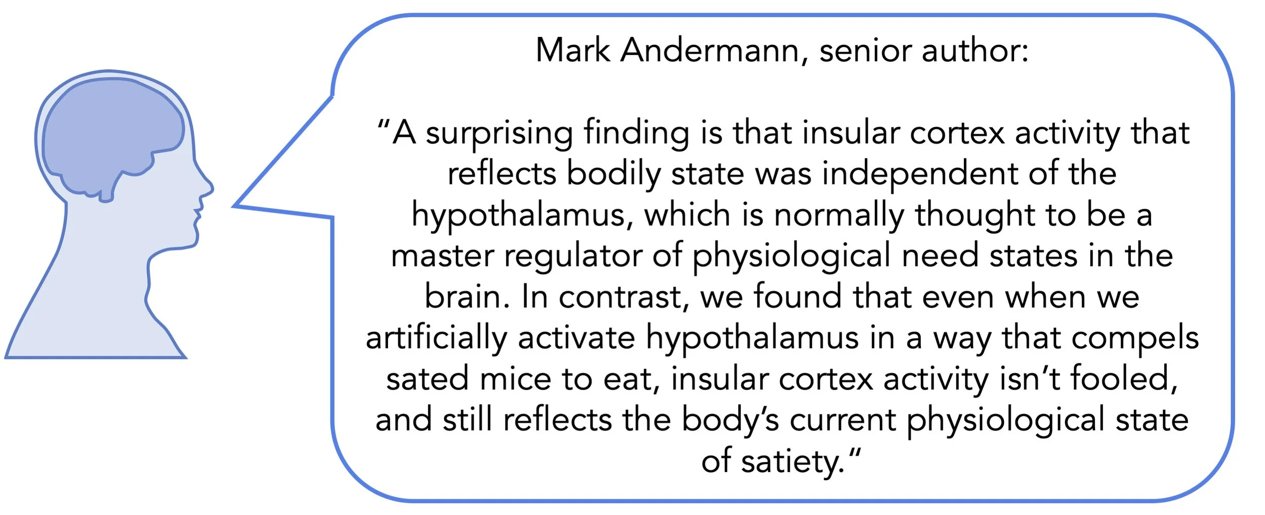
The authors also demonstrated that task events, arousal and motion could only be predicted in a small set of insular cortex neurons, suggesting that the activity of the majority of insular cortex neurons do not reflect these factors. To confirm their findings in an independent sample, the authors also classified hungry versus sated (as opposed to thirsty versus quenched) states in mice with good classification accuracy. However, they could not predict hungry versus sated states when they trained the classifier on thirsty versus quenched states from another day. They suggest that ongoing activity in insular cortex may be different for hunger vs. thirst states. The authors found that artificial activation of thirst neurons during a quenched state restored insular cortex responses to water-related cues. Similarly, artificial inhibition of hypothalamic thirst neurons reduced insular cortex response to water cues. Importantly, however, when the authors analyzed ongoing activity during artificial thirst stimulation following behavioral quenching and rehydration, they found that insular activity did not resemble the thirst-like pattern. Critically, water cues and consumption of a drop of water in the dehydrated state transiently shifted the pattern of activity towards the pattern of ongoing activity observed in the quenched state, suggesting cues led to prediction of a future quenched state

Stephanie_picture1.png

What's the impact?

The authors show that 1) ongoing activity patterns can discriminate between physiological states, and that 2) cues predicting availability of water/food may actually drive a prediction of a future satiety state in insular cortex. These findings deepen our understanding of interoceptive prediction, and will allow future studies to better understand interoceptive prediction errors.

hunger_thirst_quote_Jan21.jpg

Livneh et al. Estimation of Current and Future Physiological States in Insular Cortex. Neuron. (2020). Access the original scientific publication here.

In addition to Andermann and Livneh, authors included Arthur U. Sugden, Joseph C. Madera, Rachel A Essner, Vanessa I Flores, Jon M. Resch and Bradford B. Lowell, all of BIDMC; and Lauren A. Sugden of Duquesne University.

This work was supported by The Charles A. King Trust Postdoctoral Fellowship; Boston Nutrition Obesity Research Center P&F 2P30DK046200-26; grants from the National Institutes of Health (K99 HL144923; T32 5T32DK007516; DP2 DK105570; R01 DK109930; DP1 AT010971; R01 DK075632; R01 DK096010; R01 DK089044; R01 DK111401; P03 DK046200; and P03 DK057521); grants from the National Science Foundation (DGE1745303); Klarman Family Foundation; McKnight Foundations; Smith Family Foundation; and Pew Scholars Program.

Modeling Brain Development in Neonates

Post by Elisa Guma

What's the science?

During the perinatal period, the brain is rapidly developing, resulting in changes in size, gyrification, and contrast between tissues (as seen during brain imaging). To further complicate the situation, these changes may occur at different rates in different brain regions. This complexity makes it very challenging to accurately and reliably interpret clinical magnetic resonance imaging (MRI) data for those who may have experienced premature birth or perinatal brain injury. It is difficult to know what is abnormal for a neonatal brain given the age and clinical history of a patient. This week in Brain, O’Muircheartaigh and colleagues leveraged a large multi-contrast MRI dataset acquired during the perinatal period to model trajectories of normal brain development, and to accurately identify focal brain injury.  

How did they do it?

The authors used cross-sectionally acquired structural MRI data (T1- and T2- weighted) for 408 neonates, 189 of which were female, from the developing Human Connectome Project database, a publicly available dataset. The participants were all scanned postnatally, with post-menstrual ages ranging from 26 to 44 weeks (and gestational age at birth ranging from 23-42 weeks). A template was created from scans for 20 neonates over a wide age range based on two imaging features: T2-weighted image volume intensity and the cortical mantle. This was used to represent the midpoint for the sample’s age range. Next, the 408 scans were registered to the template, which means that the brain images were warped to match the template brain (using linear and nonlinear registration). The degree to which each voxel had to be expanded or shrunk to match the template gives us a measure of volume difference. The authors used a Gaussian process regression, which is a non-parametric approach, which they argue is a superior way to model growth curves for tissue intensity and shape at each voxel in the brain accounting for age (or degree of prematurity) and sex.

Next, the authors wanted to determine whether their model was longitudinally valid. Thus, for a subset of 46 neonates that had a second scan (excluded from the model construction), they quantified the deviation from the predicted image intensity. They then tested to see whether their model was able to detect deviations in tissue contrast that would predict the presence of punctate white matter lesions. A common brain injury associated with premature birth is a punctate white matter lesion, which is detectable using MRI. Their presence was identified in 40 neonates and manually labeled on each of the scans. Since focal abnormalities such as these lesions are reflected by deviations from typical development, the authors wanted to see if their model could accurately detect these deviations.

What did they find?

The authors were able to identify anatomically informed growth curves at each voxel in the brain, based on the MRI image intensity, with corresponding measures of variability, accounting for the degree of prematurity and sex of the infant. They observed differences in variability and shape in the growth curve based on structure; for example, the subventricular and intermediate zones were observed to have a rapid growth around term-equivalent age as they transition into white matter, whereas other white matter structures had a more linear growth, such as the sensory cortex. Interestingly, the frontal cortex had a flat curve in the perinatal period, as its development typically occurs later in life.

neonatal_img_Jan21.png

Longitudinal accuracy was also found to be high - 83% of the subset of neonates who had longitudinal scans had growth curves that matched those of the model. Accuracy, however, was not as good for younger neonates who had more intermediate structures present in their brains. The model also proved to have clinical utility as it was able to detect signal deviations due to punctate white matter lesions with high accuracy. 

What's the impact?

This study provides accurate estimates of non-linear changes in brain tissue intensity by modeling ex utero brain development over a wide age range (26 to 45 post menstrual weeks). Further, this work provides continuous growth charts for brain development based on shape and image intensity, similar to those used for height in clinical practice, providing an index that accounts for age and clinical history (i.e. prematurity). Future work may incorporate in utero MRI, and perhaps extend the postnatal period further.

Jonathan_Quote3.png

Jonathan O’Muircheartaigh et al. Modelling brain development to detect white matter injury in term and preterm born neonates. Brain (2020). Access the original scientific publication here

Surprise and Decision Making in the Anterior Cingulate Cortex

Post by Anastasia Sares

What's the science?

The anterior cingulate cortex, situated in the frontal lobe at the midline where the two hemispheres meet, is an important region of the human brain. Previous studies have connected it to error detection, cognitive control, and decision making. However, there are a few different hypotheses about what drives activity in this area. The “choice difficulty” hypothesis (CD) says that the anterior cingulate tracks difficulty—tougher decisions generate more activity. The “expected value of control” hypothesis (EVC) tracks net gain—how much reward will I get for the effort I put in? EVC predicts more activity for high reward, especially if the brain has to gear up and make an effort in order to get the reward. Finally, the “predicted response outcome” hypothesis (PRO) tracks surprise—events that violate expectations generate more activity, regardless of whether the results are perceived as “good” or “bad.” This week in Nature Human Behaviour, Vassena and colleagues pitted these three hypotheses of anterior cingulate function against each other in a speeded decision-making task.

How did they do it?

Before they started the experiment, participants learned to associate random fractal images with a certain number of points (picture 1 = 30 points, picture 2 = 80 points, etc.). They were told that the points they accumulated in the task would translate into extra money at the end of the experiment. In each trial of the task, four of the fractal images were shown on the screen. The participants had three-quarters of a second to choose either the set of images on the left or the set of images on the right. Of the images they chose, one would be randomly selected, and they would receive the equivalent amount of points. To complete this task, therefore, the participants had to be good at quickly estimating the value of the options on the right versus the options on the left. The task was completed in MRI so the response of the anterior cingulate could be measured.

Each of the hypotheses presented earlier (choice difficulty, expected value of control, and predicted response outcome) should show a different pattern of activity in the anterior cingulate during this task. CD predicts the most activity when the choice is difficult; that is when the amount of points on the left is similar to the amount on the right. EVC predicts the opposite: the anterior cingulate should be least active when the options are similar because the difference in potential reward is small compared to the effort required to distinguish between them. PRO predicts two peaks of activity, because with a large difference in value, one option is surprisingly bad, and with a small difference in value it is uncertain which option you will choose (making the choice itself “surprising”).

 What did they find?

The authors compared the activity in the anterior cingulate cortex to the three different predictions and found that the PRO model (predicted response outcome) matched most closely. This is in line with the idea that the anterior cingulate reacts to unlikely or unpredicted events (surprise). During a task like this, people are quickly able to figure out how difficult the average trial is, so when a surprisingly bad option or two very similar options appear, the anterior cingulate’s activity increases. What’s more, an extremely low or high reward at the time of feedback also activated the same area.

acc_img_Jan21.png

What's the impact?

This study tested predictions from three different hypotheses and found one to be the clear winner: the main function of the anterior cingulate is processing surprise. This is a crucial step in the scientific enterprise; after some hypotheses are generated about the natural world, they must be tested against one another in carefully designed experiments that allow us to determine which hypothesis is stronger.

ACC_quote_Jan21.jpg

Vassena et al. Surprise, value and control in anterior cingulate cortex during speeded decision-making. Nature Human Behavior (2020). Access the original scientific publication here.