The Role of Gut Microbes in Neurological Disorders

Post by Amanda McFarlan

What's the science?

Research investigating the relationship between the gut microbiota (the ensemble of microorganisms in the gut) and overall health has gained increasing popularity in recent years. Studies have shown that the gut microbiota is important for regulating many major systems of the body, including the central nervous system. This week in The Lancet Neurology, Cryan and colleagues summarized past and present research that highlights the important role of gut microbiota in regulating the central nervous system.

What do we already know?

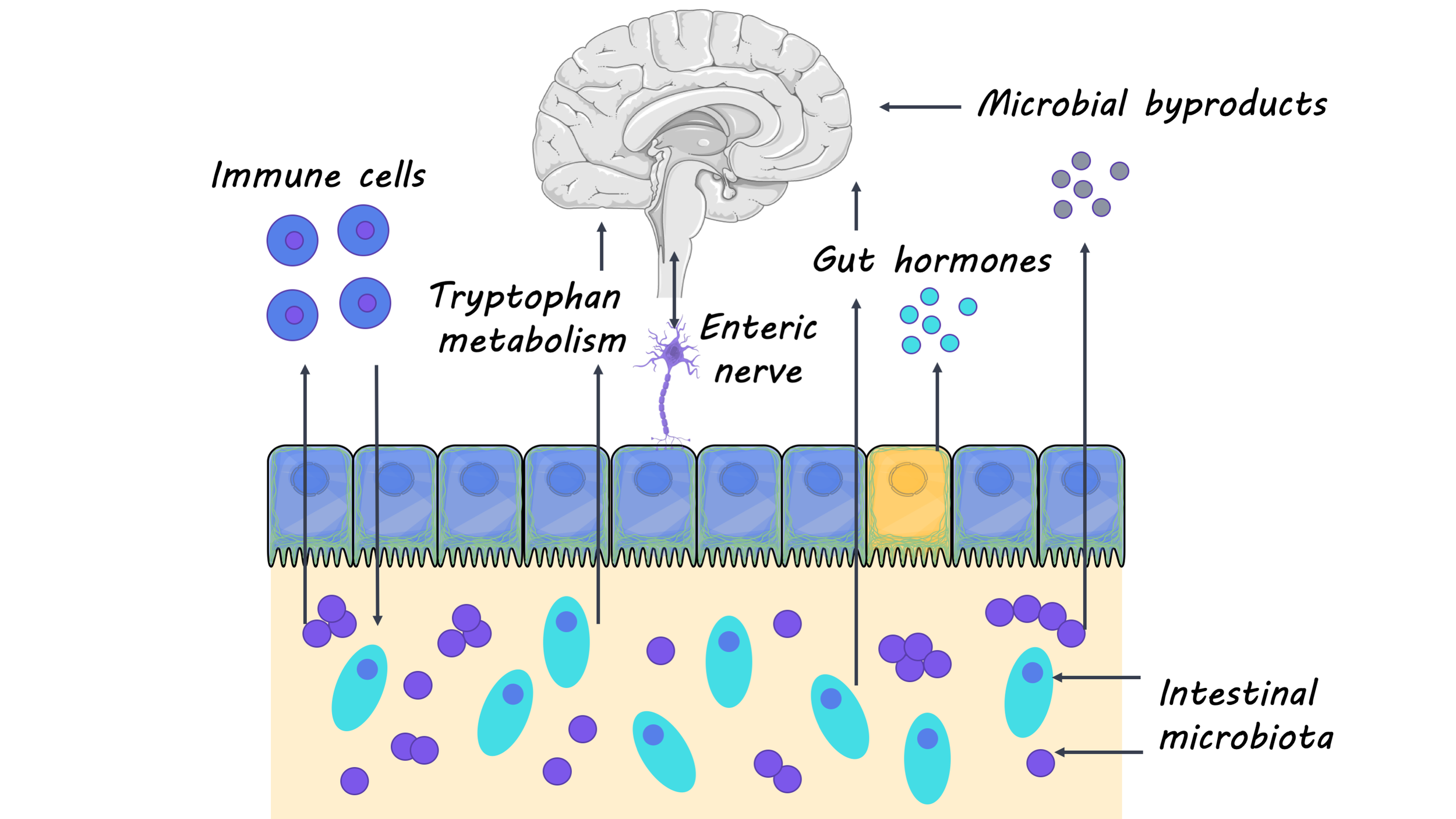
Much of our understanding of how the gut microbiota affects the central nervous system comes from studies using germ-free mice (i.e. lacking any gut microbiota). Researchers have shown that without the gut microbiota, germ-free mice have deficits in many neural processes that are critical for development and aging, including the maturation and myelination of neurons, neurogenesis and microglia activation. Additionally, germ-free mice had higher levels of neuroinflammation and an increase in the permeability of the blood-brain barrier, which are hallmarks of the immune-related neurological disease, multiple sclerosis. These studies with germ-free mice have provided researchers with important insights into how the gut microbiome is implicated in neurodevelopment, aging, and neurodegenerative disease. Researchers have continued to use animal models as well as clinical studies to further our understanding of the interaction between the gut and the brain. 

What’s new?

In recent studies, researchers have focused on how gut microorganisms can be targeted for therapeutic purposes in neurological diseases. In the case of treating multiple sclerosis, for example, researchers found that a multispecies probiotic, administered two times a day for two months, had an anti-inflammatory effect and reversed changes in gut microbiota. Moreover, a clinical trial for children with autism spectrum disorder revealed that children who received a microbiota transfer had significantly reduced gastrointestinal problems (commonly found to be co-morbid with autism spectrum disorder) as well as behavioral improvements. These findings suggest that some gut microbiota may be a potential target for the treatment of neurological disorders. Additionally, researchers have started to look at how gut microbiota may regulate the physiology and behavior of neurological disorders like Huntington’s disease, amyotrophic lateral sclerosis (ALS) and epilepsy. For example, researchers found that the gut microbiota is involved in synaptic changes that occur in brain regions that are associated with epilepsy. They also found that the therapeutic effects of a ketogenic diet on epilepsy are dependent on gut microbiota. Finally, recent studies have shown that many non-antibiotic drugs influence gut microbiota, highlighting the importance of investigating the relationship between the gut microbiome and medications (especially those that are prescribed for treating a neurological disorder).

gut_brain_Dec3.png

What's the bottom line?

The importance of gut microbiota on the development and overall health of the brain is becoming more and more evident. Studies with animal models and clinical trials have provided strong evidence for the role of the gut microbiota in neurological diseases like multiple sclerosis and autism spectrum disorder, with a growing body of research showing implications of the gut microbiota in many other diseases. Still, there is much more to be done in this field to elucidate the mechanisms by which gut microorganisms influence the brain. A better understanding of the gut-brain axis will provide insight into new therapies that may help treat or prevent a wide range of neurological disorders. 

Cryan et al. The gut microbiome in neurological disorders. The Lancet Neurology (2019). Access the original scientific publication here.

Exploring the Neurobiology of Consciousness Using the Psychedelic DMT

Post by Flora Moujaes 

What's the science? 

What is consciousness and what are the neural mechanisms that underlie it? We still don’t know the answer to these elusive questions. One exciting new avenue for studying the neurobiology of consciousness is to examine the altered states of consciousness produced by psychedelic drugs. For example, research has shown that LSD, psilocybin (the main psychoactive principle in magic mushrooms), and DMT (a naturally occurring psychedelic used in the ceremonial brew ayahuasca) consistently show broadband decreases in oscillatory power using EEG, particularly in alpha power (oscillations at approximately 8-12 Hz) which is linked to relaxation. Psychedelics have also been shown to increase the complexity or diversity of brain activity. DMT is a psychedelic of particular interest, as it produces one of the most unusual and intense altered states of consciousness, and has previously been likened to both dreaming and the near-death experience. This week in Scientific Reports, Timmermann and colleagues conducted the first ever placebo-controlled investigation of the effects of DMT on brain activity in humans at rest

How did they do it? 

To examine the effects of DMT on the brain at rest the authors collected EEG data on thirteen participants during a placebo session first and a DMT session a week later. Placebo or DMT was administered intravenously. The authors collected EEG data starting one minute prior to administration and ended 20 minutes post administration. Blood samples were collected at regular intervals throughout the EEG sessions. Three types of subjective effect measures were collected: (1) participants were required to give a real-time intensity rating of the subjective effects they experienced once every minute, (2) participants completed Visual Analogue Scales once the subjective effects had subsided, (3) the next day an independent researcher conducted a micro-phenomenological interview, designed to reduce subjective bias in first-person reports. 

What did they find? 

The authors’ primary hypothesis was that DMT would decrease oscillatory power in the alpha band and increase cortical signal diversity and that these effects would correlate with changes in conscious experience over time. Overall, their results support this hypothesis, as the authors found strong correlations between alpha and beta power decreases, real-time changes in the intensity rating of subjective effects of DMT, and DMT levels in plasma. They also found increases in delta and theta oscillations, which emerged during the peak of DMT’s effects. These findings suggest that the emergence of theta/delta rhythmicity combined with suppression of alpha/beta rhythmicity may relate to the ‘DMT breakthrough experience’, where the brain switches from processing external information to a state where processing is internally driven, which is also characteristic of dreaming during REM sleep.

DMT immersion also led to widespread increases in signal diversity which correlated with real-time changes in the intensity rating of subjective effects of DMT, and DMT levels in plasma. This is consistent with the ‘entropic brain hypothesis’ which proposes that within a limited range of states, the richness of content of a conscious state can be indexed by the entropy, or complexity, of spontaneous brain activity.  

eeg_Dec3.jpg

What's the impact?

This study is the first ever placebo-controlled investigation into the effects of DMT on brain activity in humans at rest. The finding that DMT results in decreased spectral power in the alpha/beta bands and widespread increases in signal diversity, both of which correlate with the subjective intensity of DMT’s effects, indicates that more work is needed to better understand how these findings relate to the neurobiology of consciousness. This study can also shed light on the mechanisms underlying DMT’s antidepressant effects, as depression has been linked to increased alpha power. Overall, this study advances our understanding of altered states of consciousness.

Timmermann et al. Neural correlates of the DMT experience assessed with multivariate EEG. Scientific Reports (2019). Access the original scientific publication here.

Unique Cortical-Brainstem Activity Underlies Compulsive Alcohol Drinking

Post by Lincoln Tracy

What's the science?

A key feature of alcohol use disorders is compulsive drinking—defined as continued drinking regardless of the resulting negative consequences. While most people drink alcohol at some point during their adult life, less than a third develop an alcohol use disorder. But what makes these individuals more vulnerable to compulsive drinking? Scientists currently have a poor understanding of the individual differences in behavior and neural circuitry that drive compulsion. Previous animal studies suggest that the prefrontal cortex, a brain region involved in planning and coordinating our thoughts and actions, plays a crucial role in compulsive behaviors. Prefrontal cortex activity is different in individuals who have consumed alcohol or who have a family history of alcohol use disorders. This week in Science, Siciliano, and colleagues investigated how individual differences in behavior and neural activity in the prefrontal cortex predict the development of compulsive drinking in mice.

How did they do it?

First, the authors took the mice and exposed them to a “binge-induced compulsive task” (BICT), a conditioning task comprising of three different periods. In the first period, the pre-binge, mice had been conditioned to drink from a bottle containing only alcohol. After three days, increasing amounts of the bitter-tasting quinine was added to the alcohol to act as a punishment—or negative consequence—of drinking. In the subsequent 14-day binge drinking period the mice had unlimited access to water and alcohol at certain times. Finally, the post-binge period ran similarly to the pre-binge period, where the mice were presented with alcohol alone for the first three days followed by the alcohol-quinine mix for the next four. Mice were sorted into groups based on their drinking behavior during the post-binge period. Second, the authors compared drinking behavior in the pre-binge period between the newly identified groups. Third, they used cellular-resolution calcium imaging as a proxy for neuronal activity during the BICT to examine whether the activity of the neural connections between the medial prefrontal cortex and the dorsal periaqueductal grey contributed to susceptibility of developing compulsive drinking behaviors. Fourth, they used two different light-sensitive proteins and optic fibers to determine whether mimicking endogenous neuronal activity in this cortical-brainstem pathway could alter drinking behavior. One of the light-sensitive proteins—halorhodopsin—can inhibit cellular activity, while the other light-sensitive protein—channelrhodopsin-2—helps activate cells.

What did they find?

Three groups of mice were identified based on post-binge period drinking behavior: low drinkers (low alcohol intake regardless of if quinine was present or absent), high drinkers (high alcohol intake that ceased when quinine was present), and compulsive drinkers (high alcohol intake even when quinine was present). Second, compulsive drinking mice drank more of the alcohol-quinine mix during the pre-binge drinking period compared to the other two groups. This compulsive drinking behavior was exacerbated after the binge drinking period. Third, the authors observed more inhibitory responses in the neurons connecting the medial prefrontal cortex and the dorsal periaqueductal grey in compulsive drinking mice compared to the low drinking mice. The low drinking mice also exhibited more excitatory neuronal activity between these two brain regions when consuming alcohol. Therefore, the neural response during initial alcohol exposure predicted the future development of compulsive drinking. Finally, they found that inhibiting neuronal activity between the medial prefrontal cortex and the dorsal periaqueductal grey increased quinine intake and that stimulating neuronal activity over the same neurons decreased alcohol intake. The authors concluded that light-induced inhibition prevented punishment signals being sent from the cortex to the brainstem, whereas light-induced stimulation enhanced the punishment.

alcohol_img_Nov26.png

What's the impact?

This study provides a mechanistic explanation for the individual variance in the susceptibility to compulsive alcohol drinking. These findings are particularly important as this newly discovered cortical-brainstem circuit may help guide efforts in drug discovery to prevent alcohol use disorders. Future research is needed to determine the specific mechanisms underlying the reactivity of this circuit to alcohol.

Tye_quote_Nov26.jpg

Siciliano et al. A cortical-brainstem circuit predicts and governs compulsive alcohol drinking. Science (2019). Access the original scientific publication here.