Exploring the Neurobiology of Consciousness Using the Psychedelic DMT

Post by Flora Moujaes 

What's the science? 

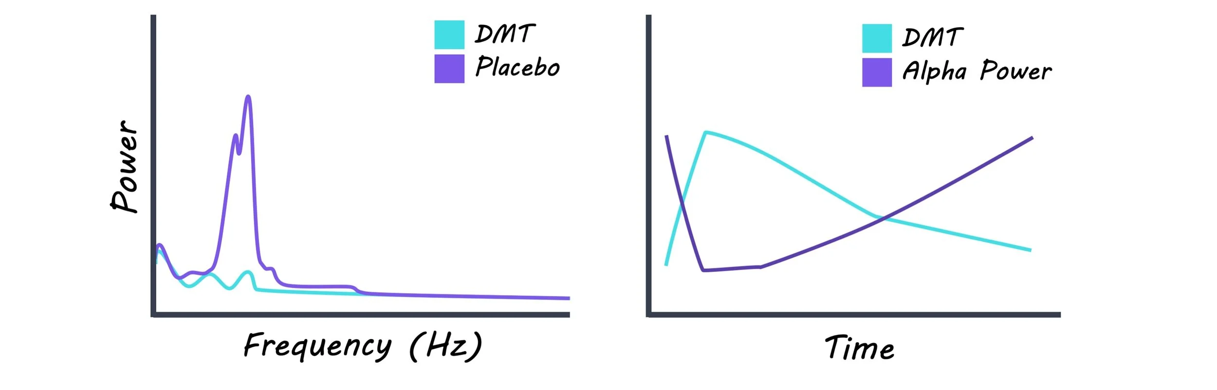
What is consciousness and what are the neural mechanisms that underlie it? We still don’t know the answer to these elusive questions. One exciting new avenue for studying the neurobiology of consciousness is to examine the altered states of consciousness produced by psychedelic drugs. For example, research has shown that LSD, psilocybin (the main psychoactive principle in magic mushrooms), and DMT (a naturally occurring psychedelic used in the ceremonial brew ayahuasca) consistently show broadband decreases in oscillatory power using EEG, particularly in alpha power (oscillations at approximately 8-12 Hz) which is linked to relaxation. Psychedelics have also been shown to increase the complexity or diversity of brain activity. DMT is a psychedelic of particular interest, as it produces one of the most unusual and intense altered states of consciousness, and has previously been likened to both dreaming and the near-death experience. This week in Scientific Reports, Timmermann and colleagues conducted the first ever placebo-controlled investigation of the effects of DMT on brain activity in humans at rest

How did they do it? 

To examine the effects of DMT on the brain at rest the authors collected EEG data on thirteen participants during a placebo session first and a DMT session a week later. Placebo or DMT was administered intravenously. The authors collected EEG data starting one minute prior to administration and ended 20 minutes post administration. Blood samples were collected at regular intervals throughout the EEG sessions. Three types of subjective effect measures were collected: (1) participants were required to give a real-time intensity rating of the subjective effects they experienced once every minute, (2) participants completed Visual Analogue Scales once the subjective effects had subsided, (3) the next day an independent researcher conducted a micro-phenomenological interview, designed to reduce subjective bias in first-person reports. 

What did they find? 

The authors’ primary hypothesis was that DMT would decrease oscillatory power in the alpha band and increase cortical signal diversity and that these effects would correlate with changes in conscious experience over time. Overall, their results support this hypothesis, as the authors found strong correlations between alpha and beta power decreases, real-time changes in the intensity rating of subjective effects of DMT, and DMT levels in plasma. They also found increases in delta and theta oscillations, which emerged during the peak of DMT’s effects. These findings suggest that the emergence of theta/delta rhythmicity combined with suppression of alpha/beta rhythmicity may relate to the ‘DMT breakthrough experience’, where the brain switches from processing external information to a state where processing is internally driven, which is also characteristic of dreaming during REM sleep.

DMT immersion also led to widespread increases in signal diversity which correlated with real-time changes in the intensity rating of subjective effects of DMT, and DMT levels in plasma. This is consistent with the ‘entropic brain hypothesis’ which proposes that within a limited range of states, the richness of content of a conscious state can be indexed by the entropy, or complexity, of spontaneous brain activity.  

eeg_Dec3.jpg

What's the impact?

This study is the first ever placebo-controlled investigation into the effects of DMT on brain activity in humans at rest. The finding that DMT results in decreased spectral power in the alpha/beta bands and widespread increases in signal diversity, both of which correlate with the subjective intensity of DMT’s effects, indicates that more work is needed to better understand how these findings relate to the neurobiology of consciousness. This study can also shed light on the mechanisms underlying DMT’s antidepressant effects, as depression has been linked to increased alpha power. Overall, this study advances our understanding of altered states of consciousness.

Timmermann et al. Neural correlates of the DMT experience assessed with multivariate EEG. Scientific Reports (2019). Access the original scientific publication here.

Unique Cortical-Brainstem Activity Underlies Compulsive Alcohol Drinking

Post by Lincoln Tracy

What's the science?

A key feature of alcohol use disorders is compulsive drinking—defined as continued drinking regardless of the resulting negative consequences. While most people drink alcohol at some point during their adult life, less than a third develop an alcohol use disorder. But what makes these individuals more vulnerable to compulsive drinking? Scientists currently have a poor understanding of the individual differences in behavior and neural circuitry that drive compulsion. Previous animal studies suggest that the prefrontal cortex, a brain region involved in planning and coordinating our thoughts and actions, plays a crucial role in compulsive behaviors. Prefrontal cortex activity is different in individuals who have consumed alcohol or who have a family history of alcohol use disorders. This week in Science, Siciliano, and colleagues investigated how individual differences in behavior and neural activity in the prefrontal cortex predict the development of compulsive drinking in mice.

How did they do it?

First, the authors took the mice and exposed them to a “binge-induced compulsive task” (BICT), a conditioning task comprising of three different periods. In the first period, the pre-binge, mice had been conditioned to drink from a bottle containing only alcohol. After three days, increasing amounts of the bitter-tasting quinine was added to the alcohol to act as a punishment—or negative consequence—of drinking. In the subsequent 14-day binge drinking period the mice had unlimited access to water and alcohol at certain times. Finally, the post-binge period ran similarly to the pre-binge period, where the mice were presented with alcohol alone for the first three days followed by the alcohol-quinine mix for the next four. Mice were sorted into groups based on their drinking behavior during the post-binge period. Second, the authors compared drinking behavior in the pre-binge period between the newly identified groups. Third, they used cellular-resolution calcium imaging as a proxy for neuronal activity during the BICT to examine whether the activity of the neural connections between the medial prefrontal cortex and the dorsal periaqueductal grey contributed to susceptibility of developing compulsive drinking behaviors. Fourth, they used two different light-sensitive proteins and optic fibers to determine whether mimicking endogenous neuronal activity in this cortical-brainstem pathway could alter drinking behavior. One of the light-sensitive proteins—halorhodopsin—can inhibit cellular activity, while the other light-sensitive protein—channelrhodopsin-2—helps activate cells.

What did they find?

Three groups of mice were identified based on post-binge period drinking behavior: low drinkers (low alcohol intake regardless of if quinine was present or absent), high drinkers (high alcohol intake that ceased when quinine was present), and compulsive drinkers (high alcohol intake even when quinine was present). Second, compulsive drinking mice drank more of the alcohol-quinine mix during the pre-binge drinking period compared to the other two groups. This compulsive drinking behavior was exacerbated after the binge drinking period. Third, the authors observed more inhibitory responses in the neurons connecting the medial prefrontal cortex and the dorsal periaqueductal grey in compulsive drinking mice compared to the low drinking mice. The low drinking mice also exhibited more excitatory neuronal activity between these two brain regions when consuming alcohol. Therefore, the neural response during initial alcohol exposure predicted the future development of compulsive drinking. Finally, they found that inhibiting neuronal activity between the medial prefrontal cortex and the dorsal periaqueductal grey increased quinine intake and that stimulating neuronal activity over the same neurons decreased alcohol intake. The authors concluded that light-induced inhibition prevented punishment signals being sent from the cortex to the brainstem, whereas light-induced stimulation enhanced the punishment.

alcohol_img_Nov26.png

What's the impact?

This study provides a mechanistic explanation for the individual variance in the susceptibility to compulsive alcohol drinking. These findings are particularly important as this newly discovered cortical-brainstem circuit may help guide efforts in drug discovery to prevent alcohol use disorders. Future research is needed to determine the specific mechanisms underlying the reactivity of this circuit to alcohol.

Tye_quote_Nov26.jpg

Siciliano et al. A cortical-brainstem circuit predicts and governs compulsive alcohol drinking. Science (2019). Access the original scientific publication here.

Different Learning Strategies Used During Pavlovian Conditioning

Post by Shireen Parimoo

What's the science?

In Pavlovian conditioning, people form associations between a neutral stimulus (e.g. a bell) and an upcoming unconditioned stimulus (e.g. food). The neutral stimulus later becomes the conditioned stimulus because it elicits the same response as the unconditioned stimulus. People can learn these associations using a value-based or an uncertainty-based strategy. In value-based learning, learning occurs based on the difference between the expected reward and the actual reward received, which is the reward prediction error. In uncertainty-based learning, people learn the probability that a conditioned stimulus will elicit a specific unconditioned stimulus, which generates the state prediction error. There are individual differences in whether people pay more attention to the conditioned stimulus (sign-trackers) or the unconditioned stimulus (goal-trackers). The neural basis of these learning strategies is not yet well understood. This week in Nature Human Behavior, Schad and colleagues used eye-tracking and functional magnetic resonance imaging (fMRI) techniques to investigate the neural substrates of learning strategies used by sign-trackers and goal-trackers.

How did they do it?

Participants were 129 male adults who completed a Pavlovian conditioning task in the fMRI scanner while their eye movements were recorded. They learned associations between visual-auditory cues that predicted monetary reward (appetitive conditioned stimulus; $1, $2), no reward (neutral conditioned stimulus: $0), or loss (aversive conditioned stimulus; -$1, -$2). The authors computed a gaze index to categorize participants as sign-trackers or goal-trackers. The gaze index is the difference between the proportion of fixations made to the unconditioned stimulus and the proportion of fixations made to the conditioned stimulus. A value of 0 indicates that participants made an equal proportion of fixations to both conditioned and unconditioned stimuli, whereas positive and negative values indicate that they made more fixations to the conditioned and the unconditioned stimulus, respectively. To identify sign-trackers, the authors examined the relationship between gaze index and the value of the conditioned stimulus. The top third of the participants who looked more frequently at the conditioned stimulus predicting monetary rewards than at the conditioned stimulus predicting losses were deemed to be sign-trackers. A similar analysis was conducted with the value of the unconditioned stimulus to identify goal-trackers. Eye movement behavior during the conditioning task, including pupil dilation and the number of fixations, was compared across the two groups for the different stimuli.

The authors used computational modeling to determine whether the eye movement patterns of sign-trackers and goal-trackers during the conditioning task reflected value-based or uncertainty-based learning strategies. Value-based learning was assessed in a reinforcement learning model that computes a reward-prediction error value. On the other hand, uncertainty-based learning was assessed in a model that produced a state prediction error value. Finally, the authors examined the neural substrates of the different learning strategies. They used a reinforcement learning model to compute reward prediction error signals in reward-processing regions like the nucleus accumbens in response to the stimuli. Uncertainty-based learning was investigated by computing the state prediction error signal at the onset of the unconditioned stimulus in regions associated with the state prediction error effect, such as the intraparietal sulcus and the lateral prefrontal cortex. The reward prediction and state prediction error effects in the brain were compared between sign-trackers and goal-trackers.

What did they find?

Sign-trackers made more fixations to the appetitive conditioned stimulus associated with a monetary reward than to the aversive conditioned stimulus associated with monetary loss, which is in line with value-based learning. Conversely, goal-trackers made more fixations to the appetitive unconditioned stimulus more than the aversive unconditioned stimulus, but over time, they looked away from the conditioned stimuli and more at the unconditioned stimuli, which is in line with uncertainty-based learning. Pupil dilation in response to both conditioned and unconditioned stimuli also differed between sign-trackers and goal-trackers. Pupil size decreased over the course of learning in goal-trackers but did not change in response to the appetitive and aversive conditioned stimuli. Among sign-trackers, there was no change in pupil size over time, but the pupils dilated in response to appetitive conditioned stimuli compared to the aversive conditioned stimuli. Computational modeling indicated that the value-based model captured pupillary changes for sign-trackers, whereas the uncertainty-based model best explained the pupil dilation in goal-trackers. Overall, these results suggest that eye movement behavior tracks the value of the stimulus among sign-trackers and the upcoming expectation state among goal-trackers.

shireen (5).png

Distinct patterns of brain activity were associated with learning strategies used by sign-trackers and goal-trackers. In reward-processing brain regions such as the nucleus accumbens, ventromedial prefrontal cortex, and amygdala, the value-based model explained more variance in brain activity for the sign-trackers than for the goal-trackers. In contrast, the uncertainty-based model better explained activity in the intraparietal sulcus of the goal-trackers than the sign-trackers. In sum, the eye-tracking and neural data indicate that sign-trackers used value-based learning strategies while goal-trackers relied more on an uncertainty-based strategy.

What's the impact?

This study is the first to demonstrate the distinct behavioral and neural profiles of learning in sign-trackers, who primarily use value-based learning strategies, and goal-trackers, who rely on uncertainty-based learning. By providing a deeper understanding of the different learning systems in humans, these findings have important implications for the treatment of disorders that involve aberrant reward learning, like addiction.

Schad_quote_Nov26.jpg

Schad et al. Dissociating neural learning signals in human sign- and goal-trackers. Nature Human Behavior (2019). Access the original scientific publication here.