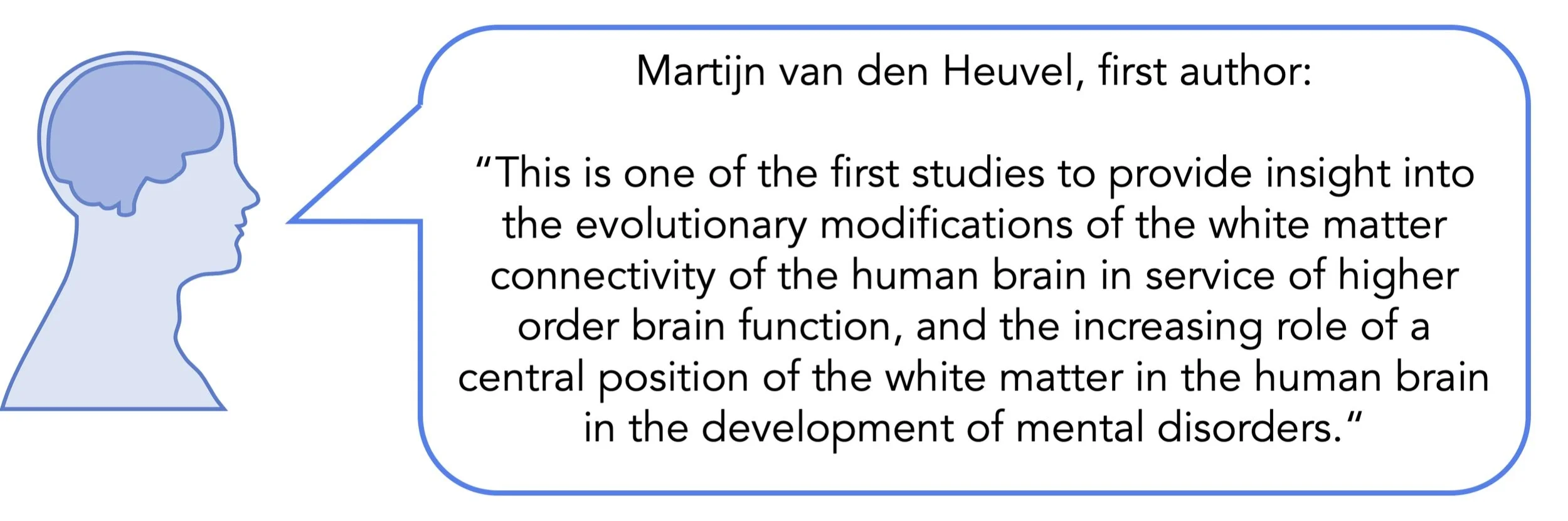
The Role of Evolution on Brain Connectivity in Schizophrenia

Post by Elisa Guma

What's the science?

Schizophrenia is a debilitating psychiatric disorder characterized by hallucinations, delusions, and cognitive dysfunction, often associated with impaired brain connectivity. The genetic origin, human-specific traits, and similar prevalence observed across societies (1% of the population is affected globally) have led to the idea that human brain evolution may have played a role in the development of the disorder. This week in Brain, van den Heuvel and colleagues aim to investigate schizophrenia-related changes in brain connectivity in the context of evolutionary changes in the human brain by comparing humans and chimpanzees.

How did they do it?

To measure brain connectivity, the authors studied diffusion-weighted imaging data (sensitive to the integrity of the brain’s white matter tracts and myelin levels) from individuals with schizophrenia and gender-matched healthy control subjects, as well as from chimpanzees. Connectome maps were created by 1) subdividing the cerebral cortex into 114 subdivisions based on a commonly used brain atlas, and then 2) calculating cortico-cortical connectivity between every subdivision or brain region and every other brain region. The measure of cortico-cortical connectivity was based on whether each pair of brain regions were interconnected by common diffusion-weighted streamlines (measuring common white matter tracts). The authors compared the connectome maps of individuals with schizophrenia to those of controls to examine differences in brain connectivity. In order to confirm whether the results they observed were schizophrenia specific, they performed the same analysis for a variety of other neuropsychiatric and neurological disorders including major depressive disorder, bipolar disorder, obsessive-compulsive disorder, autism spectrum disorder, a behavioural variant of frontotemporal dementia, and mild cognitive impairment. The authors also performed their analysis in a separate cohort of schizophrenia and control subjects to ensure their results were not biased by the data acquisition of their original sample.

To determine the human-specific connections in the brain, the authors compared the connectome maps between humans and chimpanzees. Connections that were present in at least 60% of the human subjects and 0% of the chimpanzees were classified as human-specific connections (and vice-versa for chimpanzee-specific connections). Human-chimpanzee-shared connections were classified as those present in at least 60% of humans and 60% of chimpanzees. To investigate the evolutionary nature of dysconnectivity (i.e. reduced connectivity) in schizophrenia, the authors compared human-specific connection patterns to brain dysconnectivity patterns due to schizophrenia. The authors extended their cross-species comparison to include rhesus macaques, a more distantly related primate species, to further investigate the evolutionary specialization of the human-specific connections they identified. Finally, the authors validated their findings by repeating the analysis using a different brain atlas that is designed to map homologous regions across humans and chimpanzees and subdivides cortical areas into 38 rather than 114 regions.

What did they find?

First, the authors show that many brain regions involved in higher-order processing had greater dysconnectivity in patients with schizophrenia. These findings were consistent with previous studies. Next, the authors found a 94% overlap between the human and chimpanzee cortico-cortical networks, with 3.5% human-specific connections, and 1.1% chimpanzee-specific connections. Many of the brain regions identified to be human-specific are involved in language networks and important for semantic comprehension. The authors also identified human-specific connections in regions implicated in cognitive control, social cognition, emotional processing, and bonding behaviour. Interestingly, these behaviours are often impaired in individuals with schizophrenia. In fact, the authors found high similarity between patterns of schizophrenia dysconnectivity and human-specific connections in comparison to connections shared by both humans and chimpanzees. This held true when the authors extended their cross-species comparison to include macaques, and when the data were analyzed with the alternative brain atlas. Furthermore, the human-specific connections were not associated with brain dysconnectivity patterns for any other disorders. There were trend level associations with bipolar disorder, which shares a genetic background with schizophrenia, as well as some symptoms like psychosis.

schizophrenia.png

What's the impact?

These findings provide compelling evidence for the hypothesis that human brain evolution may have played a role in the development of schizophrenia. The authors show that human-specific features of cortical connectivity are associated with patterns of cortical dysconnectivity in individuals with schizophrenia, suggesting that evolutionary pressure to develop higher-order functions may have rendered the brain vulnerable to dysfunction. In future work, it could be interesting to see how these patterns extend to other measures of brain function.

vandenHeuvel_Nov19.jpg

Van den Heuvel et al. Evolutionary modifications in human brain connectivity associated with schizophrenia. Brain (2019). Access the original scientific publication here.

Auditory Cortex Contributes to Threat Memory

Post by Sarah Hill

What's the science?

The same learning principle made famous by Pavlov and his dogs - classical conditioning - is exercised when an animal associates a neutral stimulus with a threat. For example, a mouse that has learned to associate an auditory cue with an impending aversive stimulus (e.g. a foot shock), will exhibit freezing behavior upon hearing the cue even if the cue is no longer followed by a shock. This type of aversive learning results in a threat memory, a form of memory important for future avoidance of aversive stimuli. Whether the auditory cortex, a brain region located in the temporal lobe, is involved in threat memory is unclear, as lesions to this brain region have had mixed effects on memory. This week in Neuron, Dalmay and colleagues show that the auditory cortex and other subregions of the temporal cortex contribute to threat memory acquisition and retrieval.

How did they do it?

The authors used optogenetic methods to inhibit the auditory cortex, and conditioned mice to associate an auditory cue with a foot shock. They carried out both discriminative (i.e. using a conditioned stimulus [CS+] and a neutral stimulus [CS-]) and non-discriminative conditioning (i.e. using only a CS+), presenting one set of animals with complex naturalistic auditory cues (akin to sounds heard in nature) and another with pure tones. To test threat memory, they presented mice the next day with acoustic stimuli, this time without the associated foot shock, and recorded freezing behavior as a measure of the fear response. An analogous series of experiments were then carried out to determine the contribution of neighboring brain areas to threat memory. Optogenetics techniques were similarly used to inhibit adjacent regions of the temporal neocortex, including the ventral region of the secondary auditory cortex, the temporal association cortex, and neuronal axons projecting to the amygdala, the brain region that mediates the fear response. Fear conditioning was again carried out followed by threat memory testing.     

What did they find?

Mice with auditory cortex inhibition exhibited reduced freezing behavior following presentation with complex naturalistic auditory cues, but not after presentation with pure tone cues, suggesting that the role of the auditory cortex in threat memory is dependent on stimulus complexity. This effect was observed whether the auditory cortex was inhibited during fear conditioning or during memory retrieval, as well as in the context of both discriminative and non-discriminative conditioning. Thus, the auditory cortex was shown to contribute in a stimulus-dependent manner to discriminative and non-discriminative threat memory expression. In contrast, mice with inhibition of adjacent temporal cortex regions displayed significant memory impairments regardless of stimulus complexity. Some neocortical subregions were shown to contribute more to threat memory than others — particularly the ventral region of the secondary auditory cortex and the temporal association cortex. Finally, inhibition of amygdala-projecting neurons resulted in reduced freezing behavior when paired with complex, but not pure tone, auditory cues. In other words, complex acoustic stimuli selectively activate direct information transfer between the neocortex and amygdala to elicit auditory threat memory expression.            

threat_memory_Nov19.jpg

What's the impact?

This study conclusively demonstrates a role for the temporal cortex, including the auditory cortex, in auditory threat memory. These findings are particularly important for understanding the extent to which the neocortex participates in learning and memory and the circumstances in which this form of neocortical processing occurs. 

Letzkus_Nov19.jpg

Dalmay et al. A Critical Role for Neocortical Processing of Threat Memory. Neuron (2019). Access the original scientific publication here.

Driving Brain Plasticity with Gamma Oscillations

Post by Anastasia Sares

What's the science?

The visual cortex is a classical area for studying neuron tuning, specialization, and brain plasticity. Located in the very back of the brain, the visual cortex was studied as early as the 1960s, and it was discovered that different clusters of neurons responded to moving stripes oriented at different angles. Changing the preferred orientation of some neurons is a small-scale example of brain plasticity, but it doesn’t happen all by itself. Something has to happen in the brain to change the status quo. This week in Proceedings of the National Academy of Sciences, Galuske and colleagues induced brain plasticity by pairing visual conditioning with stimulation of a brainstem area (midbrain reticular formation).

How did they do it?

The authors studied the visual cortex of cats, implanting electrodes in order to record neural activity (more specifically: electrocorticograms, multiunit activity, and local field potentials), and also performed optical imaging. They recorded neural responses to different orientations of stripes to create an “orientation map” of the cortex. Recordings took place before and after a long conditioning session, where the cats were exposed to moving stripes (also called ‘gratings’) in a single orientation. Repeatedly exposing neurons to the same stimulus (stripes at a certain orientation) usually just causes them to habituate, firing less as they get used to the stimulus. It does not typically change their preferred orientation. However, during some of the conditioning sessions, the authors additionally stimulated the midbrain reticular formation (MRF) in the brainstem. Activity in this brainstem area can enhance gamma oscillations in the visual cortex, which the authors believed would drive plasticity. This plasticity would cause greater responsiveness and attunement to the orientation presented in the conditioning session.

What did they find?

The authors succeeded in causing a plastic change in the visual neurons. After the conditioning session with the MRF being stimulated, more neurons responded to the grating that had been presented. The cells that changed the most were the ones that had originally responded to orientations 10-30 degrees away. These cells were “re-tuned” so that they preferred the orientation presented in the session. The effect lasted at least 6 hours, at which point the researchers stopped measuring it. It wasn’t just stimulation of the MRF that caused this plasticity. Only when MRF stimulation led to an increase in gamma oscillations did this effect show up.

visual_cortex.png

What's the impact?

This study shows that gamma oscillations put the brain’s cortex in a unique state that facilitates plasticity, changing how it represents the outside world. More research is needed to connect gamma oscillations to learning, but some evidence suggests that they may be related to context and prediction, providing a way for the brain to turn plasticity on or off when the need arises.

Galuske et al. Relation between gamma oscillations and neuronal plasticity in the visual cortex. Proceedings of the National Academy of Sciences (2019). Access the original scientific publication here.