Synaptic Transcription Is Driven by Circadian Rhythm While Protein Expression Is Driven by Sleep in the Mouse Forebrain

Post by Amanda McFarlan 

What's the science?

The sleep-wake cycle is involved in regulating the size of synapses and brain protein expression, so, it is not surprising that it may also be involved in regulating local synaptic transcription and protein expression. In support of this, it has been shown previously that approximately 6% of transcription in the forebrain fluctuates throughout the day, which is thought to be mediated by the sleep-wake cycle. This week in Science, Noya and colleagues investigated the role of sleep and circadian rhythms on the rhythmicity of synaptic transcript and protein expression in the mouse forebrain and found a remarkably different picture than that seen in transcripts in the cell as a whole. 

How did they do it?

The authors collected tissue from the mouse forebrain every 4 hours for one 24-hour period to assess daily rhythms in messenger RNA (mRNA) transcripts and protein expression. They used biochemical homogenization and fractionation to purify synaptoneurosomes (isolated synaptic terminals) and identify mRNAs that were present in these synapses. Then, using isolated synaptoneurosomes, they performed mass spectrometry-based proteomics to identify patterns of protein expression in the synaptic proteome (i.e. all proteins expressed in the synapse) and the total forebrain. Next, the authors investigated the role of the sleep-wake cycle in regulating transcription of synaptic mRNA and protein expression by collecting forebrain tissue every 4 hours for one 24-hour period from mice that were sleep-deprived for four hours prior to sacrifice. They assessed the effect of high sleep pressure (i.e. sleep deprivation/greater amplitude of delta oscillations) on the daily rhythmicity of mRNA and protein expression in forebrain tissue. 

What did they find?

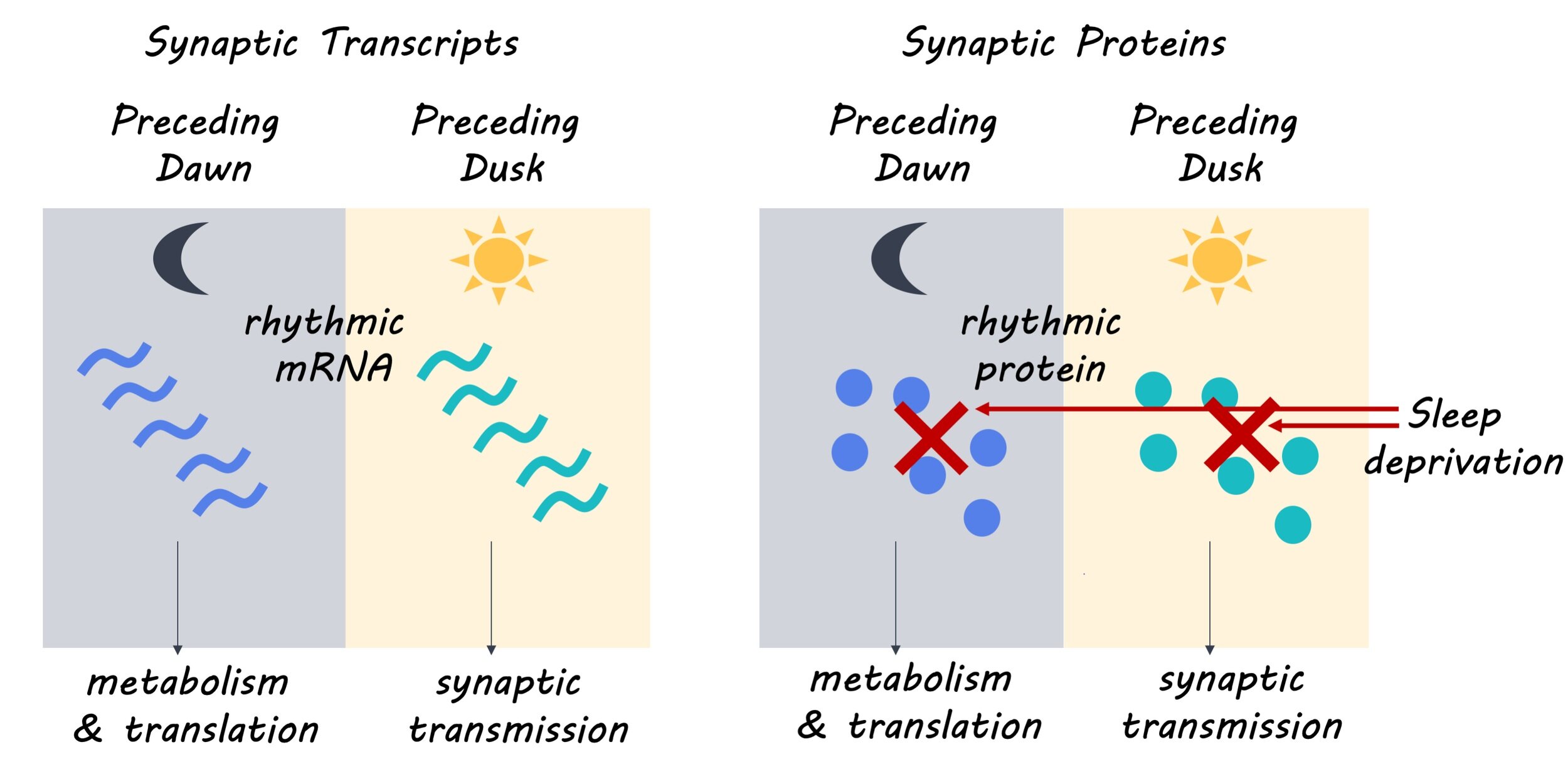
The authors found that 67% of synaptic mRNA transcripts had a rhythmic pattern of expression, with 93% of these transcripts exclusively showing rhythmic patterns of expression in synaptoneurosomes. This suggests that the oscillatory or cyclic pattern of synaptic mRNA expression may be controlled by post-transcriptional mechanisms. They also found that 11.7% of synaptic proteins and 17.2% of proteins in the forebrain showed rhythmic patterns of expression. Next, the authors determined that the rhythmic expression of synaptic mRNA formed two distinct clusters separating mRNA transcripts expressed in the day from those expressed at night. Furthermore, they found that the mRNA transcripts expressed in the day participated in biological processes including synapse organization and transmission, while mRNA transcripts expressed at night participated in biological processes such as metabolism, cell proliferation and development. These distinct patterns of expression and biological roles were also observed in the synaptic proteome, with 75% of rhythmically expressed synaptic proteins showing concomitance with their mRNA counterpart. Finally, the authors determined that the rhythmicity of synaptic mRNA transcript expression was mostly preserved with high sleep pressure, but rhythmicity in protein expression could no longer be detected with high sleep pressure, suggesting that synaptic mRNA expression may be controlled by a molecular clock while protein expression may be gated by sleep-wake pressure.

synapse_img_Oct15.jpg

What's the impact?

This is the first study to show that synaptic mRNA transcripts in the mouse forebrain have a highly rhythmic expression that is controlled post-transcriptionally. The rhythmic expression of mRNA transcripts and their related proteins was shown to be important for distinct biological processes associated with either day or night. These findings provide insight into the mechanisms by which synaptic mRNA and protein expression are regulated in the mouse forebrain. 

Brown_Oct15.jpg

Noya et al. The forebrain synaptic transcriptome is organized by clocks but its proteome is driven by sleep (2019). Access the original scientific publication here.

X-Chromosome Insufficiency Alters the Structure and Function of the Human Visual Cortex

Post by Lincoln Tracy

What's the science?

Turner syndrome is a genetic condition in females caused by the absence of a part of, or an entire, X chromosome. Women with Turner syndrome have specific cognitive deficits relating to the ability to perceive spatial relationships between objects and the ability to select and monitor goal-related behaviors. Previous research suggests that women and girls with Turner syndrome have structural changes in the parieto-occipital cortex (the part of the brain responsible for the integration of sensory information and the processing of visual information) when compared to typically developing controls. Specifically, females with Turner syndrome are reported to have a smaller volume of parieto-occipital cortex with a thicker surface. Despite knowing that these structural changes exist, it is not known whether abnormal processing of visual information occurs in females with Turner syndrome. This week in The Journal of Neuroscience, Green and colleagues used functional magnetic resonance imaging (fMRI) and population receptive field (pRF) mapping to investigate receptive field processing in Turner syndrome.

How did they do it?

The authors recruited 24 girls (aged 7-14 years) with Turner syndrome and 28 typically developing girls of the same age to act as controls. All 52 participants had an MRI scan where they watched a screen that showed a flickering black and white checkerboard pattern in either expanding rings or rotating wedges to obtain and assess data for the pRFs. pRFs can be used to generate topographic maps of the visual field (i.e. the total area an individual can see) using representations of the polar angle (the angle from the horizontal axis) and eccentricity (the distance from fixation). Data from the MRI scans were used to determine the volume, surface area, and cortical thickness of the early visual areas V1-V3. Visual field coverage (the locations within a visual field that evoke a response from voxels within a brain map) in these areas were also drawn from the MRI data. Outside of the MRI scanner the girls completed the Picture Puzzles test, one of the tests from the NEPSY-II battery, which tests visuospatial processing. The relationship between performance on the Picture Puzzles test and pRF properties was compared.

What did they find?

First, the authors found that girls with Turner syndrome and typically developing girls showed a similar organization of polar angle and eccentricity maps. These maps were then used to define visual field maps for V1-V3. Using these maps, the authors then showed that while there were no differences in total brain volume between girls with Turner syndrome and typically developing girls, the cortical volume and surface area of the V1-V3 visual field maps were smaller in girls with Turner syndrome. There were no differences in V1-V3 cortical thickness between girls with Turner syndrome and typically developing girls. Second, they found that despite Turner syndrome and typically developing participants having similar pRF size for V1-V3, participants with Turner syndrome had less pRF eccentricity in visual areas V1-V3 (meaning that their pRF was closer to the center of their gaze). Third, visual field coverage was determined by comparing how the pRFs from V1-V3 fit together in the visual field. There were no differences between participants with Turner syndrome and typically developing girls in the visual field coverage of V1-V3, nor were there differences in the polar angle and eccentricity maps. Fourth, they found that participants with Turner syndrome performed worse in the Picture Puzzles task compared to the typically developing group. Performance in the Picture Puzzles task was found to be negatively correlated with pRF size and eccentricity in girls with Turner syndrome. There was no correlation between performance in the Picture Puzzles task and the cortical volume of V1-V3 in either the participants with Turner syndrome or typically developing girls.

lincoln_turner.jpg

What's the impact?

The authors have demonstrated that Turner syndrome may negatively affect the functional properties of brain regions responsible for visual processing, which may in turn influence behavior. The findings of this study may serve as a novel approach for investigating how other regions of the brain are affected by Turner syndrome. Furthermore, the authors provide a target for interventions to improve the visuospatial deficits observed in Turner syndrome. Taken together, these results suggest a fundamental change in our understanding of how variations in sex chromosome pairings affect visuospatial development in humans.

Green_Oct15.jpg

Green et al. X-Chromosome Insufficiency Alters Receptive Fields across the Human Early Visual Cortex. Journal of Neuroscience (2019).Access the original scientific publication here.

Uncovering Neuronal Circuitry Using Computational Modelling

Post by Amanda McFarlan

What's the science?

With the advancement of technology, it is now possible to acquire recordings from large populations of neurons in anaesthetized or awake, behaving animals. These recordings generate large data sets that may be useful for understanding how neuronal activity is related to behaviour. However, interpreting and analyzing the results from a large data set can be very challenging and requires sophisticated computational methods. This week in Nature Communications, Kobayashi and colleagues investigated the efficacy of their computational model in identifying neural connectivity in both synthetic simulations and biological data sets.  

How did they do it?

The authors estimated neuronal connectivity using a computational model by cross-correlating the spike (or action potential)  times for each neuron pair. Then, they applied a generalized linear model to identify pairs of neurons with small (millisecond) differences in spike timing to determine the pairs that were likely monosynaptically connected. To determine the level of conservatism that optimally balanced the rate of detected false positives and false negatives, the authors applied their model to spike train data obtained from a synthetic neuronal network of 1000 neurons (800 excitatory, 200 inhibitory) for which the connectivity was already known. Next, the authors sampled neurons from the total population to evaluate the efficacy of their model at predicting neural connections. They generated a connection matrix with four quadrants to represent the different combinations of neural connections: inhibitory-excitatory, excitatory-excitatory, excitatory-inhibitory and inhibitory-inhibitory. Then, they applied their model to spike train data from hippocampal neurons in the CA1 region of a rat to assess its efficacy in estimating neuronal connectivity when applied to a real, biological data set. Finally, the authors established the validity of their model by comparing it to two other commonly used methods, the cross-correlation method and the jittering method, while using both synthetic and biological data.

What did they find?

The authors showed that their simulation of a neuronal network produced results that were consistent with balanced state network models. They determined that balancing the rate of false positives and false negatives was optimally achieved using a significance level of α=0.001 to determine neuronal connections. Compared to excitatory neurons, inhibitory neurons tended to have higher firing rates and exhibit more regular spiking. Additionally, the likelihood of identifying neuronal connections was positively correlated with the duration of the recording, suggesting that it may be possible to correctly identify a larger number of connections between excitatory and inhibitory neurons by extending the length of a recording. Next, the authors showed that applying their computational model to biological data in the rat hippocampus yielded similar results as applying it to synthetic data, suggesting that their model is useful for estimating neuronal connections in real-life examples. Finally, they revealed that their computational model, with only 13 false connections, was superior to the cross-correlation and jittering methods which had 88 and 27 false connections, respectively.

amanda.png

What's the impact?

This is the first study to show a new computational model that combines the generalized linear model and cross-correlations to reliably identify neuronal connections in complex neural networks. The authors have demonstrated that their model is superior to other commonly used methods and is also effective when applied to both synthetic and biological data. This computational model may be instrumental for mapping neural connections in large data sets from extracellular recordings in animal subjects and may provide insight into patterns of neuronal connectivity throughout the brain.  

quote_naturecomms.png

Kobayashi et al. Reconstructing neuronal circuitry from parallel spike trains. Nature Communications (2019). Access the original scientific publication here.

Illustration by Kai Shinomoto

Illustration by Kai Shinomoto