Hyperexcitability in Hippocampal Neurons Derived from Individuals with Bipolar Disorder

Post by Deborah Joye

What's the science?

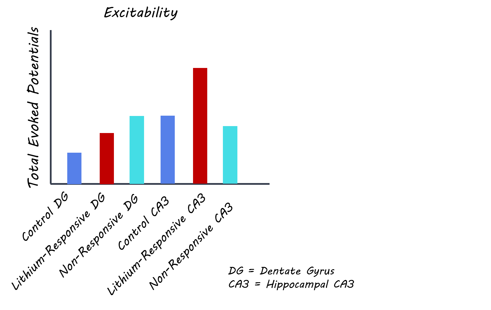
Bipolar disorder (BD) is a complex affective disorder that can be trademarked by repetitive episodes of mania and depression. One of the most common, pharmacological treatments for individuals experiencing BD is lithium. Although lithium has been used for decades to successfully treat BD, the mechanism by which lithium works to treat symptoms is not clear and not every patient responds to lithium treatment. Recently developed induced pluripotent stem cell (iPSC) technology allows researchers to take living cells (like skin cells or white blood cells) from BD patients and turn them into specific types of neurons, allowing for the investigation of cellular differences in individuals with BD versus healthy controls. Previous research using this methodology has found cellular differences in the dentate gyrus region of the hippocampus, where cells derived from BD patients are hyperexcitable, exhibiting longer durations of increased firing activity relative to cells from controls. This week in Biological Psychiatry, Stern and colleagues use induced pluripotent stem cells technology to demonstrate that CA3 hippocampal pyramidal cells can also be hyperexcitable in BD due to differences in potassium currents and that lithium treatment can reduce this hyperexcitability.

How did they do it?

The authors collected cells from 6 BD patients (3 known to respond to lithium and 3 non-responders) and 4 individuals without BD (controls). Using iPSC methodology, the authors created CA3 hippocampal cells from each participant and then used whole-cell patch-clamp electrophysiology to study the cell’s a) electrical properties, b) response to lithium and c) response to specific potassium channel blockers The authors also used quantitative polymerase chain reaction (qPCR) to investigate the expression of specific genes within each cell.

What did they find?

As with cells from the dentate gyrus, the authors found that CA3 BD neurons were hyperexcitable, but only when derived from patients that responded to lithium. This cellular hyperexcitability correlated with higher amplitude potassium currents and with faster kinetics. Faster potassium currents can result in hyperexcitability because the cell is able to recover faster from each action potential and therefore produce more action potentials for a given input. The authors also found that neurons derived from lithium-responding BD patients exhibited overexpression of genes Kcnc1 and Kcnc2, which code for subunits of voltage-gated potassium channels. When the authors applied potassium channel blockers to cells, hyperexcitability was reduced, further supporting the role of potassium channels in BD-derived CA3 cell hyperexcitability. Chronically treating cells with lithium also decreased hyperexcitability in cells derived from lithium responders, which was associated with an increase in sodium currents and a reduction in fast potassium currents. Fast potassium currents slow a cell’s ability to re-polarize after firing an action potential and increase the amount of time before another action potential can fire. While CA3 cells from lithium non-responders did not exhibit hyperexcitability, they did display altered physiology compared to healthy controls, including reduced sodium currents and increased fast and slow potassium currents as well as a unique distribution of highly excitable and very low excitability neurons. Changes to these features suggest that, in general, BD patients may have altered cellular physiologies, but that CA3 hyperexcitability was specific to lithium responders.

leigh_image_oct888.png

What's the impact?

This study uncovers neuron-specific physiological changes that can occur in the brains of individuals with BD and provides critical insight into how lithium might successfully treat bipolar disorder in a subset of patients. Due to the slow progress of BD research, treatment options for those suffering from this complex disorder have not changed much in decades. This study demonstrates important differences in hippocampal cell function in BD patients who respond to lithium, relative to both lithium non-responders and healthy controls, providing important avenues for future therapeutics.

quote_biological_psychiatry.png

Stern et al., Mechanisms underlying the hyperexcitability of CA3 and dentate gyrus hippocampal neurons derived from bipolar disorder patients, Molecular Psychiatry (2019). Access the original scientific publication here.


Brain Regulation of Emotional Conflict Predicts Antidepressant Treatment Response for Depression

Post by Elisa Guma

What's the science?

Major depression is a chronic and disabling mood disorder, for which the primary treatment is traditionally antidepressant medications. These are effective for a subset of individuals, however, for many they do not afford much improvement over placebo treatment. Given this heterogeneity, it is thought that an individuals’ neurobiological characteristics might predict their response to treatment. This week in Nature Human Behaviour, Fonzo and colleagues investigated whether an individual’s response to or regulation of emotional conflict and associated neural response could predict treatment outcome to an antidepressant, sertraline, compared to placebo in a double-blind trial.

How did they do it?

The authors used functional magnetic resonance imaging (MRI) data on an emotional conflict task from a large double-blind trial, Establishing Moderators and Biosignatures of Antidepressant Response for Clinical Care for Depression (EMBARC). In the trial, participants were randomized into an 8-week course of sertraline (an antidepressant) or placebo, and depressive symptoms were measured each week. The authors chose to focus their study on emotional conflict, as its regulation is critical to wellbeing, it engages cognitive and emotion related brain circuitry, is often dysregulated in depression, and is thought to be of relevance to the efficacy of antidepressant treatment. The task performed while undergoing functional MRI involved the presentation of an emotional face with either a fearful or happy expression, with an overlaid emotional word (i.e. fear or happy). Participants were instructed to identify the facial emotion while trying to ignore the emotional word. Stimuli were either presented as congruent (i.e. face and word match), or incongruent (emotional conflict). First, the brain regions that were more active during this task were identified, as well as the relationship between their activity and task performance. Next, the authors built a prediction model (using relevance vector machines — a type of machine learning model) to assess whether the neural response associated with emotional conflict regulation could predict treatment outcome. 

What did they find?

In keeping with other large clinical trials, the sertraline treated group had a slightly larger improvement in symptoms than the placebo group. During the task, the incongruent trials (emotional conflict trials) resulted in a slower reaction time. The neural response to this conflict was identified as a conflict response network and included activation of the dorsomedial and dorsolateral prefrontal cortices, ventrolateral prefrontal cortex and anterior insula, and deactivation of the ventromedial prefrontal cortex and anterior medial prefrontal cortex as well as the posterior cingulate, precuneus, hippocampus, and parahippocampal gyri. The authors found that better emotional conflict regulation (i.e. mitigation of the conflict effect on conflict trials that were preceded by another conflict trial) was positively associated with greater dampening of the conflict response network activation.

Elisa (2).png

In addition, the authors found that individuals who were more successful at dampening their emotional conflict response network during conflict regulation (i.e. adapting to interference when the face and word do not match) also had more symptom improvement due to sertraline rather than placebo. Interestingly, the prediction model the authors built was able to reliably predict how treatment changed depressive symptoms when the model was trained (built) using data from the group receiving sertraline, but it did not work when trained on the placebo group. This suggests that the model developed on sertraline outcome reflects a sertraline-specific signal that explains a meaningful effect of treatment, separate from the placebo response. The model was not related to or affected by depression diagnosis or clinical severity.

What's the impact?

The authors were able to identify a neural trait sensitive to medication response within a broader clinical diagnosis of depression. This trait was related to the brain’s ability to adapt its response to emotional conflict and was a better predictor of treatment outcome than either the clinical measures or the behavioural response alone. Future studies could incorporate a broader range of brain imaging modalities and behavioural assessments, and assess generalizability to other antidepressant medications. These findings highlight the heterogeneity in treatment response within a clinically defined population and suggest that individual neurobiological characteristics could predict treatment response. 

Fonzo_quote.jpg

Fonzo et al. Brain regulation of emotional conflict predicts antidepressant treatment response for depression. Nature Human Behaviour (2019). Access the original scientific publication here.

Tracking Neurons During Development to Understand How Neural Circuits Form

Post by Sarah Hill

What's the science?

Neurons never act alone, but instead, organize into coordinated cellular ensembles or 'circuits' to direct behaviors. Linking how newborn neurons are arranged into coordinated networks during development has previously been limited by available technologies, leaving an incomplete picture of how neural circuits are formed. However, recent advances in imaging and computational methods have offered insight into this process. This week in Cell, Wan and colleagues present a new imaging framework for tracking neurons from cell birth to emergence of synchronized global activity, shedding new light on how neural circuits assemble during development.                

How did they do it?

The authors developed an imaging method based on light-sheet fluorescence microscopy to simultaneously track the identities, lineages, migration, and activation of newborn neurons, and demonstrated the approach in the zebrafish spinal cord. First, they imaged a whole zebrafish embryo using cell-type-specific markers to identify neuron types, trace cell lineages, and monitor neuronal movements. They then performed functional imaging of the embryos to record neuronal activation during circuit formation. Additional experiments were carried out to establish how ensembles of neurons become coordinated in their activation along with multiple segments of the spinal cord, as well as how synchronized activity on the left and right sides of the spinal cord is established. Using the imaging data, they pieced together how early spinal cord neurons assemble into a fully functional circuit.        

What did they find?

Through this new imaging method, the authors successfully reconstructed an assembly of the spinal cord circuit at the single-cell level. Using the zebrafish as a model, they identified three key stages in spinal cord circuit development. First, nascent motor neurons (cells that execute motor movements) pair up with other neurons in the same spinal segment to form local ensembles of synchronized activity. In stage II, the local ensembles merge based on size into a globally synchronized ensemble that spans multiple segments. Local spinal microcircuits continue to merge until only two neural ensembles remain, on the left and right sides of the spinal cord. In stage III, alternating left-right activation is synchronized by commissural interneurons (cells that project to the opposite side of the spinal cord) recruited into the global ensembles relatively late in the process.   

sarah.png

What's the impact?

This is the first study to trace the development of a neural circuit at the single-cell level, from neuronal birth to emergence of a functional circuit. Importantly, the imaging framework proposed in this study can be readily translated to neural circuits beyond the spinal cord and all computational methods are open-source. 

Keller_quote.jpg

Wan et al. Single-Cell Reconstruction of Emerging Population Activity in an Entire Developing Circuit. Cell (2019). Access the original scientific publication here.