A National Experiment Reveals Where a Growth Mindset Improves Achievement

Post by Stephanie Williams

What's the science?

The term “growth mindset” reflects the belief that abilities are not hard-wired into the brain, but can be developed and improved over time. Growth mindset interventions can be effective in changing the beliefs of students who think their academic abilities are fixed. Recently, experimental work in educational settings has investigated how interventions can use ideas related to a growth mindset to intervene in cases of academic underachievement. This week in Nature, Yeager, Hanselman, and colleagues demonstrate that a growth-mindset intervention can effectively improve grades and enrollment in advanced math courses in particular student populations and school environments.

How did they do it?

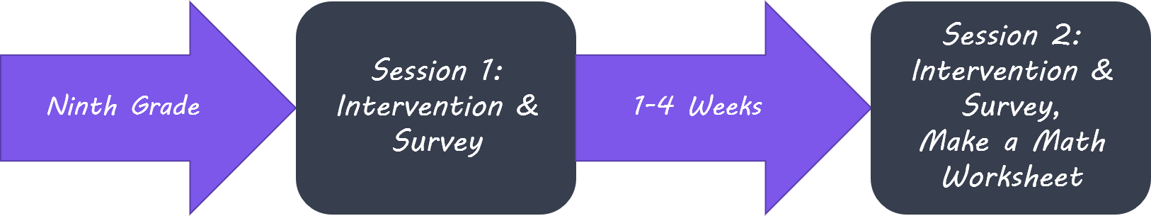
The authors analyzed the effects of a validated growth-mindset intervention in a nationally representative sample of high schools. Ninth grade students were randomized to a particular condition (intervention vs. control activity), and teachers, as well as researchers, were blind to the condition of each student. The intervention consisted of two 25-minute online modules. In 25-minute self-administered online sessions, students heard stories from both older students and adults, interacted with guided exercises, and were asked to reflect on their own learning. At the end of the second session, students participated in a math task with two options: an easy but low learning task, and a challenging but high learning task. The authors used the percentage of students in the control condition that chose the challenging problem as a metric of challenge-seeking norms for a particular school. The authors monitored the fidelity of the intervention implementation by analyzing the percentage of the module screens students viewed (97%) and analyzing the number of open-ended questions that students responded to (96%).

growth_mindset_1_of_2.png

The authors were interested in identifying which contexts were most conducive to the growth mindset intervention. They analyzed treatment effect heterogeneity by looking at the types of school contexts (availability of resources, motivation to follow an intervention, etc.), and peer contexts (supportive vs. unsupportive of taking intellectual risks) The authors point out that schools on both ends of the resource spectrum could fail to see an effect of the intervention: schools with low-quality curricula don’t offer as many learning opportunities for students, and schools with ample resources may have less of a need for the intervention. The authors assessed the effect of their intervention on the mindset of students, as well as the effect on their GPAs and advanced mathematics course enrollment. They looked at GPAs of all core courses, and also at math and science GPAs in their secondary analysis. These subjects are of particular interest because of the commonly held belief that success in math or science is innate. The authors also re-analyzed the data using a Bayesian machine learning algorithm and compared the findings from both sets of analyses.  

What did they find?

The authors found that lower-achieving students who received the intervention performed better than lower-achieving students who did not receive the intervention. This was true when the effect was quantified by assessing overall GPA as well as math-science GPA. The intervention was more effective in low and medium-achieving schools than in high-achieving schools. The authors found no significant difference in the treatment’s effectiveness in low vs. medium achieving schools, but point out that there is high variability in student performance in low-achievement schools. The intervention did still have an effect on students in high-achieving schools. For the top 25% of high achieving schools, the intervention increased the rate at which students enrolled in advanced math courses the following year by 4 percentage points. Students in school environments with peer norms that supported the adoption of intellectual challenges showed larger GPA benefits from the treatment. The authors interpret this result as suggesting that students in unsupportive environments may feel social pressure not to take intellectual risks in front of their peers. They also suggest that beliefs can affect how students react to ongoing academic challenges. The results from the Bayesian machine learning analysis confirmed these findings. 

growth_mindset_2_of_2.png

What's the impact?

A low-cost growth mindset intervention can have a substantial effect on grades and enrollment rates in advanced mathematics courses. The findings around context-specific treatment effects (e.g. school resources or peer environment) can be used to inform future types of interventions. Future interventions should target the messages students receive about learning ability from schools and assess additional challenges faced by adolescents, including social and interpersonal difficulties.

Yeager_quote.jpg

Yeager, D, Hanselman, P., et al. A national experiment reveals where a growth mindset improves achievement. Nature (2019). Access the original scientific publication here.

Cerebral Blood Flow Changes as a Potential Biomarker of Major Depressive Disorder

Post by Lincoln Tracy

What's the science?

Major depressive disorder (MDD) is a common mood disorder that is believed to be associated with changes in the brain. Researchers have used magnetic resonance imaging (MRI) techniques to identify brain-related biomarkers for other conditions such as Alzheimer’s disease. However, brain-related biomarkers have not yet been identified for use in the diagnosis or treatment of MDD. One noninvasive neuroimaging technique used to measure brain function is arterial spin labelling (ASL). In ASL a magnetic pulse labels blood before it enters the brain. The amount of labelled blood in each region of the brain can then be quantified; greater blood flow is typically indicative of greater activity in that brain region. This week in Molecular Psychiatry, Cooper and colleagues used ASL as part of a randomized, double-blind, placebo-controlled trial aiming to find brain-related biomarkers for the diagnosis and treatment of MDD.

How did they do it?

The authors recruited 200 individuals with MDD as well as 98 non-depressed individuals (to act as controls) as part of a clinical trial. All participants underwent a baseline MRI scan with an ASL sequence prior to starting their allocated treatment for the trial. The baseline MRI and ASL data was split into Discovery-Replication subgroups. The ASL-derived cerebral blood flow (CBF) data from the Discovery group was used in an exploratory manner to identify brain regions where CBF differed between MDD and non-depressed individuals. The Replication group was then used to independently confirm these CBF differences. The blood flow in regions where CBF differences replicated were then correlated with clinical features of MDD (e.g., length of illness, number of MDD episodes, age of MDD onset) to explore potential relationships between CBF and clinical features.  

What did they find?

The Discovery sample showed that there were differences in CBF between the MDD and control participants. The MDD participants had lower CBF compared to the control participants in five brain clusters, and greater CBF compared to the control participants in five brain clusters. Using the regions of interest (ROIs) from the Discovery sample, the Replication sample confirmed three clusters where CBF was lower in individuals with MDD compared to the control participants—the cerebellum and midbrain regions, the right middle temporal gyrus, and the left insula. The Replication sample also confirmed three clusters where CBF was greater in individuals with MDD compared to the control participants—the left anterior prefrontal and dorsolateral cortices, the right inferior parietal lobule, and the left inferior parietal lobule. The CBF of several replicated ROIs were correlated with the clinical features of MDD. For example, the decreased CBF within the left insula negatively correlated with the length of illness. That is, a longer length of illness in individuals with MDD was associated with lower perfusion within the left insula.

MDD_image.jpg

What's the impact?

This study is the first to discover and provide unbiased confirmation of CBF differences in a large population of individuals with MDD. These are important and exciting findings for this field of research as there has previously been a lack of discovery-replication work. These findings suggest that CBF could serve as a potential marker for the long-term effects of MDD and may also serve as a potential mediator of responses to treatment. These findings also have implications for future studies aimed at developing CBF as a biomarker for the diagnosis and treatment of MDD and other clinical populations.

Trivedi_quote.jpg

Cooper et al. Discovery and replication of cerebral blood flow differences in major depressive disorder. Molecular Psychiatry (2019).Access the original scientific publication here.

The Right Frontal Eye Fields are Causally Involved in Distractor Suppression

Post by Shireen Parimoo

What's the science?

Selective attention involves attending to the appropriate, target information and ignoring distracting, irrelevant information. The dorsal attention network, which consists of the frontal eye fields (FEF) and the intraparietal sulcus (IPS), is active during tasks that require selective attention. Specifically, these regions are thought to be involved in the control of visual attention, as they bias posterior visual areas to enhance target processing and/or inhibit distractor processing. However, it is not clear whether the FEF provides unique regulatory input or whether both regions are independently involved in attentional control of distraction. This week in the Journal of Neuroscience, Lega and colleagues used transcranial magnetic stimulation (TMS) to identify the dorsal attention network regions causally involved in distractor suppression during a visual search task.

How did they do it?

Thirty right-handed young adults completed a visual search task in two separate sessions. In the task, participants saw four pairs of triangles in each quadrant of the screen and were instructed to identify the orientation of the target pair. The target was the pair of triangles with the same orientation (i.e. both pointing up or both down). On half of the trials, a pair of differently colored triangles served as the distractor, whereas on the other half of the trials, the distractor was absent and the non-target triangles were the same color as the target. In each session, participants completed six blocks of the task while TMS was applied to the IPS, FEF, and a sham stimulation region in each hemisphere. During TMS, a coil is used to non-invasively stimulate cortical brain regions by applying pulses of magnetic stimulation. The authors stimulated both hemispheres independently to determine whether the contribution of the dorsal attention network regions comes from one side of the brain more than the other. In the sham (control) condition, the TMS coil was placed between the FEF and the IPS to prevent cortical stimulation. In the task, the search array was presented for 50ms and the authors applied three 10 Hz TMS pulses 100ms after the onset of the search array, with a 100ms gap in between each pulse. After the search array disappeared, participants had two seconds to make a response for the target orientation.

TMS.png

The authors analyzed participants’ reaction times on correct trials using linear mixed effects models, a statistical technique that accounts for variation in the data that is not explained by the TMS conditions (e.g. each participant could respond to stimulation differently). They compared differences in reaction times following TMS to the IPS and the FEF in each hemisphere and to the sham condition. The authors also computed distractor cost as the difference in reaction times between the distractor-present and distractor-absent conditions. Finally, to examine the effect of distractors on the preceding trial, they calculated distractor cost for trials that were preceded by a distractor-absent compared to a distractor-present trial. They hypothesized that in the sham condition, distractor cost would be smaller if the previous trial contained a distractor, as participants would be relatively more prepared to suppress subsequent distraction.

What did they find?

In general, participants were slower and less accurate when a distractor was present than when it was absent. In the sham condition, participants were faster on distractor trials if the distractor was also present on the preceding trial, versus when it was absent. Reaction times did not differ when TMS was applied to the FEF, IPS, or the sham region in the left hemisphere. However, distractor costs were smaller after right FEF stimulation than after left FEF stimulation, but there was no difference in distractor costs following left and right IPS or sham stimulation, suggesting that the FEF is functionally lateralized during visual attention, at least in relation to distractor inhibition. Moreover, the reduction in distractor costs following right FEF stimulation was driven by faster reaction times on distractor-present trials, particularly when the preceding trial did not contain a distractor. When a distractor was present on the previous trial, there was no difference in distractor cost in the right FEF and sham stimulation conditions, likely reflecting the attentional preparation carried over from the previous trial. These findings indicate that the dorsal attention network – particularly the FEF but not the IPS – is right-lateralized in tasks requiring top-down control of distractor suppression.

What's the impact?

This study highlights a causal role of the right frontal eye fields, but not the intraparietal sulcus, in exerting top-down control over visual attention and distractor suppression. These findings also suggest that in addition to single-trial effects, stimulation of prefrontal regions like the FEF prolong attentional control across multiple trials, providing further insight into their role in modulating visual attention in a sustained manner.

Chelazzi_quote.jpg

Lega et al. Probing the neural mechanisms for distractor filtering and their history-contingent modulation by means of TMS. Journal of Neuroscience (2019). Access the original scientific publication here.