Homeostatic Mechanisms Balance Synaptic Growth and Function

Post by Shireen Parimoo

What's the science?

Over the course of development, neural synapses (junctions where neurons communicate with their targets) grow, shrink, change in numbers, and undergo structural changes, but synaptic function or strength remains stable. Even in the presence of genetic mutations, synaptic strength often remains stable and within a narrow physiological range. It is currently not clear how synapses maintain consistent levels of activity despite the structural variability introduced during development or by mutations. This week in The Journal of Neuroscience, Goel and colleagues investigated the structural and functional neural mechanisms that help stabilize synaptic strength in mutant Drosophila (fruit flies) with disrupted synaptic growth.

How did they do it?

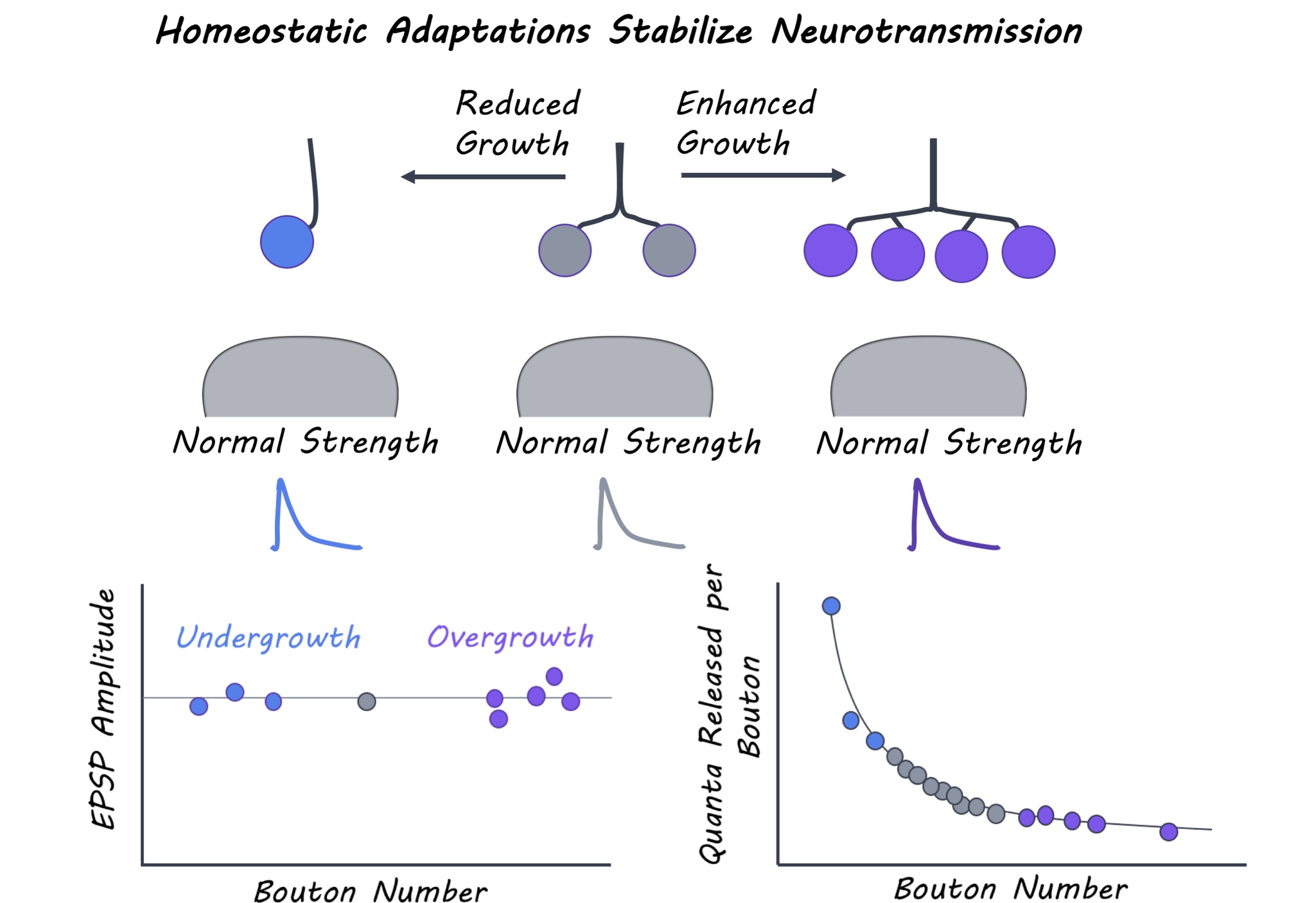
Aberrant synaptic growth and structure is associated with a variety of neural diseases. The authors first identified 300 genetic mutations linked to neural diseases and obtained Drosophila stocks that carried those mutations (mutant lines). They characterized the neuromuscular junction – a type of synapse connecting motor neurons and muscles in each of these 300 mutants. In particular, they measured synaptic growth based on a) the number of synaptic boutons (axon terminals) and b) synaptic strength by electrophysiologically recording the amplitude of excitatory post-synaptic potentials (EPSPs) evoked by stimulation. Mutant lines were selected if they had drastically altered synaptic growth but normal synaptic strength. To determine how synapses with enhanced or reduced growth maintain their strength, the authors investigated the relationship between structural and functional properties of synapses in mutant lines. Structural properties included parameters like the size of boutons and the number and area of active zones (synaptic regions where neurotransmitters are released) at a synapse. Postsynaptic parameters included the amount of postsynaptic neurotransmitter receptors. Functional properties included the quantal content of presynaptic neurons (i.e. the number of synaptic vesicles released upon stimulation) and the amplitude of the spontaneous EPSP events that occur in the absence of any stimulation. The authors systematically examined these properties in mutant lines with reduced and increased synaptic growth and compared them to wild-type Drosophila.  

What did they find?

The authors systematically probed synaptic growth and function in the 300 mutant lines and identified 12 with over-grown (too many boutons) or under-grown (fewer boutons) neuromuscular junctions, and 40 with reduced synaptic strength (EPSP amplitude) compared to wild-type Drosophila lines. For the mutant lines that had reduced EPSP amplitude, no change in synaptic growth was observed, indicating that defects in synaptic function occurred independent of altered growth. Conversely, normal synaptic strength was observed in the 12 mutants with dramatic changes in synaptic growth. The authors thus proposed a “homeostatic model” for maintenance of synaptic function, whereby neurotransmitter release at each bouton is reduced at synapses with more boutons but greater at synapses with fewer boutons. These mutants interestingly employed three main strategies to maintain normal synaptic output. When bouton numbers were massively reduced, either bouton size was enlarged to accommodate more neurotransmitter release and maintain normal synaptic strength or spontaneous EPSP amplitude and receptor levels were enhanced to increase postsynaptic sensitivity and compensate for reduced neurotransmitter release. In contrast, synaptic overgrowth mutants exhibited an increase in the total number of neurotransmitter release sites but each of these individual sites were smaller, leading to an adjustment in presynaptic release per bouton. Thus, homeostatic mechanisms stabilize overall synaptic strength while permitting substantial flexibility in synaptic growth.

JNSCI_image_april2.jpg

What's the impact?

This study is the first to illustrate the adaptive pre- and postsynaptic mechanisms that help maintain synaptic strength despite large differences in synaptic growth. These findings enhance our understanding of how neural function remains stable while the underlying synaptic structure is constantly changing during development, maturation, aging, and even disease.

neuron_quote.jpg

Goel et al. A screen for synaptic growth mutants reveals mechanisms that stabilize synaptic strength. The Journal of Neuroscience (2019). Access the original scientific publication here.

Early Skill Learning Occurs Predominantly at Rest

Post by Lincoln Tracy

What's the science?

Learning a new motor skill typically consists of short periods of active practice—physically doing the task—interspersed with short periods of rest. The combination of practice and rest strengthens the memory in our brain so we can perform the skill better. Traditionally, the consolidation of a new skill has been thought to occur over a period of hours or days. This week in Current Biology, Bönstrup and colleagues examined the time course of practice and rest in how we learn new motor skills on an unprecedented short time scale.

How did they do it?

Healthy right-handed individuals were recruited to participate in a two-day study where they trained and were tested on a procedural motor-skill task on successive days. The motor task involved pressing keys on a keypad in a specific sequence of five key presses. Participants were taught the key press sequence on the first day of the study. They used their left—or non-dominant—hand to perform the key press sequence while seeing the sequence on a computer monitor. Participants completed 36 training trials on the first day. Each trial involved a 10 second period where the participants practiced the key press sequence, followed by a 10 second rest period. Participants returned to the laboratory the next day and were tested on their performance on the task. Nine trials were performed on the testing day. Throughout both the training and testing days the brain activity of participants was recorded by magnetoencephalography (MEG) to identify potential brain mechanisms that supported skill learning.

What did they find?

The authors found that the early learning of the motor skill was driven by increases in performance between practice periods. That is, there was a greater increase in the speed at which participants completed the key press sequences between the end of one trial and the beginning of the next, rather than over the course of each trial. They also found that this learning was predicted by oscillating beta-band (16 – 22 Hz) activity in frontoparietal regions of the brain. Specifically, downregulation of this oscillating beta-band activity at rest was identified as the intrinsic neural signature that went along with performance increases in-between trials.

lincoln.png

What's the impact?

This study is the first to show that a substantial part of early skill learning occurs while we are at rest, and that these improvements can be predicted by activity in the frontoparietal region of our brains. These findings significantly change the way researchers traditionally think about the time it takes us to form a memory and learn new skills—they show this can occur over a period of seconds, rather than hours or days.

current_biology_quote.jpg

Marlene Bönstrup et al. A Rapid Form of Offline Consolidation in Skill Learning. Current Biology (2019). Access the original scientific publication here.

Dopamine Receptor D2 Hippocampal Neurons Regulate Food Intake

Post by Amanda McFarlan

What's the science?

There are a multitude of neuronal populations throughout the brain that have been identified for having a role in controlling appetite, however, it is still not known how these brain regions integrate information from past experiences and the environment to regulate feeding behaviours. The authors hypothesized that the hippocampus may be important for the integration of this information since it is well established as a brain structure that is important for spatial location and memory formation. Further, more recently, it’s its dysfunction has been shown to alter food intake. This week in the Neuron, Azevedo and colleagues investigated the role of the hippocampus is regulating food-related behaviours.

How did they do it?

The authors expressed either an inhibitory (hM4Di) or excitatory (hM3Dq) designer receptor exclusively activated by designer drugs (DREADD) in glutamatergic cells in the dorsal hippocampus of wild type mice. They activated either the inhibitory or excitatory DREADDs by infusion of clozapine-N-oxide (CNO) and observed changes in food intake over a 24-hour period. Next, the authors used PhosphoTrap (which can profile gene expression based on changes in neuronal activation) to measure hippocampal gene expression in fasted mice that underwent a behavioural task where food was presented in a context-specific manner. Next, the authors investigated the role of hippocampal dopamine receptor D2 (Drd2) neurons in food intake by performing bilateral injections of a virus containing either a Cre-dependent inhibitory or excitatory DREADD in the dentate gyrus (region of the hippocampus) in Drd2-Cre mice. They activated the inhibitory and excitatory DREADDs with infusion of CNO and measured food intake acutely (24Hr) and chronically (8 days). Then, they used multiple viral constructs (anterograde and retrograde tracers) to identify synaptic inputs onto hippocampal Drd2 neurons as well as synaptic outputs from hippocampal Drd2 neurons. Next, they targeted Channelrhodopsin-2 (a light-gated cation channel) to glutamatergic neurons in the lateral entorhinal cortex (which projects to hippocampal Drd2 neurons) and implanted optical fibers above the lateral entorhinal cortex terminals in the dentate gyrus (where Drd2 neurons are located). Similarly, they targeted the expression of either Channelrhodopsin-2 or Archaerhodopsin (an inhibitory light-gated ion channel) to hippocampal Drd2 neurons and implanted optical fibers above neuronal terminals located in the septal area. Finally, they measured changes in food intake in response to activation of synaptic inputs onto hippocampal Drd2 neurons or activation/inhibition of synaptic outputs from hippocampal Drd2 neurons.

What did they find?

The authors found that chemogenetic inhibition of hippocampal neurons increased food intake over a 24-hour period, while chemogenetic activation of hippocampal neurons decreased food intake. These data suggest that the dorsal hippocampus contains a population of glutamatergic neurons that are involved in regulating food intake in mice. Next, the authors determined that fasted mice presented with food showed significant enrichment of Drd2 in neurons located in the dentate gyrus compared to mice that were not presented with food, suggesting that hippocampal Drd2 neurons may be involved in food-related behaviours. The authors determined that in fasted mice, both acute and chronic inhibition of Drd2 hippocampal neurons increased food intake, while acute and chronic activation of these neurons decreased food intake. Together, these findings suggest that Drd2 in the hippocampus is important for regulating food intake. The authors found that Drd2 hippocampal neurons receive inputs from neurons in the superficial and middle layers of the lateral entorhinal cortex and send projections to the septal area (medial and lateral septum). Finally, they determined that optical activation of neurons in the lateral entorhinal cortex (projecting to hippocampal Drd2 neurons) and hippocampal neurons (project to septal area) decreased food intake in fasted mice, while optical inhibition of hippocampal Drd2 neurons increased food intake in fed mice. Altogether, these findings suggest that the activation of the lateral entorhinal cortex-hippocampal Drd2 pathway plays an important role in suppressing food intake.

amanda (2).png

What's the impact?

This is the first study to identify that food cues increase the neuronal activity of hippocampal Drd2 neurons. Additionally, the authors revealed that hippocampal Drd2 neurons receive inputs from the lateral entorhinal cortex and send projections to the septal area, forming a circuit that is important for regulating the suppression of food intake. Together, these findings provide a better understanding of the brain circuitry that is critical for mediating food-related behaviours.

Azevedo et al. A Role of Drd2 Hippocampal Neurons in Context-Dependent Food Intake. Neuron (2019). Access the original scientific publication here.