Early Skill Learning Occurs Predominantly at Rest

Post by Lincoln Tracy

What's the science?

Learning a new motor skill typically consists of short periods of active practice—physically doing the task—interspersed with short periods of rest. The combination of practice and rest strengthens the memory in our brain so we can perform the skill better. Traditionally, the consolidation of a new skill has been thought to occur over a period of hours or days. This week in Current Biology, Bönstrup and colleagues examined the time course of practice and rest in how we learn new motor skills on an unprecedented short time scale.

How did they do it?

Healthy right-handed individuals were recruited to participate in a two-day study where they trained and were tested on a procedural motor-skill task on successive days. The motor task involved pressing keys on a keypad in a specific sequence of five key presses. Participants were taught the key press sequence on the first day of the study. They used their left—or non-dominant—hand to perform the key press sequence while seeing the sequence on a computer monitor. Participants completed 36 training trials on the first day. Each trial involved a 10 second period where the participants practiced the key press sequence, followed by a 10 second rest period. Participants returned to the laboratory the next day and were tested on their performance on the task. Nine trials were performed on the testing day. Throughout both the training and testing days the brain activity of participants was recorded by magnetoencephalography (MEG) to identify potential brain mechanisms that supported skill learning.

What did they find?

The authors found that the early learning of the motor skill was driven by increases in performance between practice periods. That is, there was a greater increase in the speed at which participants completed the key press sequences between the end of one trial and the beginning of the next, rather than over the course of each trial. They also found that this learning was predicted by oscillating beta-band (16 – 22 Hz) activity in frontoparietal regions of the brain. Specifically, downregulation of this oscillating beta-band activity at rest was identified as the intrinsic neural signature that went along with performance increases in-between trials.

lincoln.png

What's the impact?

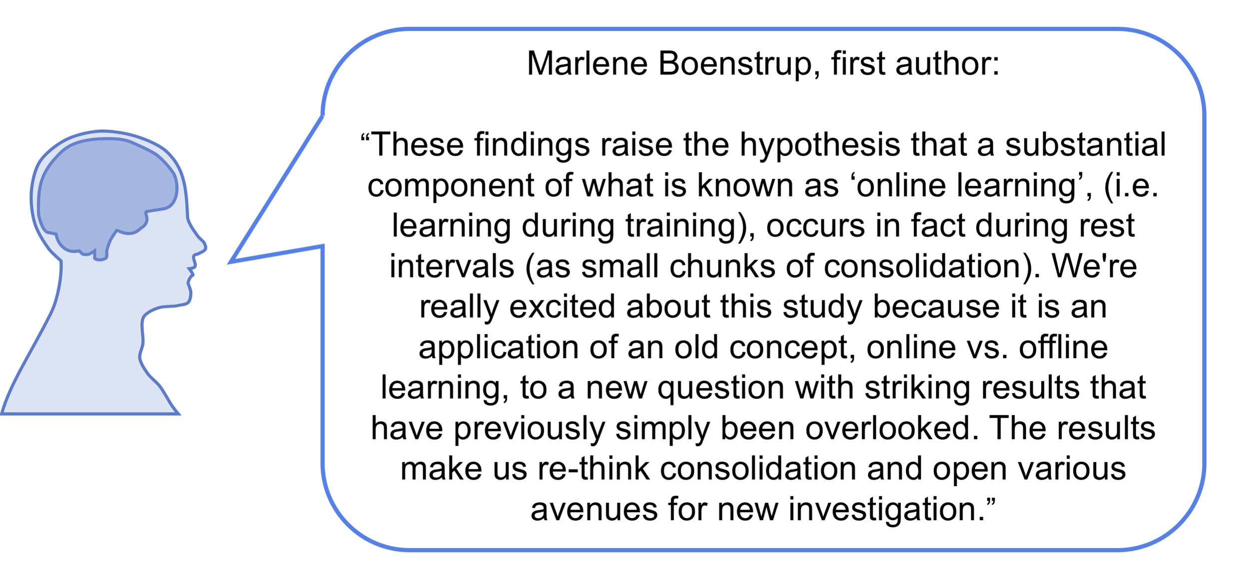
This study is the first to show that a substantial part of early skill learning occurs while we are at rest, and that these improvements can be predicted by activity in the frontoparietal region of our brains. These findings significantly change the way researchers traditionally think about the time it takes us to form a memory and learn new skills—they show this can occur over a period of seconds, rather than hours or days.

current_biology_quote.jpg

Marlene Bönstrup et al. A Rapid Form of Offline Consolidation in Skill Learning. Current Biology (2019). Access the original scientific publication here.

Dopamine Receptor D2 Hippocampal Neurons Regulate Food Intake

Post by Amanda McFarlan

What's the science?

There are a multitude of neuronal populations throughout the brain that have been identified for having a role in controlling appetite, however, it is still not known how these brain regions integrate information from past experiences and the environment to regulate feeding behaviours. The authors hypothesized that the hippocampus may be important for the integration of this information since it is well established as a brain structure that is important for spatial location and memory formation. Further, more recently, it’s its dysfunction has been shown to alter food intake. This week in the Neuron, Azevedo and colleagues investigated the role of the hippocampus is regulating food-related behaviours.

How did they do it?

The authors expressed either an inhibitory (hM4Di) or excitatory (hM3Dq) designer receptor exclusively activated by designer drugs (DREADD) in glutamatergic cells in the dorsal hippocampus of wild type mice. They activated either the inhibitory or excitatory DREADDs by infusion of clozapine-N-oxide (CNO) and observed changes in food intake over a 24-hour period. Next, the authors used PhosphoTrap (which can profile gene expression based on changes in neuronal activation) to measure hippocampal gene expression in fasted mice that underwent a behavioural task where food was presented in a context-specific manner. Next, the authors investigated the role of hippocampal dopamine receptor D2 (Drd2) neurons in food intake by performing bilateral injections of a virus containing either a Cre-dependent inhibitory or excitatory DREADD in the dentate gyrus (region of the hippocampus) in Drd2-Cre mice. They activated the inhibitory and excitatory DREADDs with infusion of CNO and measured food intake acutely (24Hr) and chronically (8 days). Then, they used multiple viral constructs (anterograde and retrograde tracers) to identify synaptic inputs onto hippocampal Drd2 neurons as well as synaptic outputs from hippocampal Drd2 neurons. Next, they targeted Channelrhodopsin-2 (a light-gated cation channel) to glutamatergic neurons in the lateral entorhinal cortex (which projects to hippocampal Drd2 neurons) and implanted optical fibers above the lateral entorhinal cortex terminals in the dentate gyrus (where Drd2 neurons are located). Similarly, they targeted the expression of either Channelrhodopsin-2 or Archaerhodopsin (an inhibitory light-gated ion channel) to hippocampal Drd2 neurons and implanted optical fibers above neuronal terminals located in the septal area. Finally, they measured changes in food intake in response to activation of synaptic inputs onto hippocampal Drd2 neurons or activation/inhibition of synaptic outputs from hippocampal Drd2 neurons.

What did they find?

The authors found that chemogenetic inhibition of hippocampal neurons increased food intake over a 24-hour period, while chemogenetic activation of hippocampal neurons decreased food intake. These data suggest that the dorsal hippocampus contains a population of glutamatergic neurons that are involved in regulating food intake in mice. Next, the authors determined that fasted mice presented with food showed significant enrichment of Drd2 in neurons located in the dentate gyrus compared to mice that were not presented with food, suggesting that hippocampal Drd2 neurons may be involved in food-related behaviours. The authors determined that in fasted mice, both acute and chronic inhibition of Drd2 hippocampal neurons increased food intake, while acute and chronic activation of these neurons decreased food intake. Together, these findings suggest that Drd2 in the hippocampus is important for regulating food intake. The authors found that Drd2 hippocampal neurons receive inputs from neurons in the superficial and middle layers of the lateral entorhinal cortex and send projections to the septal area (medial and lateral septum). Finally, they determined that optical activation of neurons in the lateral entorhinal cortex (projecting to hippocampal Drd2 neurons) and hippocampal neurons (project to septal area) decreased food intake in fasted mice, while optical inhibition of hippocampal Drd2 neurons increased food intake in fed mice. Altogether, these findings suggest that the activation of the lateral entorhinal cortex-hippocampal Drd2 pathway plays an important role in suppressing food intake.

amanda (2).png

What's the impact?

This is the first study to identify that food cues increase the neuronal activity of hippocampal Drd2 neurons. Additionally, the authors revealed that hippocampal Drd2 neurons receive inputs from the lateral entorhinal cortex and send projections to the septal area, forming a circuit that is important for regulating the suppression of food intake. Together, these findings provide a better understanding of the brain circuitry that is critical for mediating food-related behaviours.

Azevedo et al. A Role of Drd2 Hippocampal Neurons in Context-Dependent Food Intake. Neuron (2019). Access the original scientific publication here.

Brain Stimulation Using Light and Sound Reduces Alzheimer’s Pathology and Improves Cognition

Post by Deborah Joye

What's the science?

Alzheimer’s disease is a progressive form of dementia characterized by build-up of amyloid-beta (Aβ) protein plaques between nerve cells and tau protein ‘tangles’ inside of cells. Treatment options are limited, but promising research shows that neural activity can be manipulated to reduce Alzheimer’s pathology. Specifically, Alzheimer’s patients show a reduction in fast electrical ripples throughout the brain called gamma waves. Gamma waves can be induced non-invasively using a light programmed to flicker at a frequency of 40 Hz (gamma entrainment using sensory stimuli or GENUS). Exposure to the flickering light can produce gamma waves in the primary visual cortex, but it also reduces aggregates of Aβ protein, and activates microglia (the brain’s defense cells) to get rid of Aβ protein. But can GENUS be induced by other sensory systems? And can GENUS improve cognition in a model of Alzheimer’s disease? This week in Cell, Martorell and colleagues demonstrate that GENUS can be induced through both auditory and visual stimuli, reduce pathological Aβ and tau proteins, impact brain regions beyond primary sensory regions, and improve cognitive function in a mouse model of Alzheimer’s disease.

How did they do it?

The authors first checked to ensure that GENUS could be induced using an auditory tone by exposing mice to trains of tones repeating at various frequencies while simultaneously recording neural activity in the auditory cortex, hippocampus, and prefrontal cortex. To test whether auditory GENUS could improve recognition and spatial memory in a mouse model of Alzheimer’s disease, the authors employed several behavioral tasks that test hippocampus-dependent memory function: the novel object location and novel object recognition tests, which probe memory for identity or placement of an object, and the Morris Water Maze, which tests spatial memory for the location of a hidden platform. To investigate how GENUS might alter various Alzheimer’s-related proteins and cell-specific characteristics, the authors used immunohistochemistry to label Aβ and tau proteins, as well as other proteins involved with changes to astrocytes and microglia.

What did they find?

The authors found that exposure to a 40 Hz auditory tone induced GENUS in the auditory cortex, hippocampus, and prefrontal cortex. Critically, mice exposed to auditory GENUS performed better than control mice in three distinct hippocampal-dependent memory tasks, suggesting that GENUS can boost hippocampal function. Auditory GENUS also reduced the amount of Aβ protein and the pathological spreading of tau in the auditory cortex and hippocampus. Similar to effects of visual GENUS, auditory GENUS activated microglia, which increased in size and took up more Aβ. Finally, auditory GENUS increased the number of astrocytes, widened blood vessels, and increased a protein which helps to clear Aβ from the brain. When the two types of GENUS were combined, the authors found that microglia clustered around Aβ plaques, generally a precursor to phagocytosis and pathogen removal. Interestingly, auditory plus visual GENUS also reduced amyloid pathology throughout the neocortex, suggesting that inducing gamma oscillations with multiple sensory modalities can result in widespread reduction in Aβ throughout the brain.

Cell_image_March26.png

What's the impact?

This study is the first to show that gamma waves can be induced in regions of the brain using trains of auditory tones. This study builds on previous work, demonstrating that GENUS can be induced using both auditory and visual stimuli. The authors show that auditory GENUS can improve hippocampus-dependent cognitive function in a mouse model of Alzheimer’s pathology, suggesting that GENUS could be used as a non-invasive treatment to improve cognition in Alzheimer’s patients. This research demonstrates that both auditory and visual GENUS can be used to decrease AD pathology, and that multi-sensory GENUS results in unique microglial responses to Aβ protein plaques that might increase their removal from the brain. 

Tsai_quote_March26.jpg

Martorell et al., Multi-sensory Gamma Stimulation Ameliorates Alzheimer’s-Associated Pathology and Improves Cognition. Cell (2019). Access the original scientific publication here.