The Choice to Socialize Prevents Drug Addiction in Rats

Post by Deborah Joye

What's the science?

There are very few  treatment options for human addiction, despite the large amount of animal research on addiction mechanisms. One reason might be because the impact of social cues on drug use is not fully considered. For example, we know that rodents and monkeys will choose a good-tasting food over drugs. But for humans, social rewards are more likely than food rewards to protect against drug use. Research shows that rodents housed in groups are less likely to take drugs in the first place, and less likely to relapse if they have taken drugs in the past. Typically, social environments and drug use in animal studies are controlled by the experimenter. If the rats could control their choice between taking drugs or socializing, what would they choose? This week in Nature Neuroscience, Venniro and colleagues use operant conditioning to demonstrate that rats will choose a social reward over drug use regardless of the type of drug, the strength of drug, how long they’ve been taking drugs, and how long they’ve been abstinent.

How did they do it?

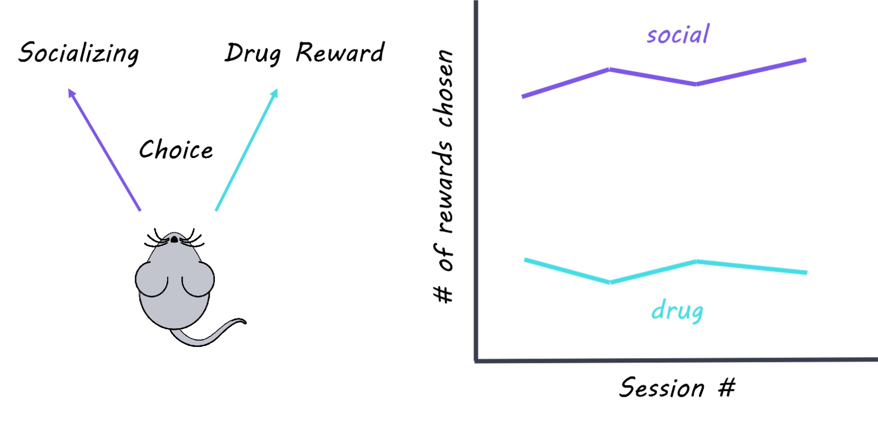
The authors used operant conditioning to train rats so that pressing a certain lever resulted in social time with another rat, while pressing a different lever resulted in an infusion of drug (methamphetamine or heroin depending on the experiment). They also measured how often rats hit an “inactive” lever which didn’t do anything, to make sure that rats weren’t randomly hitting levers. The choice between drugs or socializing was mutually exclusive, meaning that rats could only choose one or the other but not both. The authors performed several manipulations to see how the rats’ choices might change: they administered different drug dosages, progressively increased the time delay between lever pressing and socializing; punished social choice with a small shock; and compared rats based on their motivation to seek the drug (addiction score: high, medium, low). The authors then either removed access to drugs (forced abstinence) or let rats choose (i.e. voluntary) between drug and alternative non-drug rewards (either food or social ) then tested if rats were more or less likely to relapse into drug use. In relapse tests, rats were abstinent from drugs for some period, and then reintroduced to the environment they associated with drug use. Generally, the longer a rat had not had drugs, the more drug-seeking behavior they showed in a drug-related environment, a phenomenon called “incubation of craving.” Finally, to probe the underlying neural mechanisms, the authors tested whether different forms of abstinence (forced v. voluntary) were associated with changes in neuronal activity in brain regions involved in drug-seeking and relapse, such as the amygdala and insular cortex. Specifically, they used immunohistochemistry (which labels proteins) and in situ hybridization (which labels RNA) to visualize which cells were activated during a relapse test.

What did they find?

The authors found that rats trained to take drugs would consistently choose a social reward over drugs regardless of drug dosage and addiction score. Rats returned to taking drugs only when the social reward was delayed or punished. Rats that chose socializing over drug use showed less drug-seeking behavior in a relapse test, even if social choice and access to drugs were removed for a month. This suggests that social reward can be protective against incubation of craving and future relapse. Rats that were given the food reward choice showed more drug-seeking behavior in a relapse test than rats given the social reward choice, suggesting that socializing is more rewarding than food. Lastly, the authors found that rats who chose socializing over drugs had increased activation of inhibitory cells in the lateral portion of the central amygdala (protein kinase C-delta (PKCδ)-expressing cells), and decreased activation of the insular cortex. In contrast, rats that were forced into abstinence showed increased activation of different populations of cells associated with drug craving (somatostatin-expressing cells in the lateral central amygdala and output neurons in the medial central amygdala). These findings suggest that cells in the amygdala are recruited to block activity associated with drug craving and relapse while rats are socializing.

addiction1800.png

What's the impact?

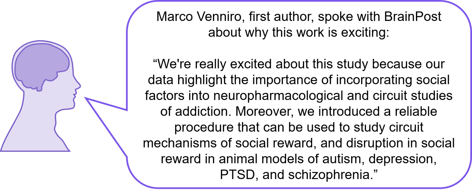
This study is the first to show that when rats are given the choice between socializing or using drugs, rates of drug abstinence are almost 100%. These findings highlight the necessity of considering social factors when investigating the neuroscience of addiction and suggest that social reward is a better drug-alternative model than food reward. Finally, this study demonstrates that positive social interactions protect against addictive behaviors and addiction-related changes in the brain, which has significant implications for clinical treatment options for human addicts.

addictionquote1800.png

Venniro et al. Volitional social interaction prevents drug addiction in rat models. Nature Neuroscience (2018). Access the original scientific publication here

Internal Versus External Processing: How Do Our Brains Make the Switch?

Post by Anastasia Sares

What's the science?

The “default-mode” network is a brain network which is more active at rest than during attention-demanding tasks. It is also known to be involved in internally-directed brain processes, like daydreaming or thinking about past experiences. Another brain network called the dorsal attention network is ‘anti-correlated’ with the default mode network; that is, it tends to be active when the default mode network is not, and vice versa. However, since most of the research on these networks is done using functional MRI (which is not the most temporally precise brain imaging method) the exact timing of activation of these networks remains uncertain: does the default mode deactivate before the dorsal attention network comes online, or after? This week in The Journal of Neuroscience, Raccah and colleagues used intracranial electrical recordings to find out when different networks become active, and whether certain network changes precede others in time.

How did they do it?

This study made use of intracranial recordings, a method using electrodes implanted directly on the surface of the brain, to record activity. These recordings only happen when a patient already has the electrodes in place because they are about have brain surgery (in most cases, surgery for epilepsy). The signal from the electrodes corresponds to the firing of many neurons (in this case, high-frequency broadband power was measured). The authors enrolled six epileptic patients who had electrodes covering parts of the default mode network (DMN) and the dorsal attention network (DAN), so they could track the activation of both network simultaneously. They administered a task in which participants were required to answer “True” or “False” to memory questions (like “I ate fruit yesterday”) and math questions (like “48 + 9 = 57”). Since memory questions activate internally-focused processing, and math questions activate networks for outward attention and problem-solving, the questions were assumed to tap into the DMN and DAN respectively. After discarding the signal from bad electrodes, the authors identified some electrodes that responded preferentially to the math or memory questions, some that responded to both, and some that deactivated while answering math questions. They compared the timing of electrode responses to the True-False questions, noting when they began to respond and when the electrodes reached their peak activity.

What did they find?

Even though participants took longer to respond to the math questions, the neurons in the DAN responded much more quickly than other neurons. The time to peak neuron firing for the dorsal attention network was around 450-500 milliseconds after seeing the True-False question, while the DMN seemed to reach its peak around 650-800 milliseconds after seeing the question. These results show that the DAN becomes active prior to any response from the DMN. This pattern was replicated in both the onset of the response and the peak of the response. The results suggest that the DAN activation precedes the DMN deactivation in time.

omri1800.png

What's the impact?

This study found that changes in the DMN occur after changes in the DAN using intracranial recordings, which are more temporally precise than functional MRI. This means that activity in the DMN (which drives internally driven processes) does not cause activity in the DAN, but the reverse may be true. These results suggest that your internally focused brain network decreases in activity as you pay attention. Understanding the role of the DMN will be crucial for understanding how it can affect cognitive function, and disorders of cognition.

omriquote1800.png

Raccah et al. Direct Cortical Recordings Suggest Temporal Order of Task Evoked Responses in Human Dorsal Attention and Default Networks.The Journal of Neuroscience (2018). Access the original scientific publication here

Chronic Sleep Disruption Worsens Alzheimer’s Pathology and Cognition in Mice

Post by Thomas Brown

What’s the science?

Tau, a protein which accumulates in the brain in Alzheimer’s, is one of the primary pathologies of Alzheimer’s disease and correlates well with disease progression. Previous studies have shown that tau inclusions often start in particular areas of the brain, eventually spreading throughout the brain over time. One of these brain regions is the locus coeruleus (LC) (a small brainstem nucleus) known to regulate wakefulness and stress by releasing norepinephrine in the brain. Locus coeruleus neurons are also known to be damaged during chronic sleep disruption, however, the role of chronic sleep disruption in tau protein aggregation in the brain remains unknown. This week in the The Journal of Neuroscience Zhu and colleagues assessed whether chronic sleep deprivation in a mouse model of tauopathy leads to an increased progression of tau pathology.

How did they do it?

The authors used P301S mice, a strain of transgenic mice that express human tau. In this mouse, a human version of the MAPT disease-associated P301S mutation is inserted into chromosome 3 of the mouse, which results in human P301S tau being expressed. In P301S mice, the authors induced one of two models of chronic sleep disruption. The first model involved placing the mice in a new environment during a lights-on period (a period when the mouse normally sleeps) while the second involved placing the mouse cage upon a rotor, repeatedly nudging mice awake resulting in increasing arousals. The use of two different methods allowed the authors to determine whether mice were simply reacting to a new environment versus responding to sleep deprivation. The chronic sleep disruption mice were compared to a control group of “rest” mice. After chronic sleep deprivation the mice were given a number of behavioural tests over time to assess motor impairment and spatial memory. The locus coeruleus and amygdala (the brain’s ‘fear centre’) were then examined for tau protein accumulation, gliosis, and signs of neuronal death.

What did they find?

The authors found that by 7 months of age (after 3 months of chronic sleep disruption) P301S mice with an irregular sleep pattern performed worse than P301S “rest” mice which had been able to rest. This motor impairment worsened over time between 5 and 7 months. In an open field test, where the locomotor activity of mice in a novel environment is monitored, mice with sleep disruption showed heightened activity in the first two minutes, which could denote anxiety. The authors also utilized the ‘Novel object recognition test’ as a spatial memory test, in which a mouse is habituated to an object, and then presented with a familiar and an unfamiliar (i.e. novel) object. A healthy mouse will normally inspect the novel item. P301S mice that underwent chronic sleep disruption had a reduced preference for the novel object, indicating that their memory was impaired compared to rested mice. Immunohistochemical analysis showed an increase in tau protein accumulation in mice with chronic sleep disruption, including soluble and tangled tau, as well as increased neuronal death in both the amygdala and the locus coeruleus. Additionally, there was also increased activation of glial cells in the mice with disrupted sleep at 7 months, which is a common finding in many neurodegenerative conditions. Taken together, these results indicate that in a model of tauopathy, chronic sleep disruption earlier in life can lead to increased accumulation of tau as well as motor and cognitive impairments.

sleep1800.png

What’s the impact?

This study presents evidence that all aspects of tauopathy are worsened by chronic sleep disruption in early life. Future work is need to understand whether these findings can translate to humans. Understanding the effects of sleep on neurodegenerative conditions could inform potential lifestyle interventions, or identify groups of high risk individuals. Further, studying the link between sleep disruption that occurs in some disorders (e.g. sleep apnea) and disease later on, is important for understanding vulnerability to neurodegenerative diseases.

sleepquote1800.png

Zhu et al. Chronic Sleep Disruption Advances the Temporal Progression of
Tauopathy in P301S Mutant Mice. The Journal of Neuroscience (2018).Access the original scientific publication here