In Vivo Imaging of REM Sleep Behavior Disorder

What's the science?

Rapid Eye Movement (REM) Sleep Behavior disorder often precedes the development of Parkinson’s disease, which is characterized by the accumulation of the protein alpha-synuclein inside of neurons. Recent research suggests that alpha-synuclein first accumulates in peripheral autonomic neurons (in the gut) and in the olfactory bulb (in the brain). Alpha-synuclein is hypothesized to then spread to the brain via autonomic nerve fibres, causing further damage via neuron-to-neuron transmission from peripheral neurons through the spinal cord and brainstem to the brain. This week in The Lancet Neurology, Knudsen and colleagues imaged pathology in the periphery and in the brain of multiple affected systems in patients with REM Sleep Behavior Disorder to better understand how alpha-synuclein might progress prior to the onset of the classical motor symptoms in Parkinson’s disease. 

How did they do it?

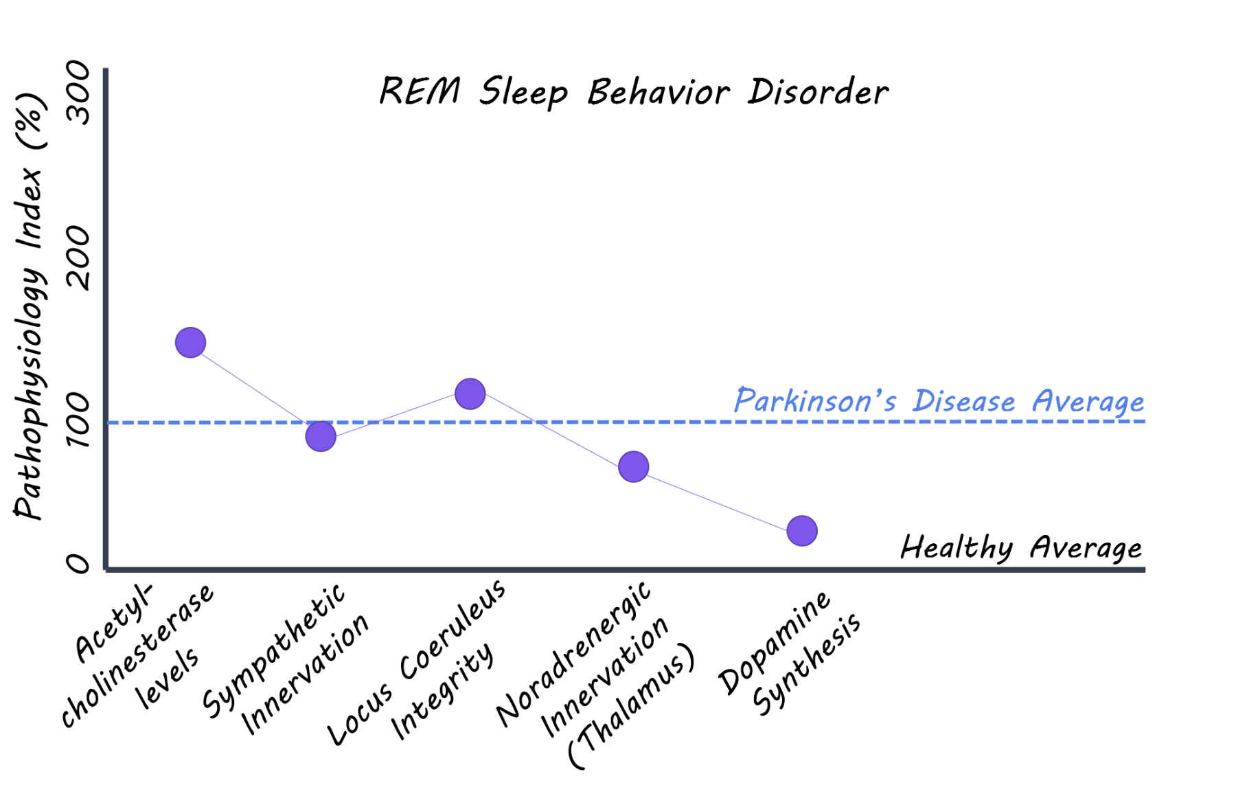
Patients with REM sleep behavior disorder (with no signs of parkinsonism or dementia), patients with diagnosed Parkinson’s disease, and a healthy control group all aged 50-85 years were recruited for the study and underwent a series of imaging scans. 11C donezepil (a radiotracer that measures acetylcholinesterase concentrations) PET imaging was used to assess the level of cholinergic innervation in the gut reflecting, in part, parasympathetic nerve fibres. 123I-MIBG (radiotracer measuring cardiac innervation) scintigraphy was used to measure the level of sympathetic innervation of the heart. Neuromelanin sensitive MRI was used to evaluate the integrity of neurons in the locus coeruleus (brainstem nucleus). 11C-methylreboxetine (a radiotracer that binds to noradrenaline transporters) was used to measure the level of noradrenergic nerve terminals in the locus coeruleus projections to the thalamus. Lastly, 18F-DOPA PET (a radiotracer that measures dopamine synthesis) was used as a measure of dopamine neuron integrity in the brain. Imaging measures between REM sleep behavior disorder and control groups were compared. Imaging multiple systems — from the peripheral autonomic nervous system through to the brainstem — allowed for an assessment of the degree of damage to these important neuronal systems.

What did they find?

Patients with REM sleep behavior disorder had significantly less cholinergic innervation in the small intestine and the colon compared to the healthy control group, while there was no significant difference between REM sleep behavior disorder and Parkinson’s disease patients. Patients with REM sleep behavior disorder also showed significantly lower cardiac innervation compared to controls and lower neuron integrity in the locus coeruleus (brainstem), while again no difference was seen when compared to the Parkinson’s disease group. Locus coeruleus neuron innervation of the thalamus was lower in REM sleep behavior patients compared to controls, but no difference was seen in any other comparison. In contrast, the dopamine synthesis capacity measured by 18F-DOPA PET was normal in 70% of REM sleep behavior patients, while all patients with Parkinson's disease show markedly reduced dopaminergic function in the striatum (the cause of their motor symptoms). Combined, these observed differences support the hypothesis that the autonomic nerve fibres of the gut and other internal organs are affected many years prior to the Parkinson's disease diagnosis. The finding also supports that the initial alpha-synuclein damage in Parkinson's disease may originate in the gut and spread via the autonomic nervous system to the spinal cord and brainstem. 

REM Sleep Behavior Disorder

What's the impact?

This is the first comprehensive imaging study of patients with REM sleep behavior disorder (prodromal Parkinson’s disease) to assess damage in vivo to the autonomic nervous system and multiple brainstem systems. The study supports the hypothesis that alpha-synuclein pathology may initially form in peripheral organs and spread neuron-to-neuron via the autonomic nervous system to the brainstem. If Parkinson's disease pathology truly originates in nerve terminals of peripheral organs, different strategies of neuro-protection might be possible, including the use of drugs which do not cross the blood-brain barrier or modification of the intestinal microbiome. 

remquote1800.png

Knudsen et al., In-vivo staging of pathology in REM sleep behaviour disorder: a multimodality imaging case-control study. The Lancet Neurology (2018). Access the original scientific publication here.
 

One Salience Network, Two Functions?

What’s the science?

The anterior insula and dorsal anterior cingulate cortex (dACC) are two brain regions that are often active together as part of the brain’s ‘salience network’. A ‘salient’ stimulus is one that is able to capture our attention easily. The anterior insula is active during many cognitive and emotional processes - this has been assumed to be due to its role in orienting attention towards salient stimuli. However, the role of the anterior insula and dACC in specific aspects of the attention-capturing process in the anterior insula and dACC have not actually been delineated. This week in Cerebral Cortex, Han and colleagues performed two experiments using both emotional and emotionally neutral events to assess the functional roles of these two brain regions in salience and emotion.

How did they do it?

Experiment 1: Fifteen healthy young adults participated. While performing functional magnetic resonance imaging (fMRI), the authors had participants look for ‘target’ images of a dining or living room which were presented in rapid succession (sequentially) among other (distraction) pictures on a screen (144 images total per trial, 135 trials). Participants were asked to press one of two buttons when they saw a living room or dining room. In a few trials, a 10-second movie was presented instead of distraction pictures. This movie was either emotional (e.g. of a person in pain or a spider on an arm), or emotionally neutral (e.g. waves or swirls). This task can help to measure the brain’s involvement in processing salient and behaviourally relevant stimuli.

Experiment 2: Fourteen healthy young adults participated. While performing fMRI, a stream of digits was presented in rapid succession, interspersed with letters indicating to the participant had to perform one of two tasks: either to judge either whether the number was odd or even or to judge whether the digit was smaller or bigger than 4 (indicated with a button press). Other letters were also interspersed between digits to indicate whether to stay on the same task (‘hold’ cue) or switch to the other task (‘switch’ cue). This task can help to measure the brain’s involvement in attention switching.

In both experiments, the authors compared brain activation between conditions/tasks.

What did they find?

Experiment 1: As expected, trials in which video clips were presented lowered performance (reaction time), likely because they captured attention and distracted from the task (finding the target - living room or dining room scene). When the fMRI response was examined, the authors found that both the anterior insula and dACC were active at the onset and offset of the emotionally neutral and emotional stimuli (video clips). However, the anterior insula only was also active throughout the presentation of the emotional clips. This indicates that the anterior insula is not simply active during relevant changes in the environment (at the beginning and end of the clip). A smaller sustained response was also found in two other brain regions; the thalamus and putamen. Overall, the authors suggest that the anterior insula and dACC are active when attention is captured by behaviourally significant events.

Experiment 2: The authors found that the dACC was more active during switch cues than hold cues, while there was no such difference for the anterior insula. Therefore, the dACC may be involved in attention set switching/goal directed behaviour (i.e. updating), while the anterior insula is involved in detection of behaviourally relevant events. In a whole brain analysis, another brain region activated by ‘switch’ cues was the medial superior parietal lobe.

fMRI signal  in anterior insula and anterior cingulate cortex

What’s the impact?

This study demonstrates, via fMRI, the different functional roles of the anterior insula and dACC in fine grain detail. The anterior insula is active during behaviourally relevant events - and the dACC is active during attention switching. We now know more about how these regions function within the brain’s salience network.

saliencequote1800.png

S.W. Han et al., Functional Fractionation of the Cingulo-opercular Network: Alerting Insula and Updating Cingulate. Cerebral Cortex (2018). Access the original scientific publication here.

A Molecular Network for Cognitive Decline in the Human Prefrontal Cortex

What's the science?

There are currently no therapies available to treat or prevent Alzheimer’s disease. This may be due to the complexity and heterogeneity of the disease. Although we know that the accumulation of beta-amyloid peptides and tau proteins occurs in the brain in Alzheimer’s disease and that certain susceptibility genes are involved, we do not understand the sequence of events that lead from genetic risk to brain pathology and cognitive decline. Brain gene expression data and network based analysis can  potentially gather nuanced information about the complex interactions among genes that lead to brain pathology. This week in Nature Neuroscience, Mostafavi and colleagues use network-based analysis of gene expression data from the aging prefrontal cortex to elucidate a network involved in aging and Alzheimer’s disease. 

How did they do it?

Prefrontal cortex RNA-sequencing (RNA-seq) data (gene transcript levels representing gene expression) from participants in longitudinal datasets of aging called the Religious Orders Study (ROS) and the Memory and Aging Project (MAP) were used. These datasets also contain post-mortem brain pathology data and longitudinal measures of cognitive performance. They ran a standard association analysis to look for genes whose expression levels associate with Alzheimer’s disease and cognitive decline. They then used an approach called gene module-trait network analysis that links key networks of genes that are co-expressed (related expression patterns) to cognitive decline and Alzheimer’s disease traits, and then selects the most strongly and directly associated networks. The goal of this was to gain more information on gene networks and their associated biological pathways than can be obtained from analyzing single gene-disease associations.

What did they find?

The known risk variant for Alzheimer’s disease in the APOE gene (strongest Alzheimer's risk gene) only explained 2.2% of the variance in Alzheimer’s disease (heterogeneity) and 5.1% of cognitive decline. When examining 21 other known Alzheimer’s risk variants, they only explain 2.1% of disease variance and 7.6% of cognitive decline, emphasizing that these genes alone do not explain disease heterogeneity and decline. In the gene module-trait network analysis, 47 modules of genes (from the RNA-seq gene expression data) were identified representing related networks of genes. 11 of these modules were associated with cognitive decline or Alzheimer’s disease related traits (beta-amyloid or tau), and they found that these modules replicated in their association (with Alzheimer’s disease) in an independent gene expression dataset.

Connections between gene networks and Alzheimer’s disease traits

They then used Bayesian network inference to determine direct gene module-trait associations while accounting for the heterogeneity of cell types in the brain (neurons, astrocytes etc). One module (module number 109) consisting of 390 genes involved in regulation of cell cycle and chromatin modification was most strongly associated with cognitive decline. They found that this module was strongly associated with beta-amyloid pathology. They then selected key genes from this module that were strongly expressed in neurons and astrocytes and showed the strongest gene-disease associations. They performed a knockdown experiment on these genes in astrocytes and stem cell derived neurons and found that knocking down 2 of these genes, INPPL1 (involved in lipid signalling) and PLXNB1 (involved in synaptic plasticity) significantly lowered beta-amyloid levels. INPPL1 explained 5.5% of variance in cognitive decline, while PLXNB1 explained 4.4%. They confirmed that these two genes drive a significant proportion of the effect of the m109 module (which explained 8.5% of cognitive decline) on beta-amyloid load, indicating that these two genes may be important for amyloid pathology and cognitive decline.

What's the impact?

This is the first study to use a network-based approach to identify direct gene network associations with Alzheimer’s disease traits and cognitive decline. This study identified a network of genes strongly associated with beta-amyloid load and cognitive decline, which are important measures of disease progression. This study demonstrates that a network-based approach can provide more information on networks of genes associated with cognitive decline that single gene associations might miss.

philquote1800.png

Mostafavi et al., A molecular network of the aging human brain provides insights into the pathology and cognitive decline of Alzheimer’s disease. Nature Neuroscience (2018). Access the original scientific publication here.