Mild Traumatic Brain Injury Increases Risk of Parkinson’s Disease

What's the science?

Mild traumatic brain injury, also referred to as a concussion, is defined as a loss of consciousness or confusion due to head injury that lasts less than 30 minutes. Mild traumatic brain injury is especially common among the elderly, athletes and in the military, and is increasingly associated with risk for psychiatric and neurodegenerative diseases. The relationship between mild traumatic brain injury and risk for Parkinson’s disease is not well understood. This week in Neurology, Gardner and colleagues assess the risk of Parkinson’s disease after mild traumatic brain injury in a large sample of patients.

How did they do it?

Participants (18 years or older) from the Veterans Health Association (military veterans) with a diagnosis of traumatic brain injury were included and were age matched to a random sample of participants without brain injury (325,870 total participants). They diagnosed Traumatic brain injury using detailed clinical assessments or criteria from the Department of Defense (IC-9 codes). They also assessed other diseases at baseline and Parkinson’s disease at least one year after baseline in this longitudinal study. They then used a Cox Proportional Hazards model (a standard statistical model for assessing longitudinal risk) to determine risk of developing Parkinson’s disease with and without traumatic brain injury, including assessment of risk specifically after mild traumatic brain injury. They controlled for confounding variables including medical conditions, psychiatric disease and demographics: age, sex, ethnicity, education and level of income.

What did they find?

A total of 1462 participants developed Parkinson’s disease over the course of the study (average 4.6 years follow-up), 65% of whom had previously experienced traumatic brain injury. Participants with previous traumatic brain injury were significantly more likely to develop Parkinson’s disease than those without traumatic brain injury (hazards ratio: 1.71). Even mild traumatic brain injury was associated with increased risk (56%) of developing Parkinson’s disease after adjusting for all confounding variables.

Relationship between Traumatic Brain Injury and Parkinson’s disease

What's the impact?

This is the first large-scale, nationwide study to demonstrate that mild traumatic brain injury is associated with an increased risk of Parkinson’s disease. We now know that focusing on the prevention of mild traumatic brain injury will be important for reducing risk of Parkinson’s disease.

itbiquote.png

R. Gardner et al., Mild TBI and risk of Parkinson disease: A Chronic Effects of Neurotrauma Consortium Study. Neurology (2018). Access the original scientific publication here.

Stimulating an Entorhinal Cortex Circuit is Antidepressive

What's the science?

Major depressive disorder can lead to many structural and functional changes in  the hippocampus, including slowed neurogenesis (new neuron formation). Stimulation of the entorhinal cortex (which sends information to the hippocampus) can improve learning and memory. It is possible that entorhinal cortex stimulation could also relieve depression. Current depression therapies, including transcranial magnetic stimulation and electroconvulsive therapy, often have side effects, so more treatment options are needed. This week in Nature MedicineYun and colleagues test whether stimulating an entorhinal circuit is antidepressive in mice and uncover the mechanisms involved.  

How did they do it?

They knocked down a protein subunit (TRIP8b) of a protein channel (hyperpolarization channel) using a viral-mediated approach which reduces the number and sensitivity of these protein channels found on neurons. This increases the excitability of neurons in the entorhinal cortex. The hypothesis was that increased excitation in the entorhinal cortex would increase neurogenesis in the hippocampus, and in turn reduce depressive behavior in mice. They performed several experiments: They tested whether psychosocial stress (a model of depression) increases TRIP8b levels in neurons. Using viral-mediated TRIP8b knockdown, they confirmed the increased excitability of entorhinal cortex neurons that project to the hippocampus (dentate gyrus). They measured neurogenesis in the hippocampus (dentate gyrus) in TRIP8b knockdown and controls. They then tested whether antidepressive behavior and memory were changed in knockdown vs. controls and whether this was dependent on neurogenesis. Lastly, they used gene transfer and transgenic mice combined with chemogenetics to activate glutamatergic neurons (i.e. excitatory neurons) in the entorhinal cortex, in order to observe the effects on antidepressive behavior.

What did they find?

They found that psychosocial stress in mice resulted in increased levels of TRIB8b in entorhinal neurons that project to the dentate gyrus. After knockdown of TRIP8b, they found increased excitability of entorhinal cells and increased neurogenesis in the connected hippocampus (in the dentate gyrus), confirming the hypothesis that increased activity in the entorhinal cortex results in neurogenesis in the hippocampus. TRIP8b knockdown in the entorhinal cortex also resulted in antidepressive-like behavior and improved memory in mice. To test whether this was dependent on neurogenesis in the hippocampus, they used X-ray irradiation to ablate new neurons and found that antidepressive behavior was dependent on hippocampal neurogenesis. Using chemogenetics to chronically stimulate glutamatergic neurons in the entorhinal cortex, they found that glutamatergic neurons drove neurogenesis in the hippocampus and were responsible for antidepressive behavior.

Dentate gyrus neurons

What's the impact?

This is the first study to show that activity in the entorhinal-hippocampal circuit results in both the formation of new neurons in the hippocampus and antidepressive behaviors in mice. Altering activity in this entorhinal cortex circuit - previously appreciated only as a memory circuit - by stimulating it could be a new way to reduce symptoms of depression in humans.

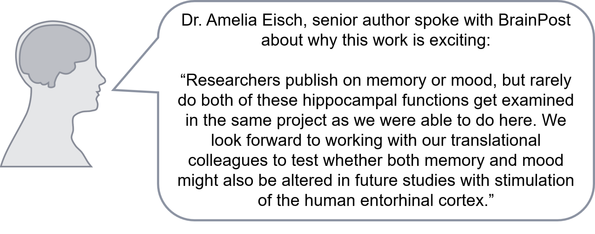
ihippoquote.png

S. Yun et al., Stimulation of entorhinal cortex–dentate gyrus circuitry is antidepressive. Nature Medicine (2018). Access the original scientific publication here.

Dopamine Synthesis Predicts Treatment Response in Patients with Psychosis

What's the science?

One type of medication that can help patients with schizophrenia and other forms of psychosis is dopamine antagonists (medications that block the neurotransmitter dopamine), however, not all patients respond well to this medication. Whether or not a patient responds may be related to dopamine synthesis capacity, whereby patients with high levels of dopamine may respond while those with low levels of dopamine do not. This week in Molecular Psychiatry, Jauhar and colleagues studied patients experiencing a first episode of psychosis, to understand whether differences in dopamine synthesis capacity were related to future treatment response.

How did they do it?

Twenty-six patients who had recently experienced a first episode of psychosis and were diagnosed with a psychosis disorder participated, along with 14 healthy controls. Psychosis symptoms were assessed using the Positive and Negative Syndrome Scale (PANSS) before treatment, 4 weeks into treatment, and at 6 months follow-up. Response was defined as a 50% reduction in PANSS score from baseline. Participants underwent a positron emission tomography (PET) scan at baseline after injection of 18F-DOPA, in order to measure dopamine synthesis capacity in the striatum (using the ‘striatal influx constant’).

What did they find?

At baseline, the striatal influx constant in the associative striatum (a region of the striatum involved in cognitive function) was higher in responders compared to non-responders and healthy controls, indicating dopamine synthesis capacity was higher in this group. Dopamine synthesis capacity was positively correlated with percent change in PANSS score, indicating those with higher synthesis capacity were more likely to experience fewer psychosis symptoms after treatment. Higher dopamine synthesis capacity was also found in responders in two specific parts of the associative striatum: in the caudate (compared to healthy controls & non-responders) and in the putamen (compared to non-responders).

Brain, Servier Medical Art, image by BrainPost, CC BY-SA 3.0

Brain, Servier Medical Art, image by BrainPost, CC BY-SA 3.0

What's the impact?

This study is the first to find that dopamine synthesis capacity (i.e. dopamine level) in the striatum is higher in individuals who respond well to treatment after a first episode of psychosis. PET imaging to measure dopamine synthesis capacity could be used to help predict who will respond well to treatment for psychosis.

ipsychosisquote.png

S. Jauhar et al., Determinants of treatment response in first-episode psychosis: an 18F-DOPA PET study. Molecular Psychiatry (2018). Access the original scientific publication here.